Home » Anatomy » Page 4

Anatomy

Iliocostalis – Functional Anatomy

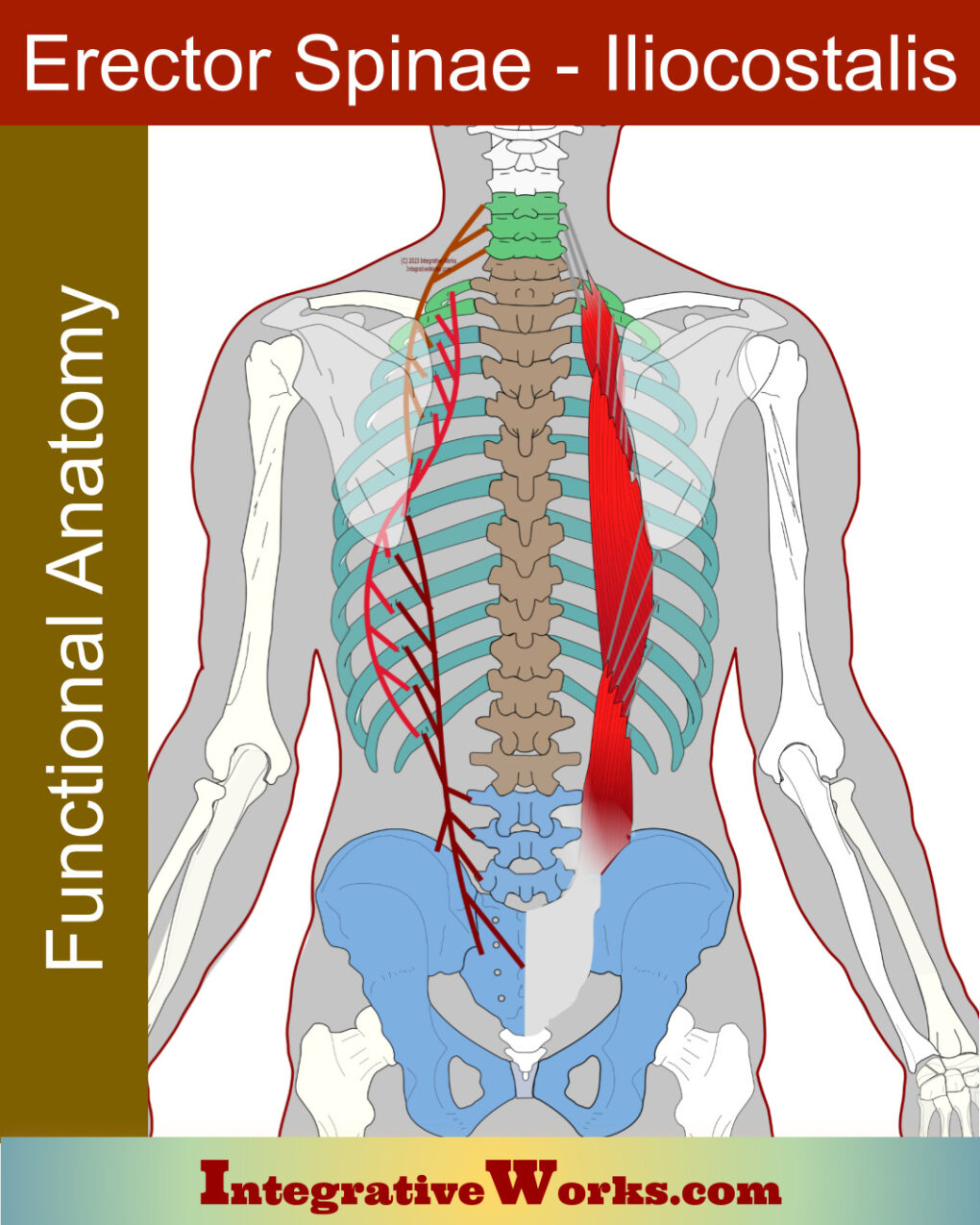
Overview of anatomy Iliocostalis is part of the erector spinae group, which also includes longissimus and spinalis. Iliocostalis, like the others, has 3 sections. The anatomy is often simplified. That’s understandable as it is both complex and variable. All 9 sections of the erector spinae are gathered into a fascial compartment that extends along the […]

Iliocostalis – Functional Anatomy Read More »

Anatomy for Massage and Bodywork

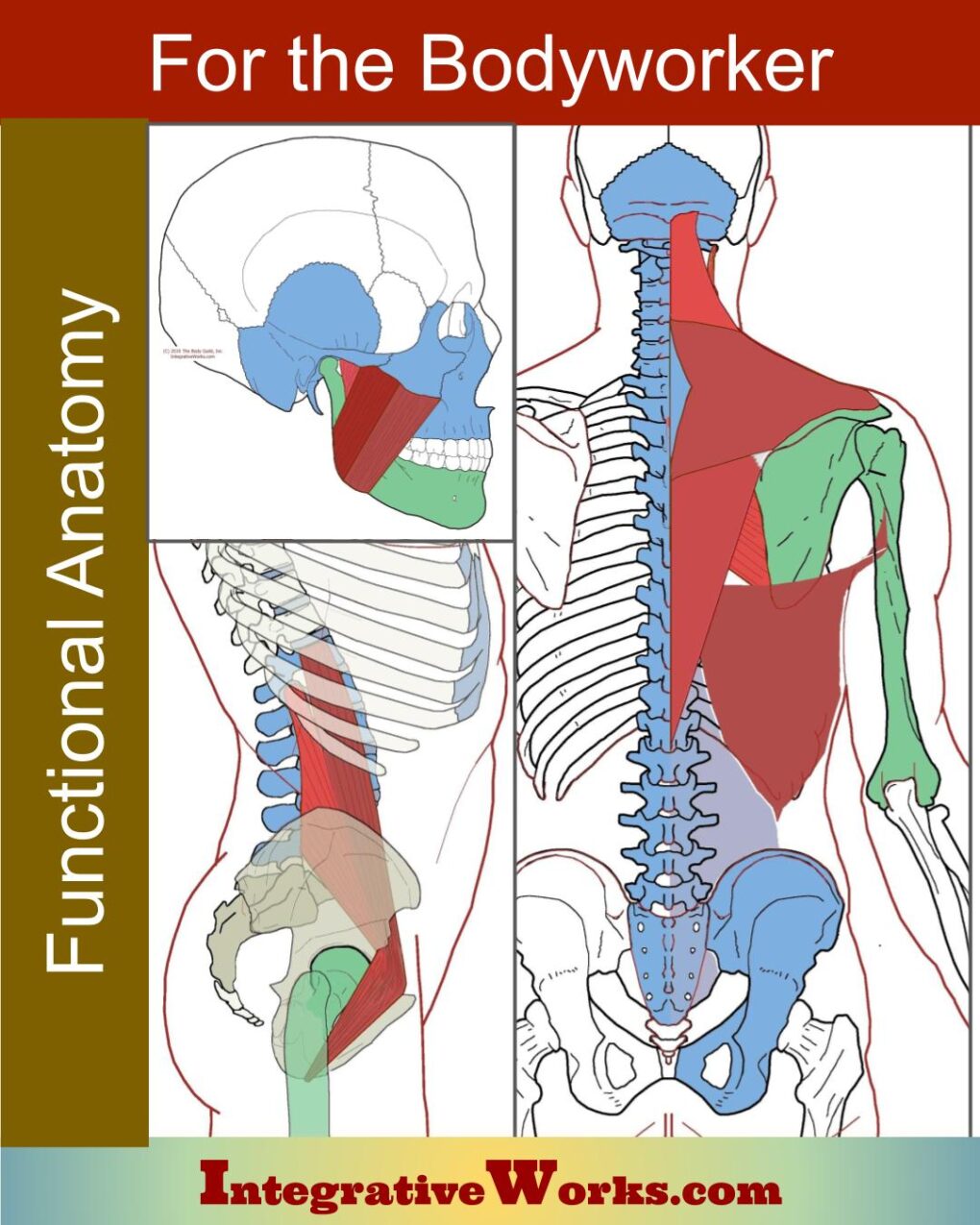
Anatomy for Massage and Bodywork is a collection of posts with illustrations that focus on relationships that interest the massage and bodywork practitioner. Understanding the relationship between muscles and their local joints is critical in effective Integrative Bodywork. Each muscle has illustrations that highlight the bones of attachments, and the bones trapped between the attachments.

Anatomy for Massage and Bodywork Read More »

Occipitalis – Functional Anatomy

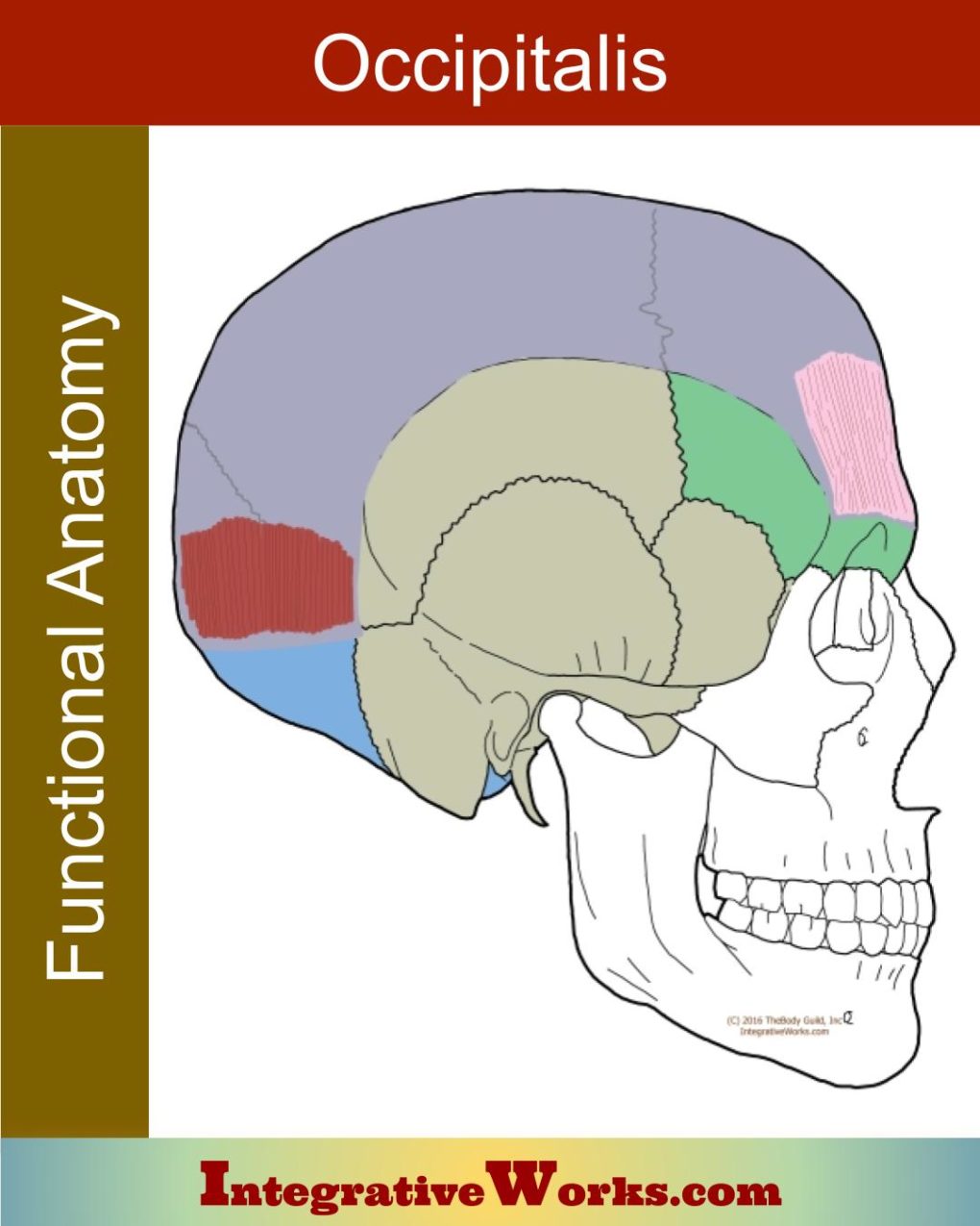
Overview Occipitalis is part of a larger structure called the epicranius or occipitofrontalis muscle. The epicranius consists of the frontalis muscle and the occipitalis muscle, which are joined by a fascial structure called the galea aponeurotica. Origin Insertion Function Innervation The occipitalis retracts the scalp, which pulls the hairline back and raises the eyebrows without

Occipitalis – Functional Anatomy Read More »

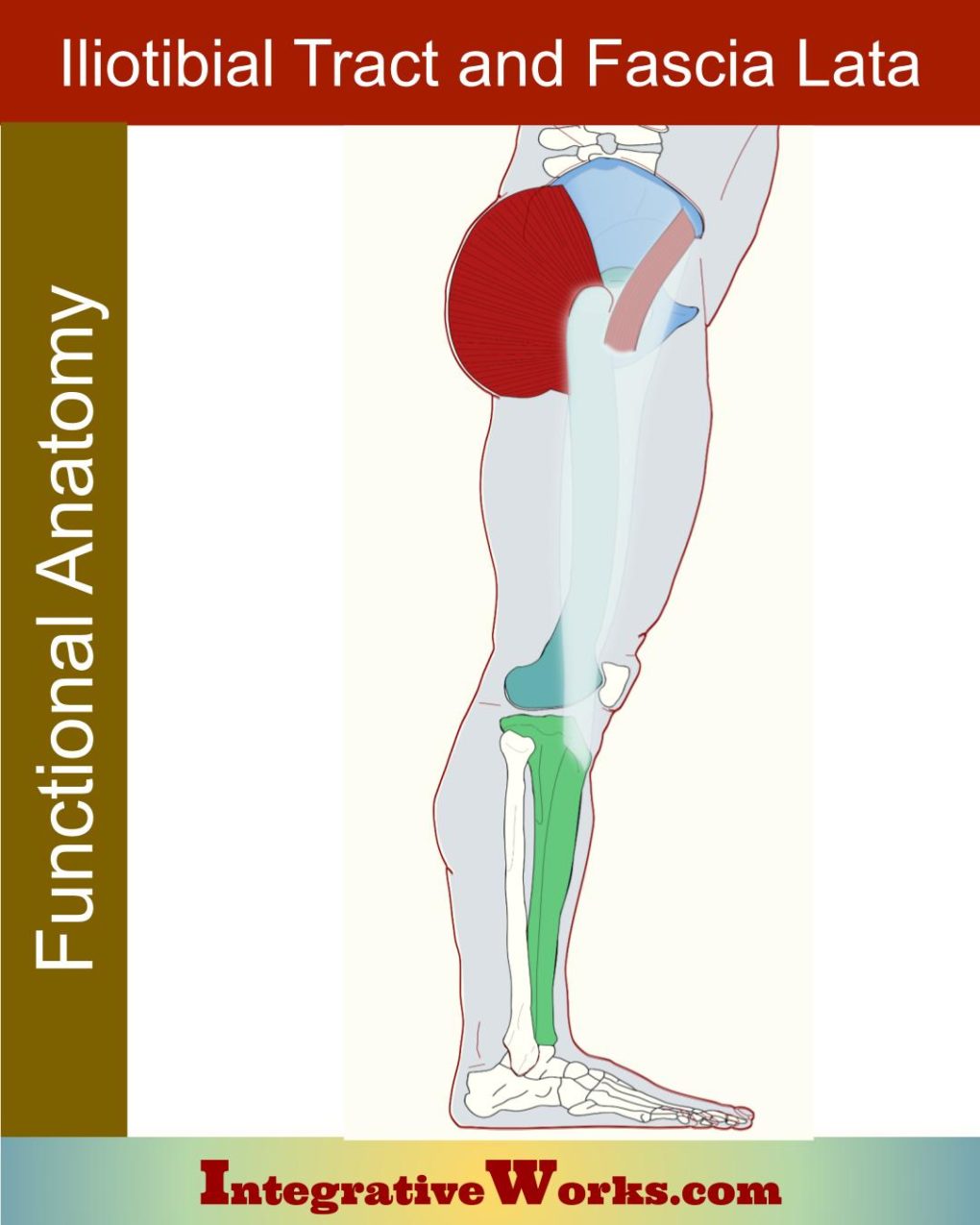
Iliotibial Tract and Fascia Lata – Functional Anatomy

Fascia Lata The fascia lata is the deep fascia that wraps the outer thigh. This thick fascial envelope attaches superiorly to the crest of the Ilium and inguinal ligament. The fascia continues onto the pubic rami, ischial tuberosity, sacrotuberous ligament, coccyx, and sacrum. Distally, it thickens as it attaches to the proximal tibia. It offers

Iliotibial Tract and Fascia Lata – Functional Anatomy Read More »

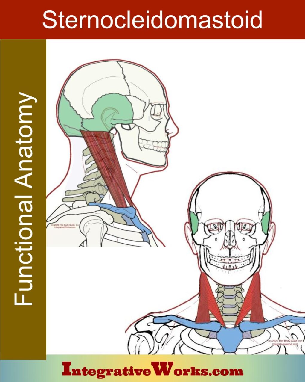
Sternocleidomastoid – Functional Anatomy

Sternocleidomastoid Anatomy. The sternocleidomastoid is a two-headed muscle that extends obliquely along the lateral neck. Origin: The sternal head attaches to the superior aspect of the manubrium. The clavicular head attaches to the medial third of the clavicle. Insertion: Both heads merge and attach to the mastoid process of the temporal bone and lateral third

Sternocleidomastoid – Functional Anatomy Read More »

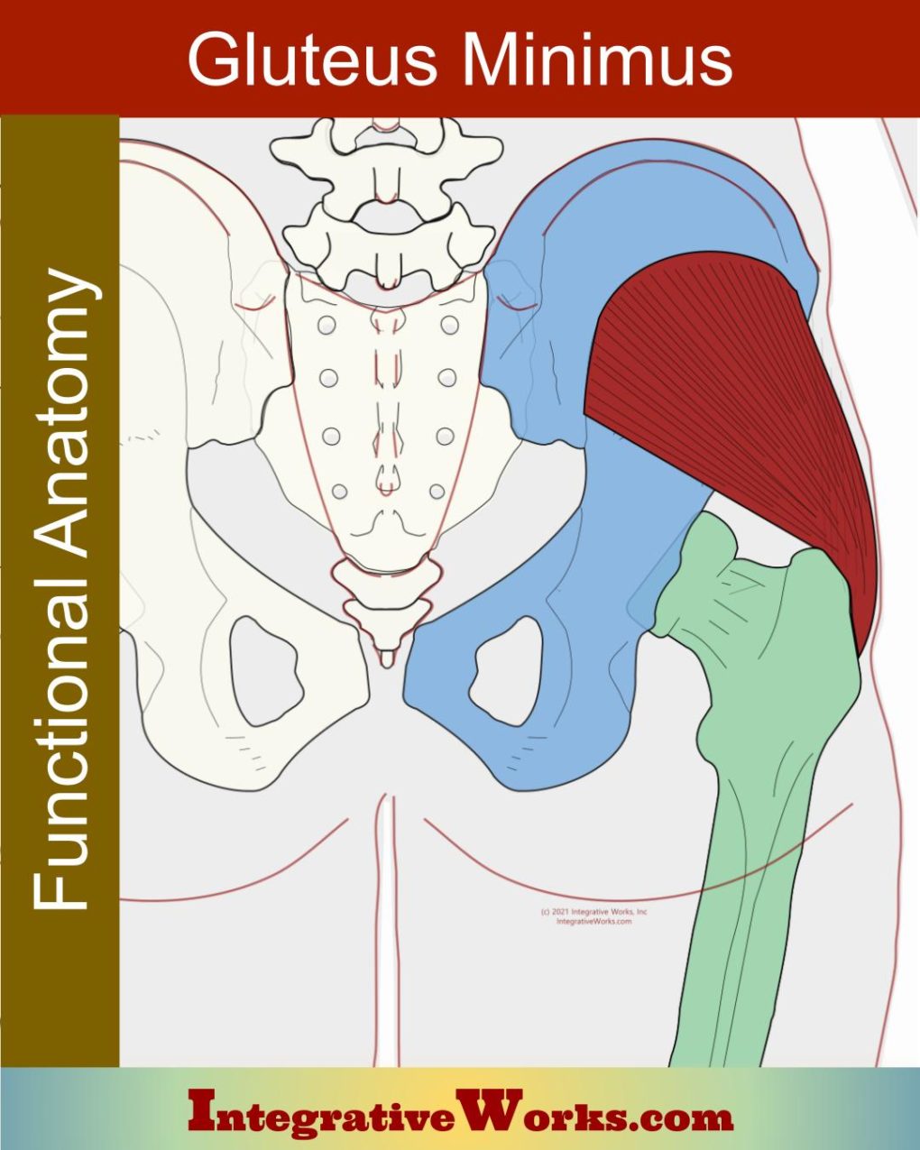
Gluteus Minimus – Functional Anatomy

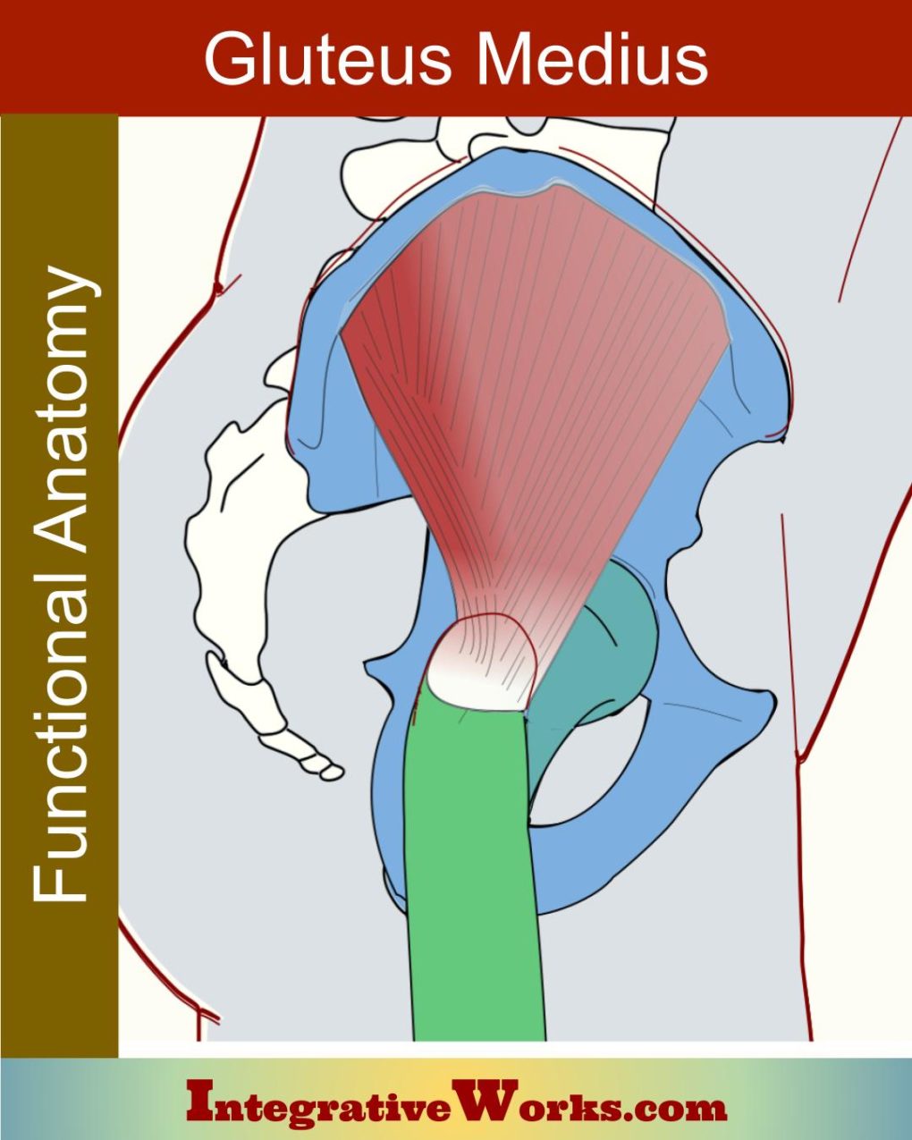
Here, you will find an overview of the anatomy of the gluteus minimus muscle and links to related posts. It has two distinct heads. Origin Insertion Function It has a strong relationship with adductors in stabilizing the pelvis’s lateral motion, rotation, and elevation. Innervation Piriformis and gluteus maximus trap the os coxae between the femur

Gluteus Minimus – Functional Anatomy Read More »

Pelvic Ligaments – Functional Anatomy

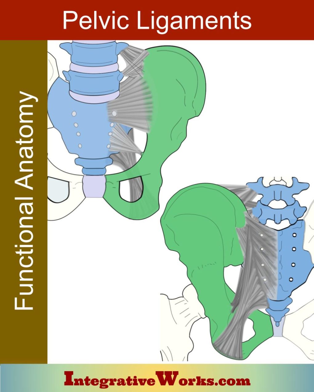
Here, I explore the anatomy of the pelvic ligaments, their structure, attachments, and how they mature through the decades of a person’s life. Overview These pelvic ligaments bind the ox coxae of the lower extremity to the axial skeleton. They consist of: Progression of Fusion A complex fascial structure wraps the major bony structures of

Pelvic Ligaments – Functional Anatomy Read More »