Home » Anatomy » Page 8

Anatomy

Coracobrachialis – Functional Anatomy

  The coracobrachialis is a long muscle on the upper, inside edge of the upper arm. Origin Insertion Function Innervation The coracobrachialis is typically a long single belly but does have clinically significant variations. The coracobrachialis is one of three muscles that attach to the coracoid process of the scapula. It usually attaches to the […]

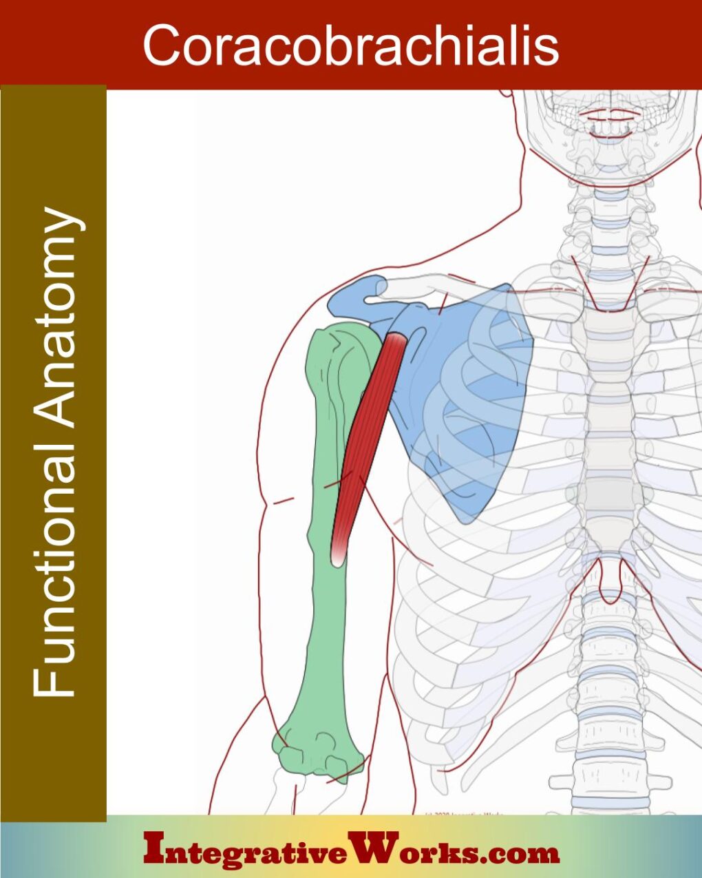
Coracobrachialis – Functional Anatomy Read More »

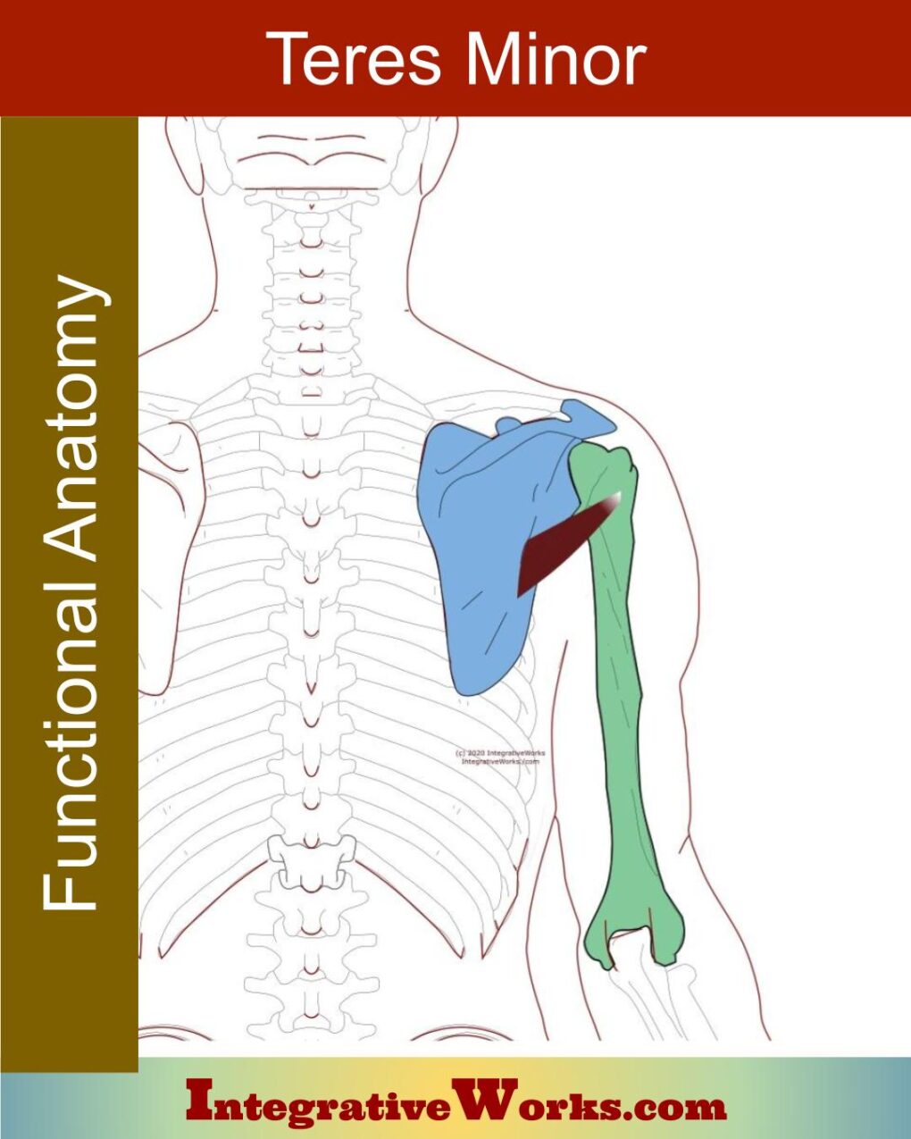
Rotator Cuff Muscles – Functional Anatomy

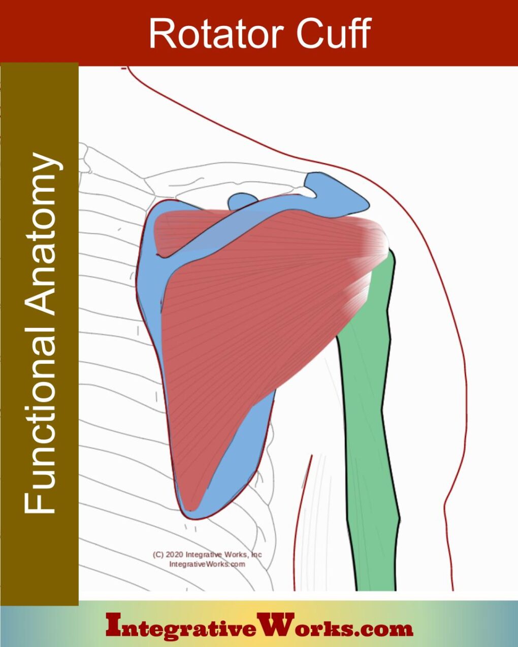
Overview Rotator cuff muscle anatomy is both simple and complicated in their anatomy. Simply put, they secure and refine movements of the head of the humerus. However, each muscle has complications in structure and function. Origin Insertion Function Innervation As a whole, they complete each other in the balancing act of securing the humeral head.

Rotator Cuff Muscles – Functional Anatomy Read More »

Infraspinatus – Functional Anatomy

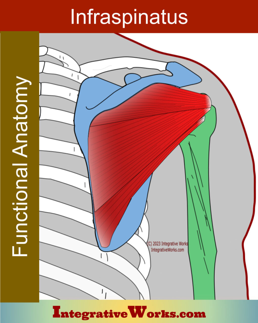
The anatomy of the infraspinatus is a bit more complicated than typically explained. A more detailed understanding is helpful for effective neuromuscular massage. The trigger point locations, which produce different patterns, are in different bellies. Overview of Anatomy Overview The infraspinatus is a flat rotator cuff muscle located in the infraspinous fossa of the scapula.

Infraspinatus – Functional Anatomy Read More »

Subscapularis – Functional Anatomy

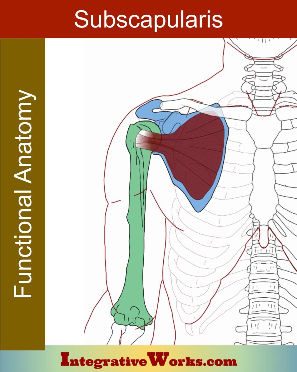
The anatomy of the subscapularis muscle is more complex than at first glance. For example, it has multiple sections and a variable attachment. The subscapularis is a thick, fan-shaped muscle on the underside of the shoulder blade. By far, it is the largest and strongest rotator cuff muscle. Also, it is the largest shoulder muscle.

Subscapularis – Functional Anatomy Read More »

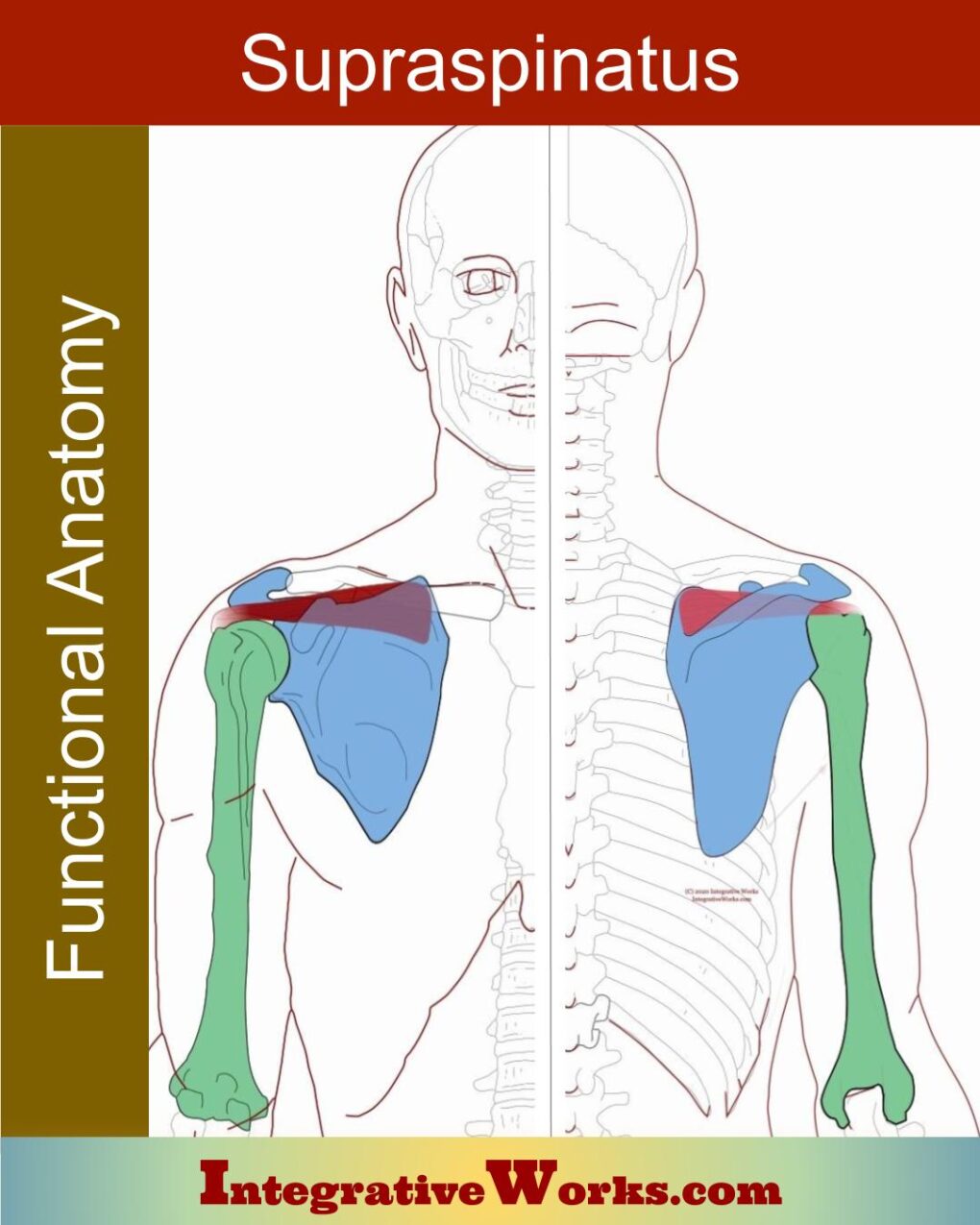
Supraspinatus – Functional Anatomy

Overview Supraspinatus, in the supraspinous fossa, is the most superior of the rotators cuff muscles, Origin Insertion Function Nerve Supraspinatus anatomy presents as a simple strip of muscle. It attaches to the top of the scapula and inserts on the head of the humerus. However, Shoulder impingement and tendinopathy prompted more extensive studies. Actually, it

Supraspinatus – Functional Anatomy Read More »

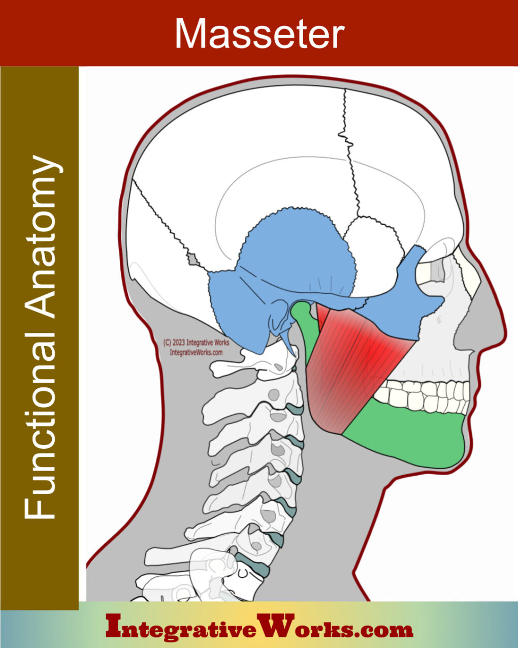
Masseter – Functional Anatomy

Overview The masseter anatomy is similar to other three-bellied muscles with a strong fascial covering, like gluteus medius and infraspinatus. Also, like those muscles, it balances and stabilizes a joint that needs both strength and flexibility. Pound for pound, it is considered to be the strongest muscle in the body. The masseter is a three-bellied

Masseter – Functional Anatomy Read More »

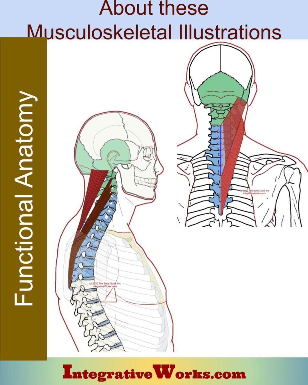
Musculoskeletal Illustrations

Years ago, I started creating my own illustrations. I wanted my manuals to display illustrations that struck a balance between simple and clear. After studying other illustrators’ work, I began to see the advantages and disadvantages of different styles (realistic, line drawings, photos, etc.). However, I also found that even very respected illustrators like Frank Netter

Musculoskeletal Illustrations Read More »

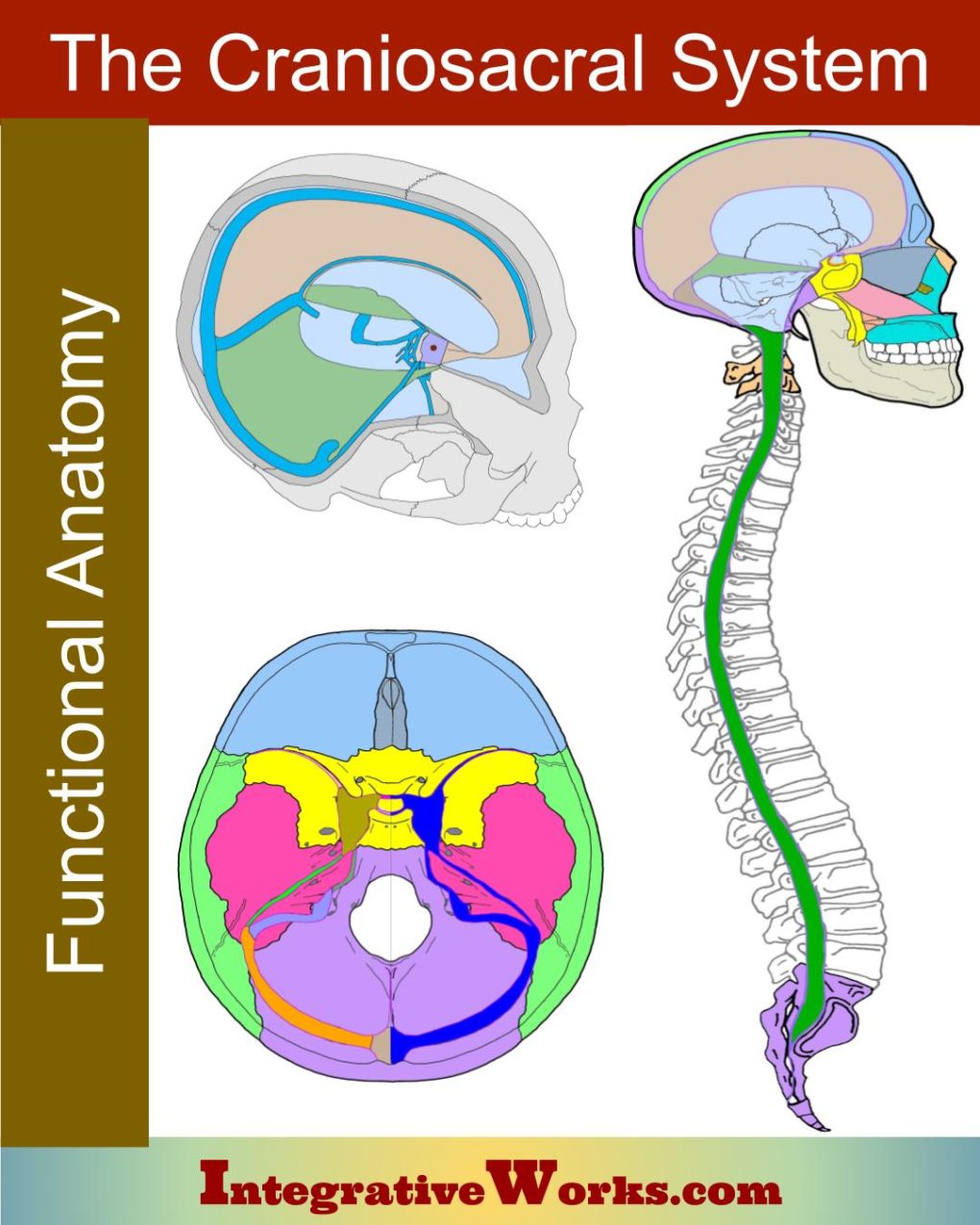
Craniosacral System Overview

Many of my clients have asked me for a simple explanation of the Craniosacral System. Here is a basic overview of how it is seen in the bio-mechanical model: The word “craniosacral” comes from the membrane systemthat surrounds the central nervous systemand connects the cranium to the sacrum.  The craniosacral system consists of: The Cranium The cranium

Craniosacral System Overview Read More »