Home » Anatomy

Anatomy

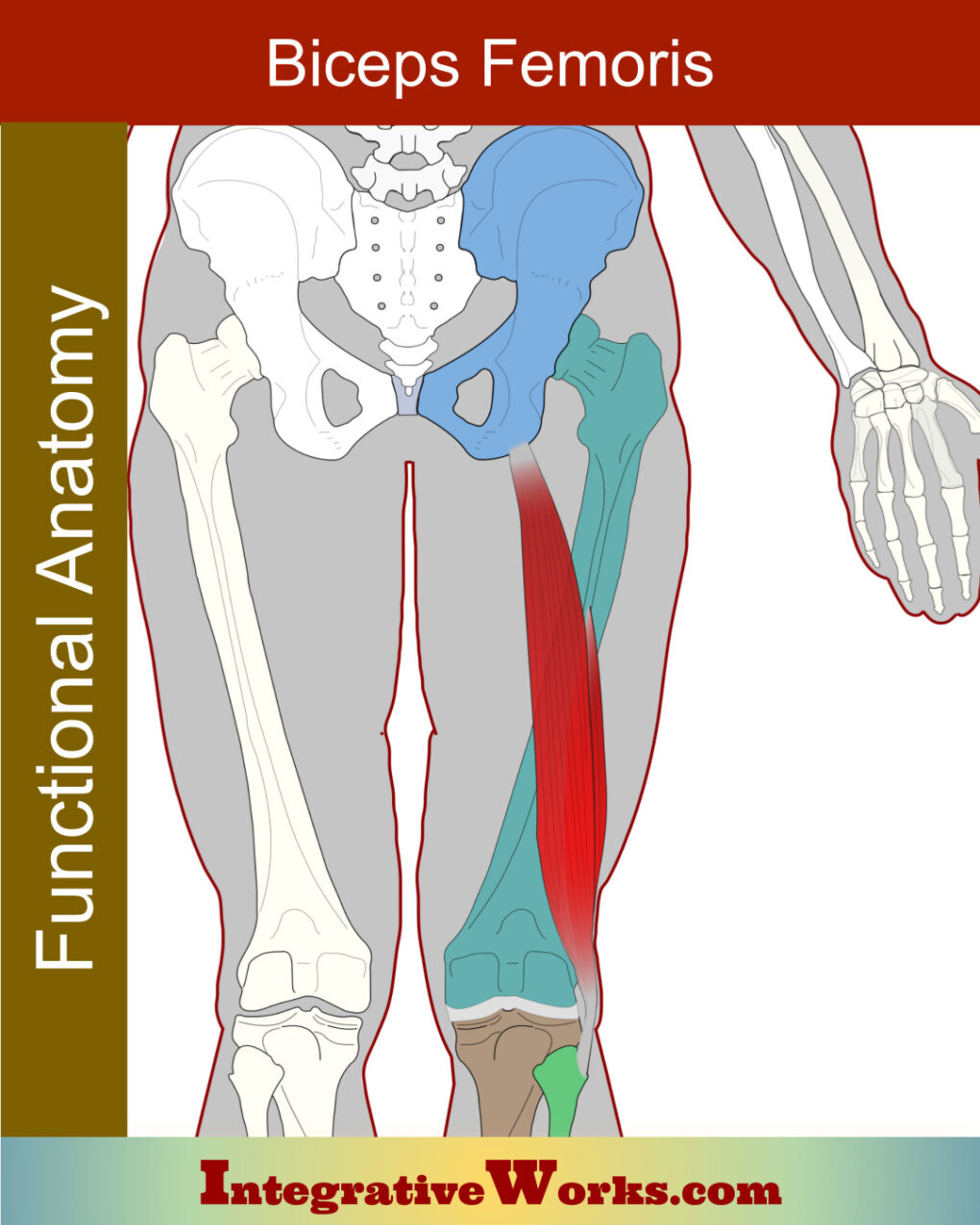
Biceps Femoris – Functional Anatomy

Overview Here, you will find the basic anatomy and Physiology of the biceps femoris muscle. The biceps femoris is a two-headed muscle on the lateral thigh. It belongs to the hamstring group. Origin Insertion Function Nerve Involvement in Exercise There are a multitude of studies about hamstring function, especially related to exercise. This meta-study is

Biceps Femoris – Functional Anatomy Read More »

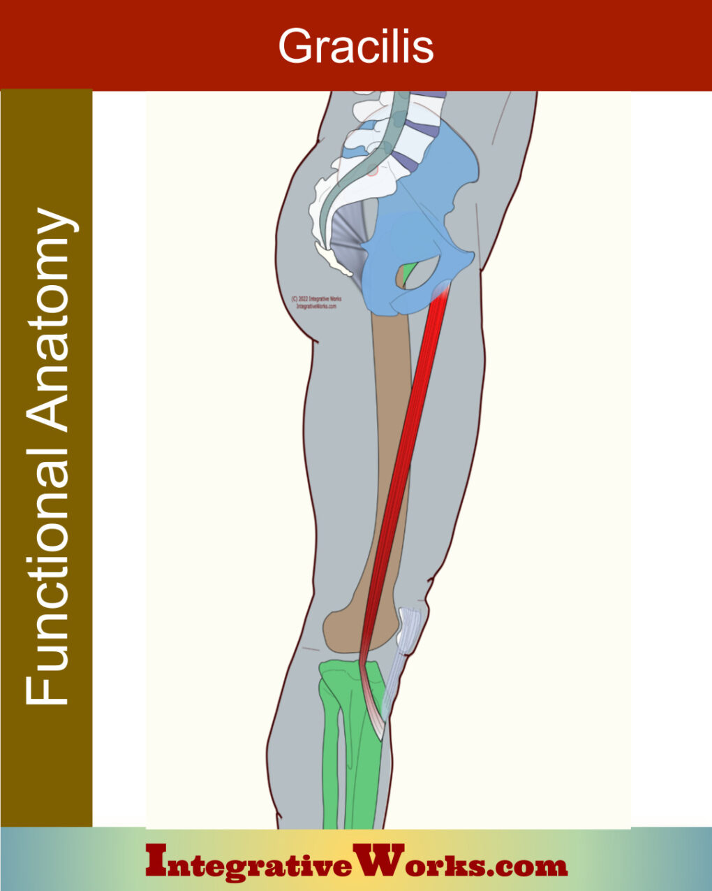
Gracilis – Functional Anatomy

Overview Gracilis is a long, thin muscle on the medial aspect of the thigh. Origin Insertion Function Nerve Gracilis is a weaker adductor. Consequently, sudden overload commonly injures this muscle. Typically, this involves groin pulls and adductor problems related to sports and accidental injury. Studies like this show that variations typically involve the arrangement of

Gracilis – Functional Anatomy Read More »

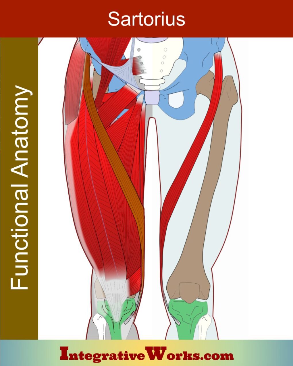
Sartorius- Functional Anatomy

Overview The functional anatomy of the sartorius muscle is complicated and often argued. The muscle is used when adopting a cross-legged position associated with a tailor’s sewing posture. Consequently, its name comes from the Latin sartor, meaning ‘tailor.’ Notably, it is the longest muscle in the body. Origin Insertion Function Nerve In discussions of sartorius’ anatomy

Sartorius- Functional Anatomy Read More »

Sacroiliac Joint – Functional Anatomy

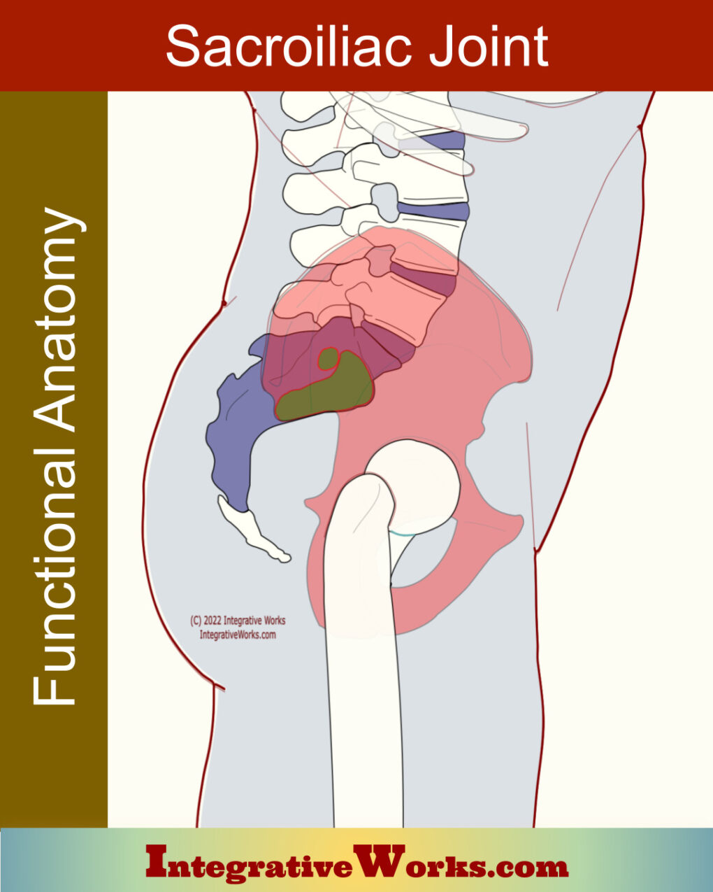
Overview Here, you will find a description of the anatomy, physiology, and progression of the sacroiliac joint. Perspectives on the pelvis and sacroiliac joint have developed over decades, even centuries. There is a long explanation of that development with many supporting links in this meta-study. Extensive research has defined pain mechanisms and the evolutionary transition

Sacroiliac Joint – Functional Anatomy Read More »

Sacroiliac Ligament – Functional Anatomy

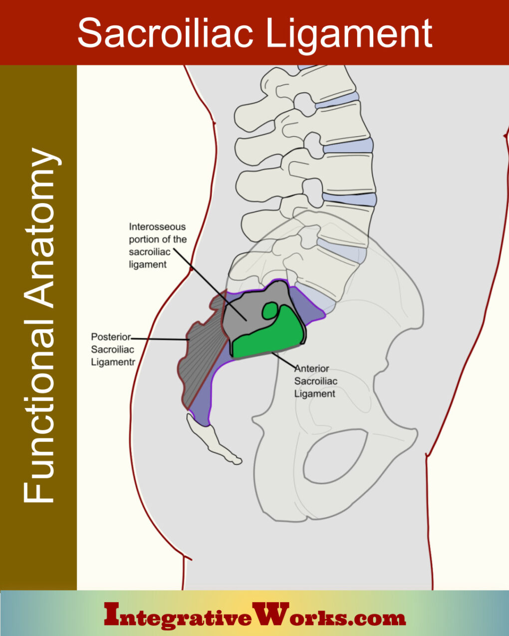
Overview Here, you will find a bodyworker’s perspective on the anatomy of the sacroiliac ligament. The sacroiliac ligament has three sections – anterior, interosseous, and posterior. Each section has a unique structure. Additionally, they impact the stability of the sacroiliac joint very differently. Sacroiliac Joint The sacroiliac joint is a complicated and highly variable structure.

Sacroiliac Ligament – Functional Anatomy Read More »

Tibialis Posterior – Functional Anatomy

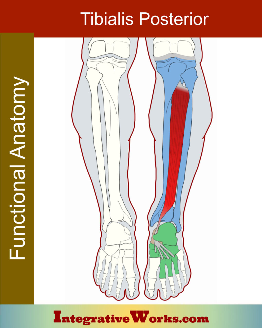
Overview The tibialis posterior is a long muscle extending along the low leg’s deep posterior compartment. The complexity and variability of the insertion tendon complicate its anatomy. That insertion usually includes most of the bones in the foot’s arch. Origin Insertion Function Nerve The tibialis posterior is deep to all the posterior low leg muscles.

Tibialis Posterior – Functional Anatomy Read More »

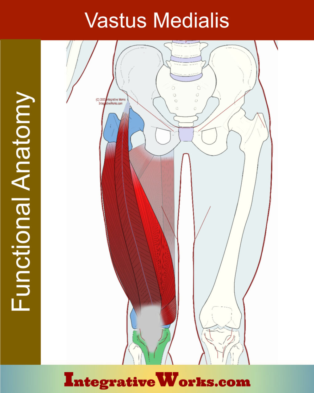
Vastus Medialis – Functional Anatomy

Overview of Anatomy The functional anatomy of the vastus medialis is a bit more complicated than the other vasti muscles. It has two distinct sections; vastus medialis longus and vastus medialis obliquus. Origin Vastus Medialis Longus Vastus Medialis Obliquus Insertion Function Nerve Patella Femoral Syndrome became a more prevalent diagnosis, based on some studies in

Vastus Medialis – Functional Anatomy Read More »

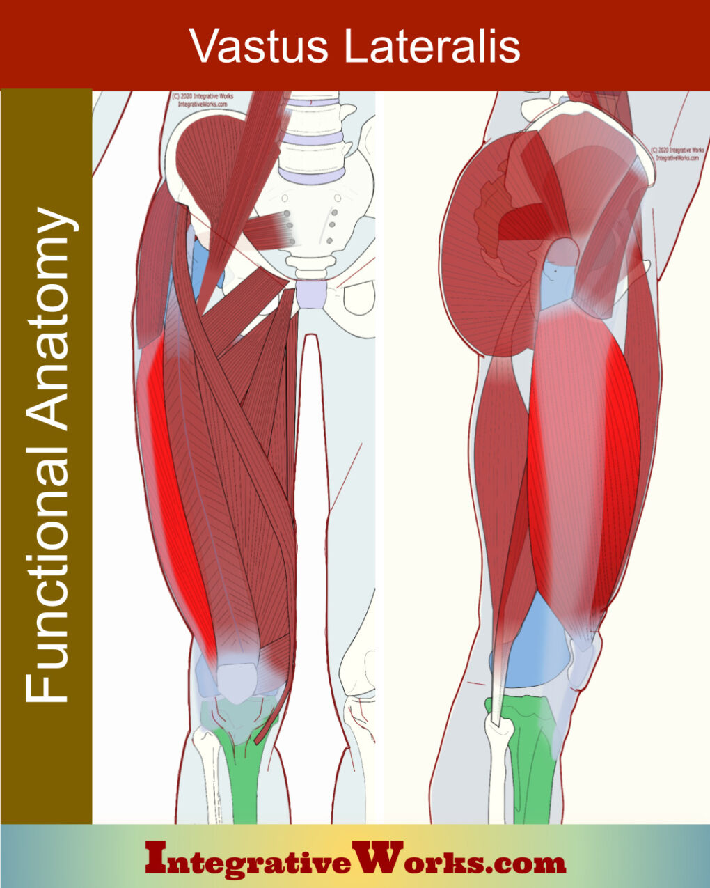
Vastus Lateralis – Functional Anatomy

Overview of Anatomy The functional anatomy of the vastus lateralis is largely uncomplicated. However, it has frequent and subtle variations. The fascia lata thickens and forms the iliotibial tract, along the lateral aspect of the vastus lateralis. Origin Insertion Function Nerve The vastus lateralis is the largest and strongest quadriceps muscle. Primarily, it extends the

Vastus Lateralis – Functional Anatomy Read More »