Lower Extremity Muscles

Vastus Lateralis – Functional Anatomy

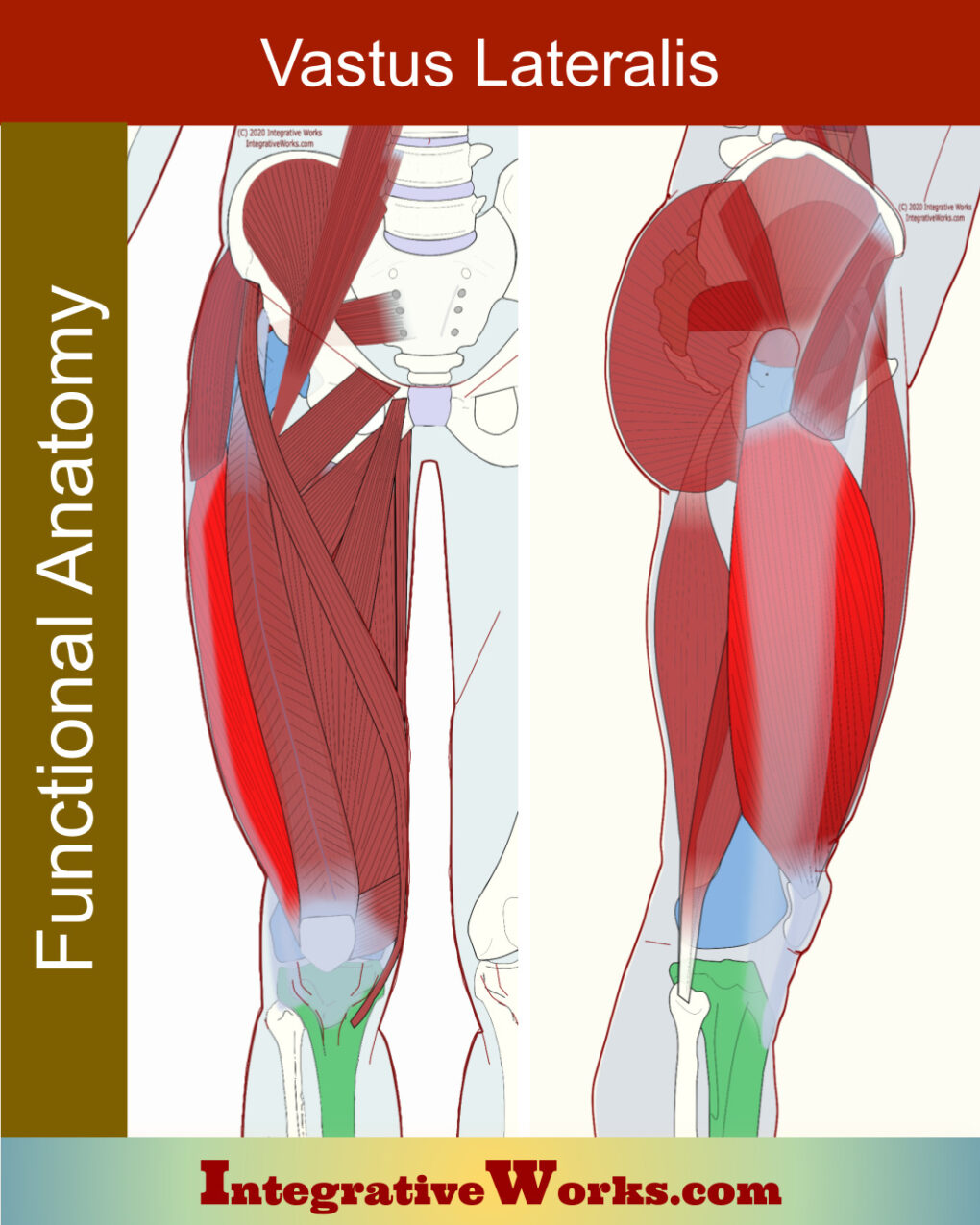
Overview of Anatomy The functional anatomy of the vastus lateralis is largely uncomplicated. However, it has frequent and subtle variations. The fascia lata thickens and forms the iliotibial tract, along the lateral aspect of the vastus lateralis. Origin Insertion Function Nerve The vastus lateralis is the largest and strongest quadriceps muscle. Primarily, it extends the […]

Vastus Lateralis – Functional Anatomy Read More »

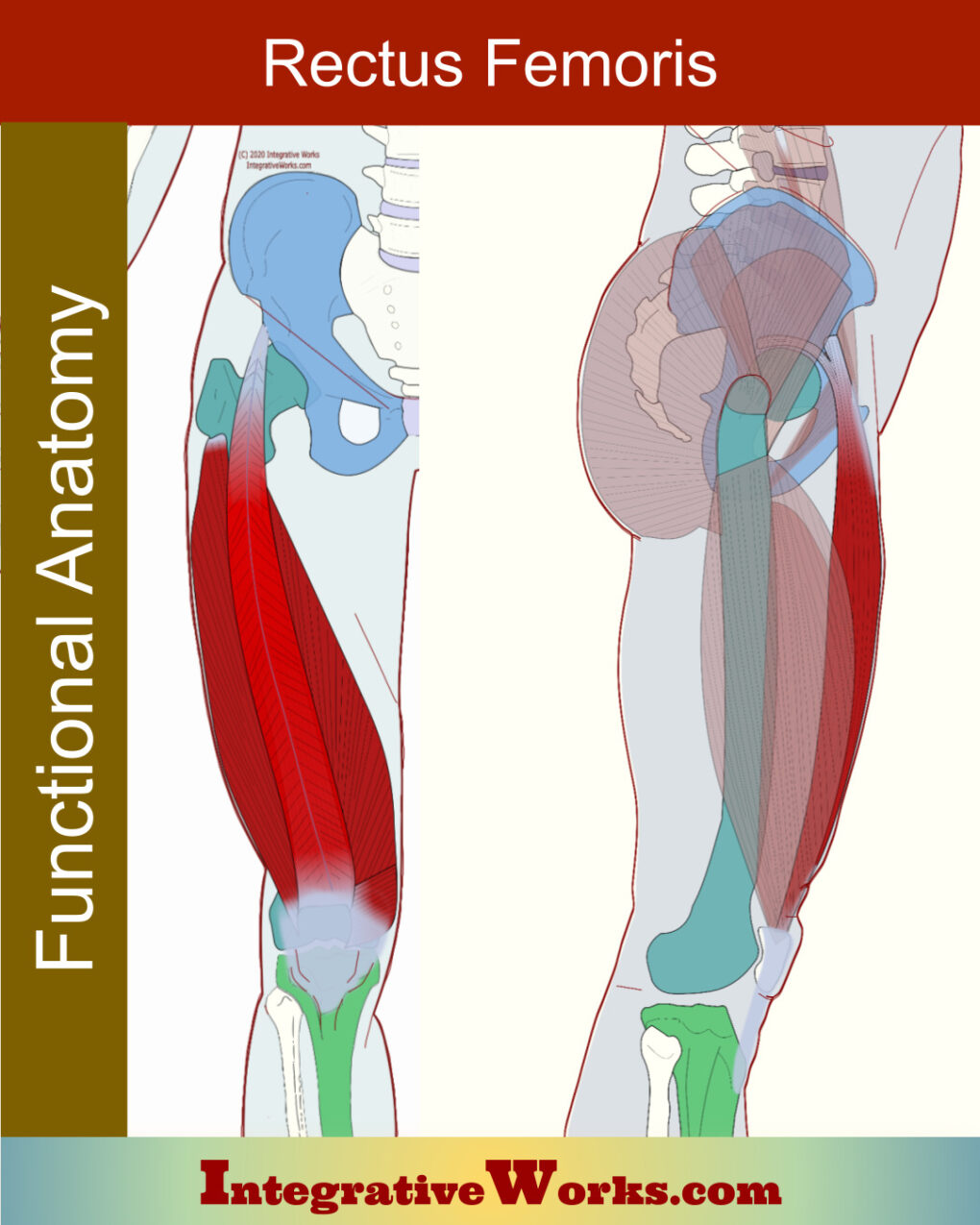
Rectus Femoris – Functional Anatomy

Overview of Anatomy The rectus femoris is a superficial muscle on the anterior thigh. However, the functional anatomy of the rectus femoris can be more complex than expected. It is part of the quadriceps group. Notably, unlike the other quadriceps muscle, it attaches above the hip joint. Origin Insertion Function Nerve Rectus Femoris Anatomy Evolution

Rectus Femoris – Functional Anatomy Read More »

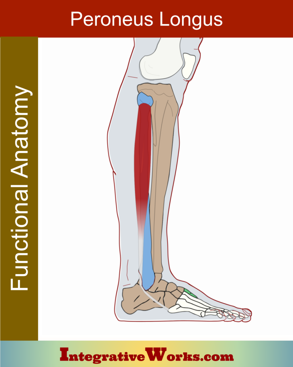
Peroneus Longus – Functional Anatomy

Overview Peroneus longus, also called fibularis longus, is a lateral low leg muscle. It wraps around the lateral foot and attaches to the inferior surface. The anatomy s complicated by variations in the attachments. Origin Insertion Function Nerve Peroneus longus, along with peroneus brevis, and with some stabilization from the tibialis posterior, evert the foot.

Peroneus Longus – Functional Anatomy Read More »

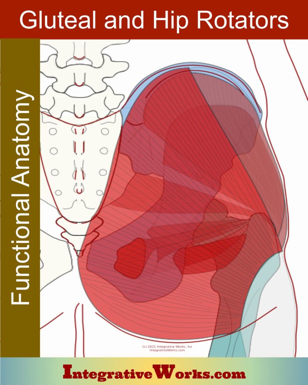
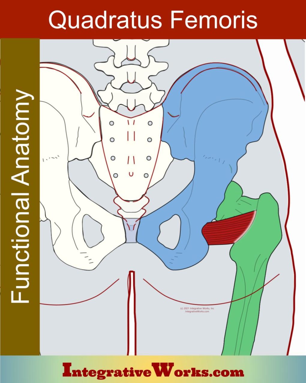
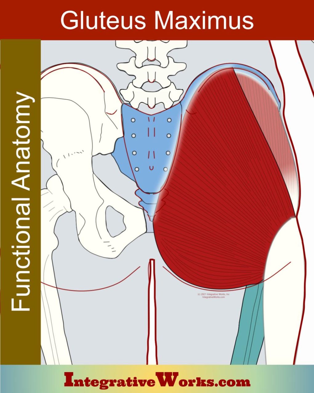
Gluteal and Lateral Hip Rotator Muscles – Functional Anatomy

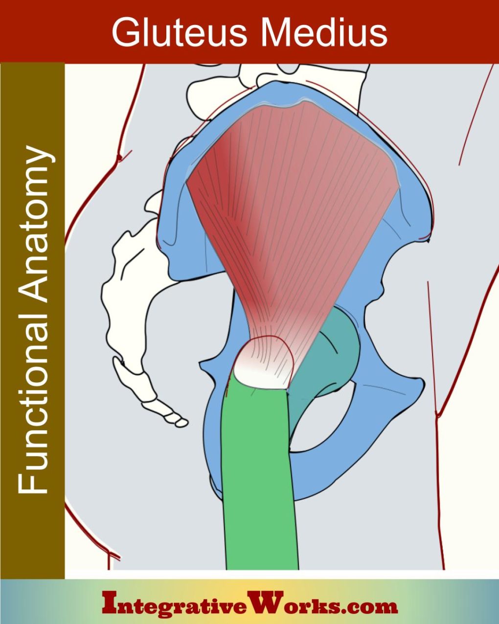
Overview Gluteal muscles form the fullness of the hip. They originate on the pelvis and insert on the femur. The gluteus maximus also attaches to the tibia via the iliotibial band. Primarily, these muscles extend and abduct the hip joint. In addition, the lateral glutes are vital in supporting the pelvis while the opposite leg

Gluteal and Lateral Hip Rotator Muscles – Functional Anatomy Read More »

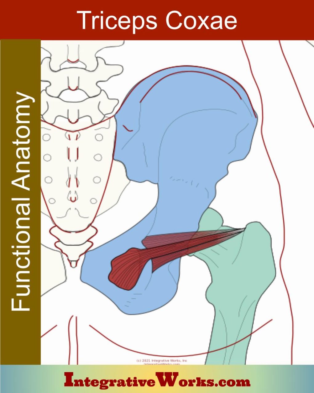
Triceps Coxae

Here, you will find anatomy for the triceps coxae, which consists of the obturator internus, gemellus superior, and gemellus inferior. These three muscles were originally grouped as a single muscle. Like the triceps brachii, they have a common insertion and function. Later, they were separated because of different origins and innervation. According to one study,

Triceps Coxae Read More »

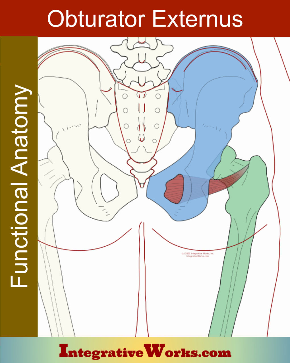
Obturator Externus – Functional Anatomy

Overview The obturator externus is a flat, triangular muscle that extends from the lower pelvis to the superior femur. The muscle has a superior belly and the main belly. Its name comes from its attachment to the external surface of the obturator foramen. Its counterpart, obturator internus, originates from the internal surface. Origin Insertion Function

Obturator Externus – Functional Anatomy Read More »