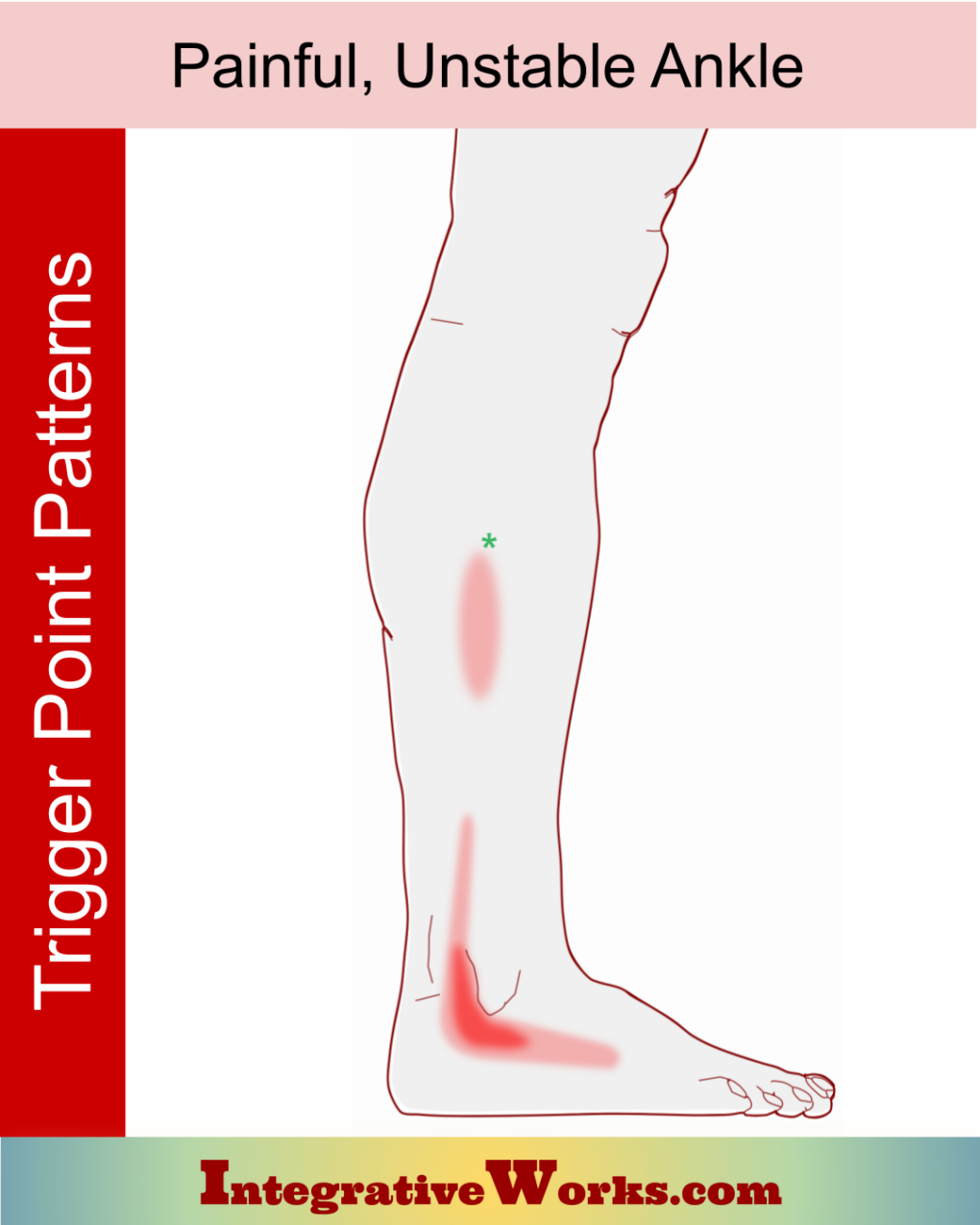
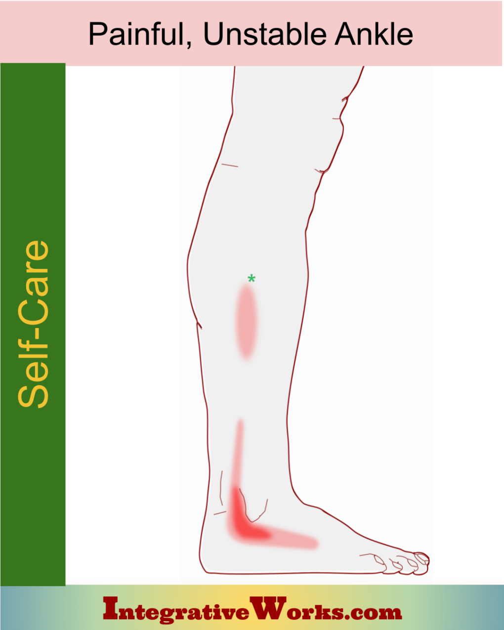
Overview
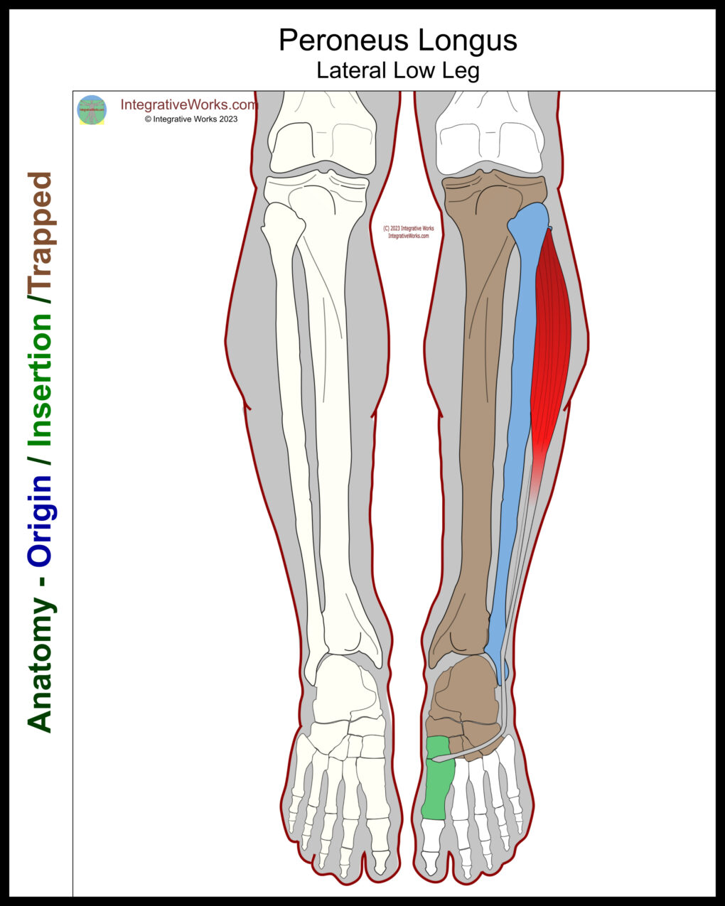
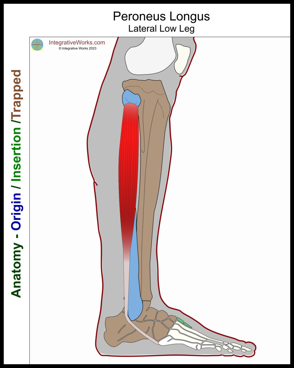
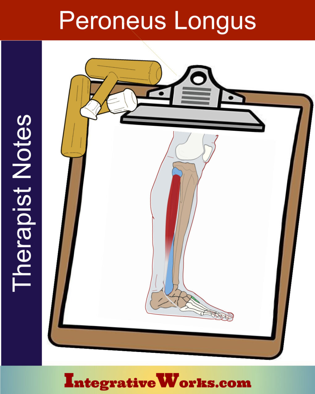
Peroneus longus, also called fibularis longus, is a lateral low leg muscle. It wraps around the lateral foot and attaches to the inferior surface. The anatomy s complicated by variations in the attachments.
Origin
- head and proximal third of the lateral tibia
Insertion
- base of the first metatarsal
- medial cuneiform
Function
- eversion of the foot
- supports the arch of the foot
Nerve
- superficial fibular nerve, sacral plexus, (L4-S3)
Functional Considerations
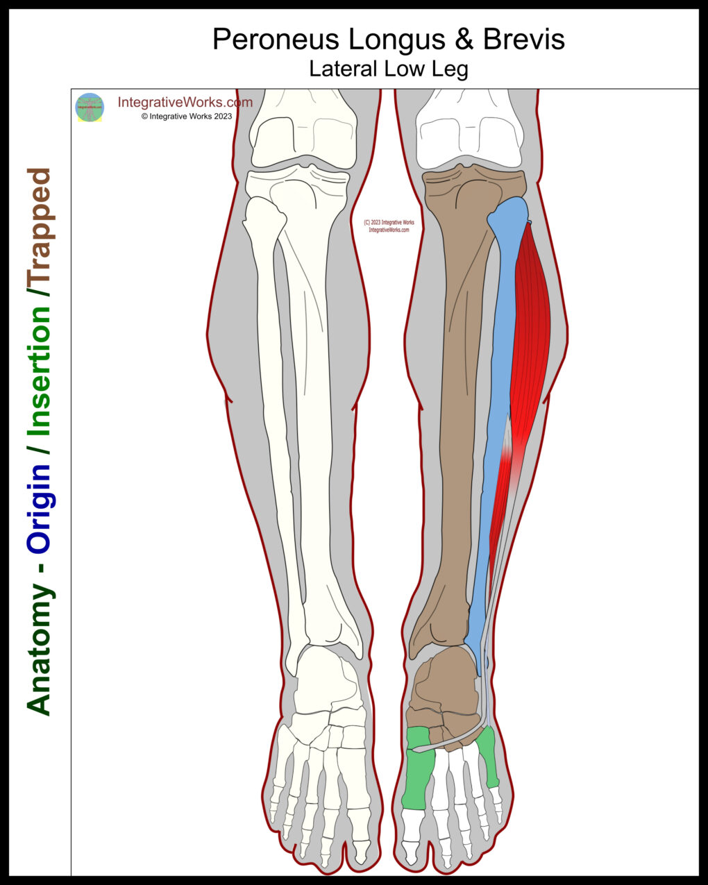
Peroneus longus, along with peroneus brevis, and with some stabilization from the tibialis posterior, evert the foot. This movement is a combination of plantarflexion while turning the plantar surface laterally.
Its position creates complexities in its functional roles.
- This muscle, along with the tibialis anterior, creates a stirrup that supports the lateral arch of the foot.
- It is vital to maintain balance while on one foot.
- The attachment on the base of the first metatarsal helps to stabilize and depress the great toe and stops the foot from collapsing medially.
Anomalies, Etc.
The anatomy of peroneus longus varies significantly. The upper attachment of this muscle often has a slip that attaches to the tibial condyle. However, the distal attachments have frequent variations in the tendonous slips. This study reports:
- The base of 1st metatarsal: 30 specimens, 100%
- Medial cuneiform: 26 specimens, 86.6%
- The neck of the 1st metatarsal: 3 specimens, 10%
- The base of 2nd metatarsal: 6, 20%
- The base of the 4th metatarsal: 5, 16.6%
- The base of 5th metatarsal: 7, 23.3%
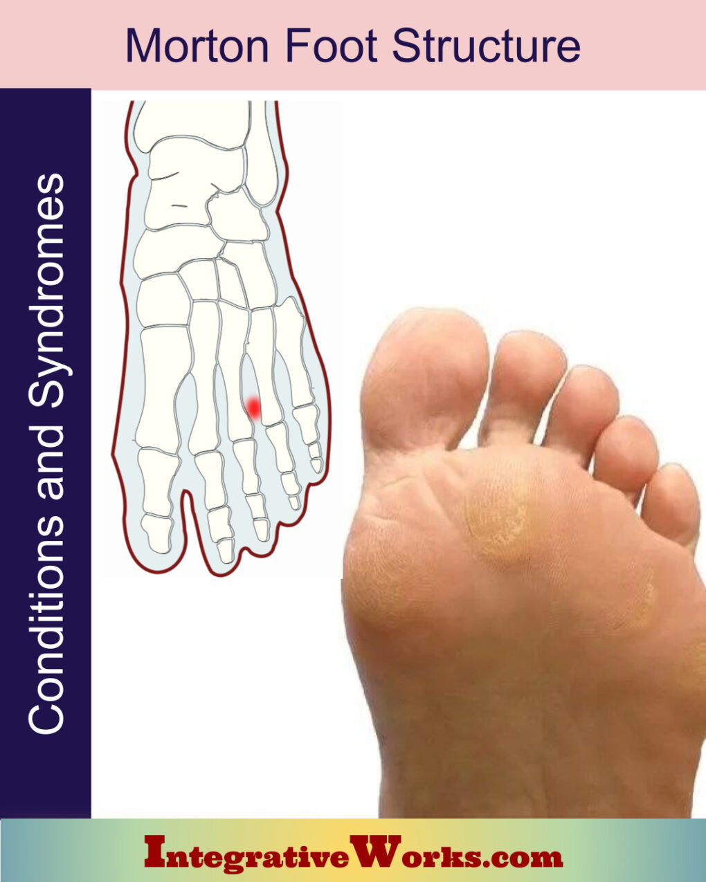
Morton’s Foot Structure, and The Wobbly Foot
Feet invert, which correlates with supination and turning the arch inward. Conversely, feet also evert, which correlates with pronation and turning the arch outward.
Morton’s Foot Structure creates a foot prone to invert and evert excessively. This is because the longer second metatarsal acts as a pivot that strikes before the first metatarsal. This structure causes the foot to wobble. Comparatively, other feet tend to strike on the first metatarsal in a stride with less foot rocking.
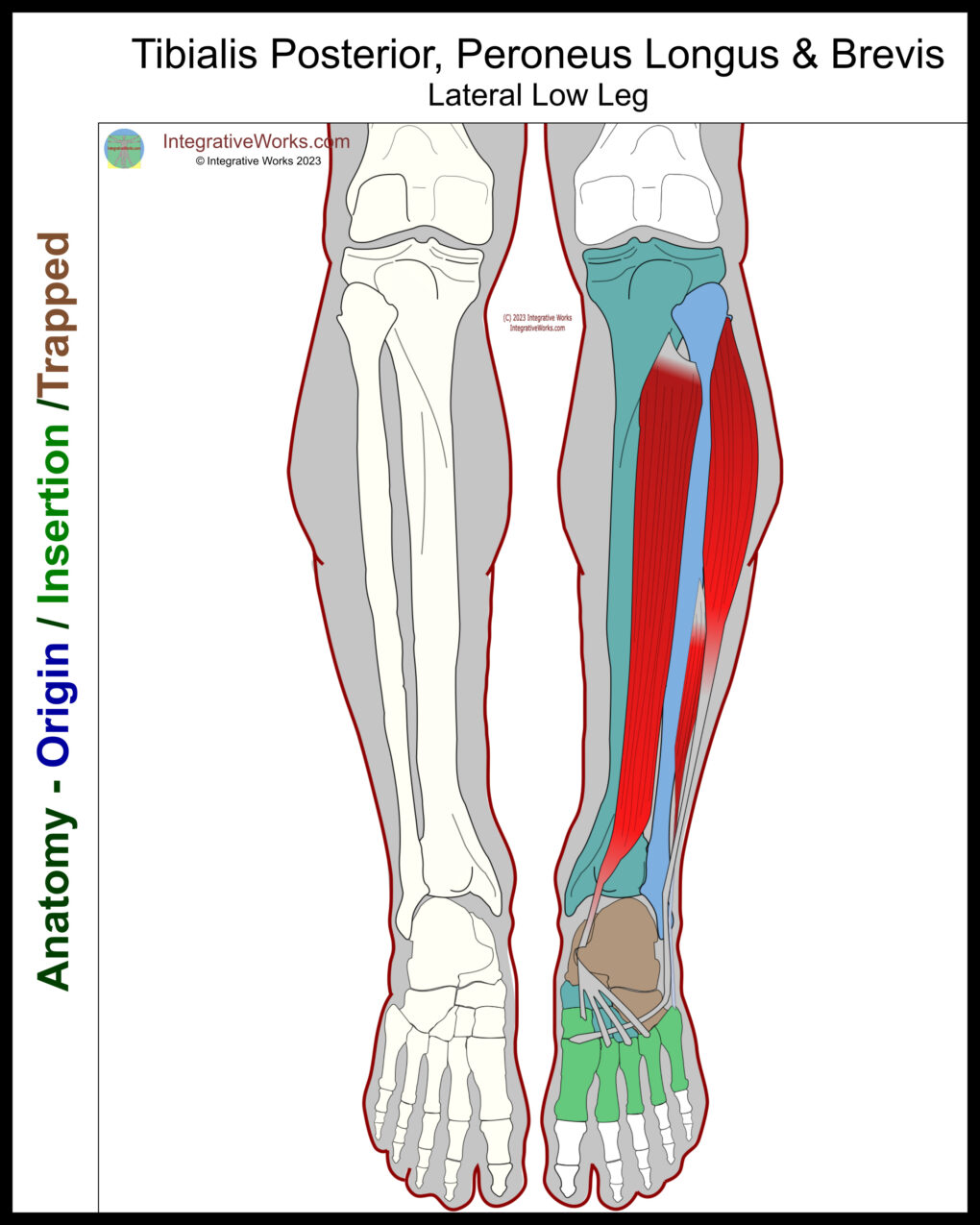
Muscles of Inversion and Eversion
The tibialis posterior is the primary inverter of the foot. It wraps around the medial ankle and attaches to most of the bones of the arch. Additionally, flexors of the toes loop through the medial ankle and assist with these functions.
At the same time, the peroneus longus is the primary everter of the foot. It wraps around the lateral ankle and attaches to the medial bones of the foot at the base of the first metatarsal and medial cuneiform. Additionally, the peroneus brevis everts the ankle.
Perpetuating ankle pain
The posterior tibialis and peroneus longus are most directly involved in stabilizing the wobbly foot. Consequently, they both create pain patterns around the ankle that are harder to resolve with Morton’s Foot Structure.
While walking, strong inverters and everters stabilize the foot. However, when those stabilizers have trigger points, Morton’s Foot Structure aggravates those muscles and generates more pain.
Related Posts

Support Integrative Works to
stay independent
and produce great content.
You can subscribe to our community on Patreon. You will get links to free content and access to exclusive content not seen on this site. In addition, we will be posting anatomy illustrations, treatment notes, and sections from our manuals not found on this site. Thank you so much for being so supportive.
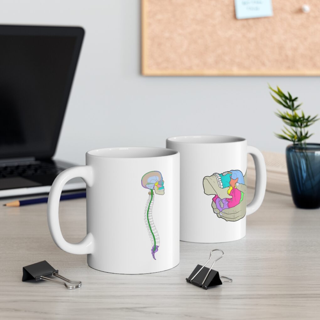
Cranio Cradle Cup
This mug has classic, colorful illustrations of the craniosacral system and vault hold #3. It makes a great gift and conversation piece.
Tony Preston has a practice in Atlanta, Georgia, where he sees clients. He has written materials and instructed classes since the mid-90s. This includes anatomy, trigger points, cranial, and neuromuscular.
Question? Comment? Typo?
integrativeworks@gmail.com
Interested in a session with Tony?
Call 404-226-1363
Follow us on Instagram

*This site is undergoing significant changes. We are reformatting and expanding the posts to make them easier to read. The result will also be more accessible and include more patterns with better self-care. Meanwhile, there may be formatting, content presentation, and readability inconsistencies. Until we get older posts updated, please excuse our mess.
Pingback: Lateral Low Leg - Neuromuscular Protocol - Integrative Works