Fascia Lata
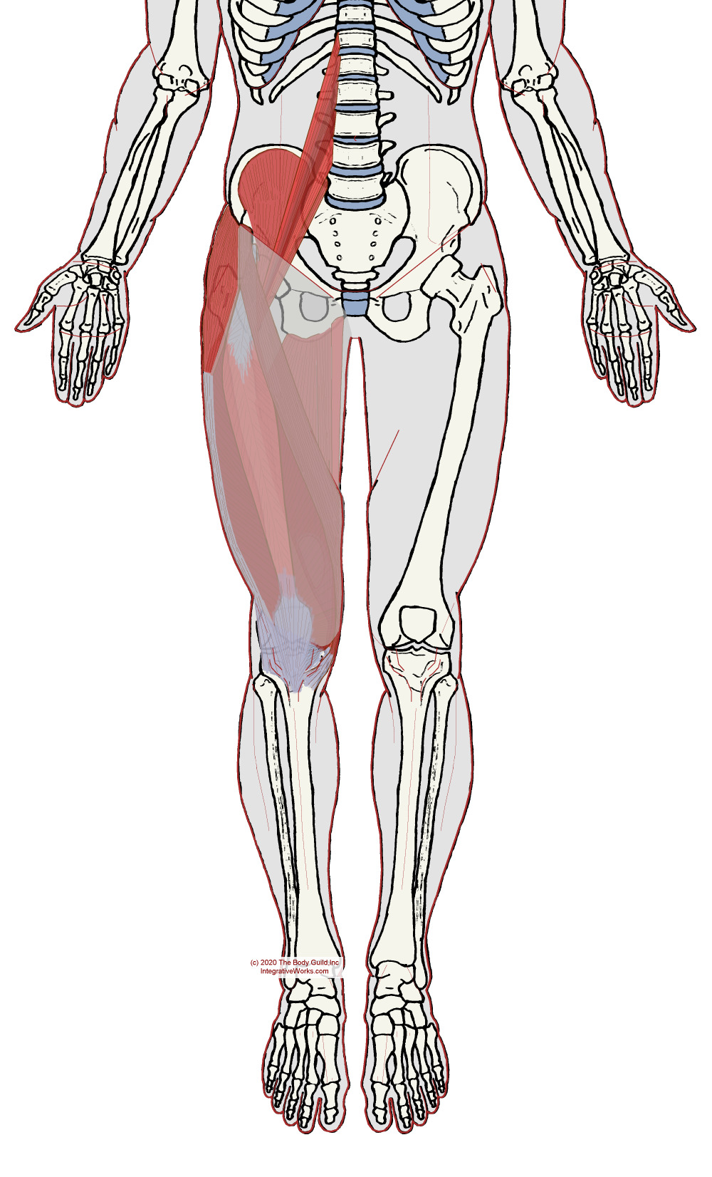
The fascia lata is the deep fascia that wraps the outer thigh.
This thick fascial envelope attaches superiorly to the crest of the Ilium and inguinal ligament. The fascia continues onto the pubic rami, ischial tuberosity, sacrotuberous ligament, coccyx, and sacrum. Distally, it thickens as it attaches to the proximal tibia.
It offers elastic stability, much like a compression stocking or kinesiotape. The fascia thickens along the iliac crest, where it connects the gluteus maximus and to the low back via a thick continuation of the fascia. This area along the iliac crest blends into the gluteal tuberosity. It is also thick around the knee and thinner around the adductor muscles.
Iliotibial Tract
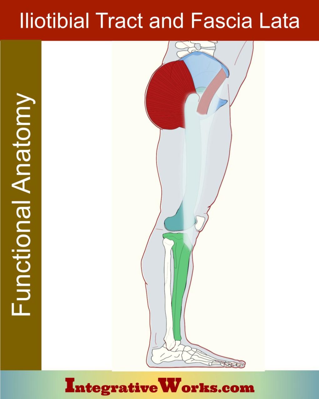
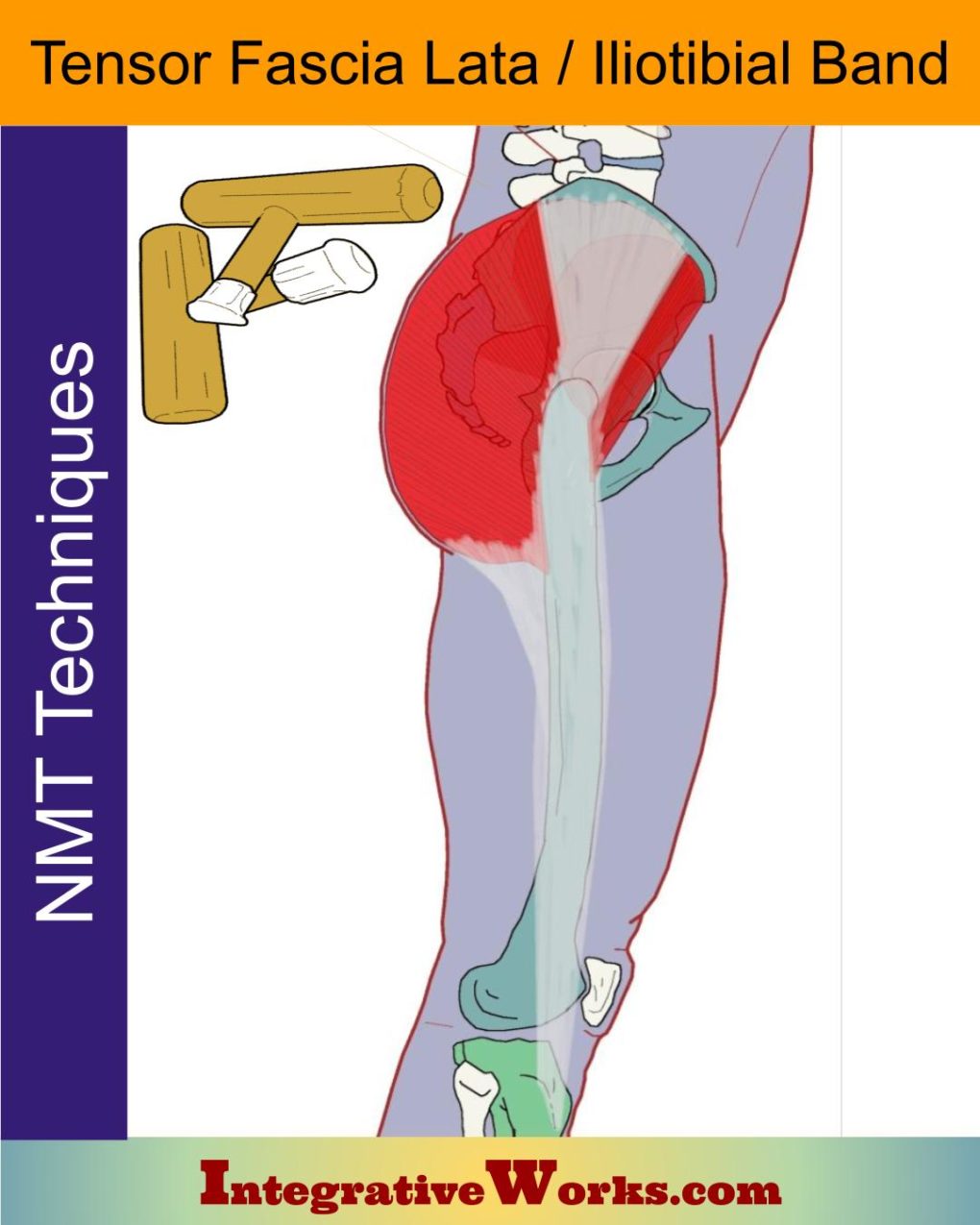
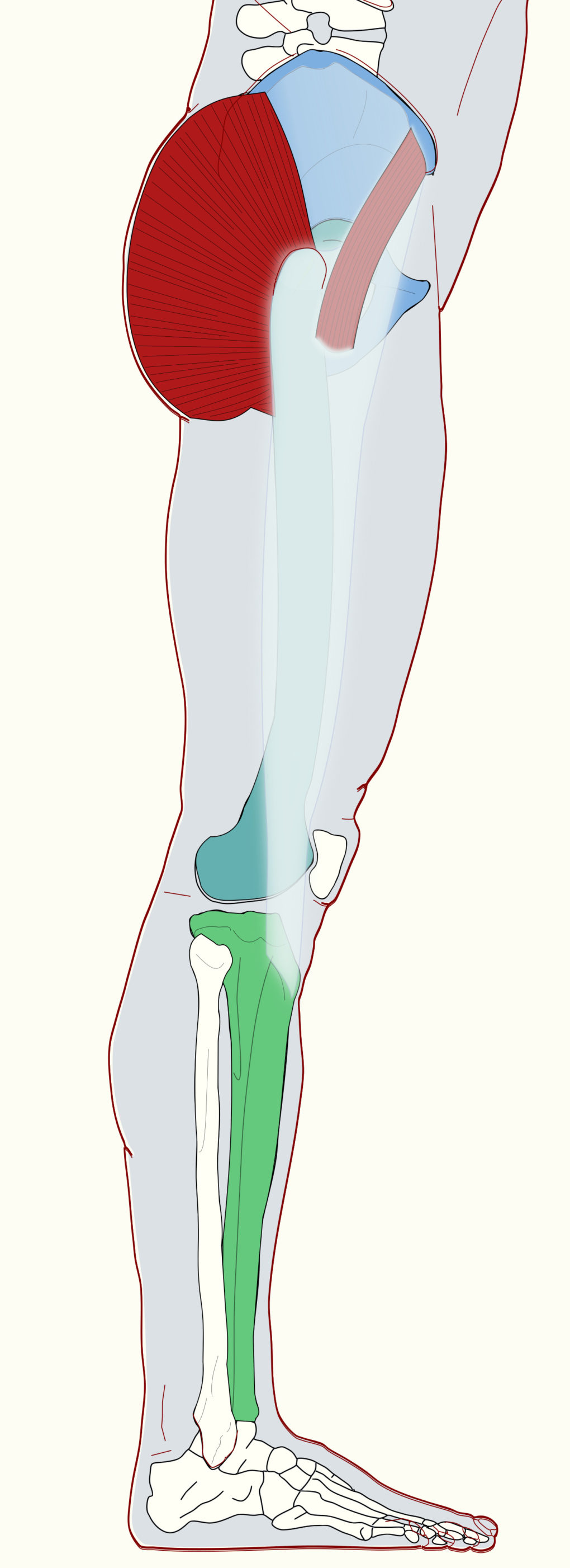
The Iliotibial Band, iliotibial tract, or IT band is a thickening of the fascia lata along the lateral thigh. It attaches to the iliac tubercle on the lateral hip and extends along the lateral thigh to the lateral tibial condyle.
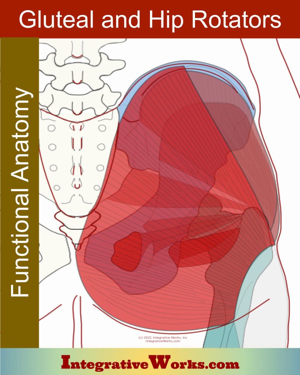
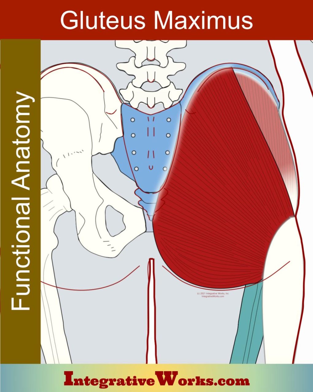
A portion of the gluteus maximus and tensor fascia lata invest into the IT band. About 25% of the superficial fiber of the gluteus maximus attaches to pull the IT band back like a bowstring.
The IT band helps to hold the femur in the acetabulum. It also extend, abduct and rotate the hip joint.
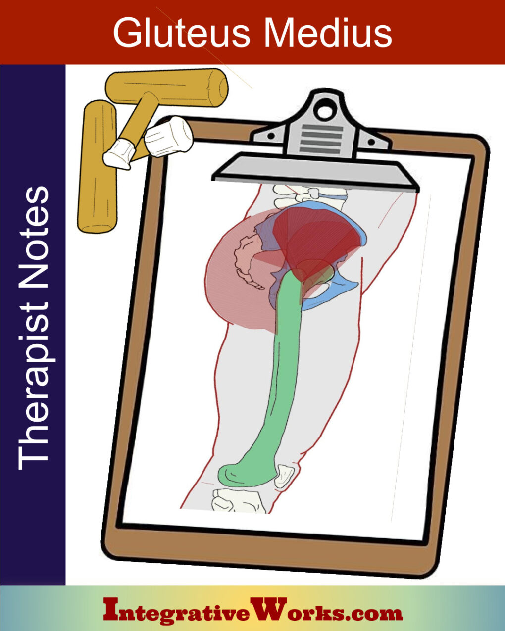
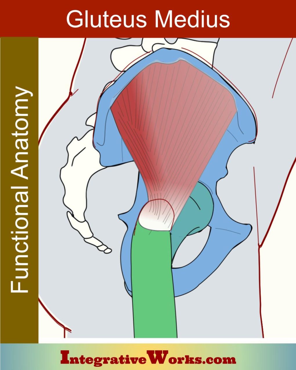
The Gluteal Aponeurosis is a thickening of the fascia lata. It originates from the iliac crest. The fascia covers and anchors the anterior two-thirds of the gluteus medius. As well, it extends inferiorly between the gluteus medius and the superficial fascia. Its anterior border inserts along the IT band and gluteal tuberosity of the femur, In that same area, it attaches to the gluteus maximus on its inferior border.
The differences in anatomical presentations are contradictory and confusing. So, most anatomical illustrations show the IT band diverging with fibers that blend toward the upper fibers of the gluteus maximus and other fibers that run along the posterior Edge of the TFL. The IT band is usually described, however, as inserting on the iliac tubercle with the gluteal aponeurosis filling the space between the iliac tubercle and the border of the gluteus maximus.
A study of 40 cadavers shows it to have 3 layers:
- The superficial layer is superficial to the TFL
- The middle layer attaches to the ilium, deep to the TFL
These layers merge at the distal end of the TFL - The deep layer originates from the supraacetabular fossa between the capsule of the hip joint and the rectus femoris’s attachment.
The deep layer extends to join with the other layers distal to their merger interior to the TFL.
Related Posts
Gluteal and Lateral Hip Rotator Muscles – Functional Anatomy
Gluteus Maximus – Functional Anatomy
Gluteus Medius – Massage Therapy Notes
Gluteus Medius- Functional Anatomy
Neuromuscular Protocol – Iliotibial Band
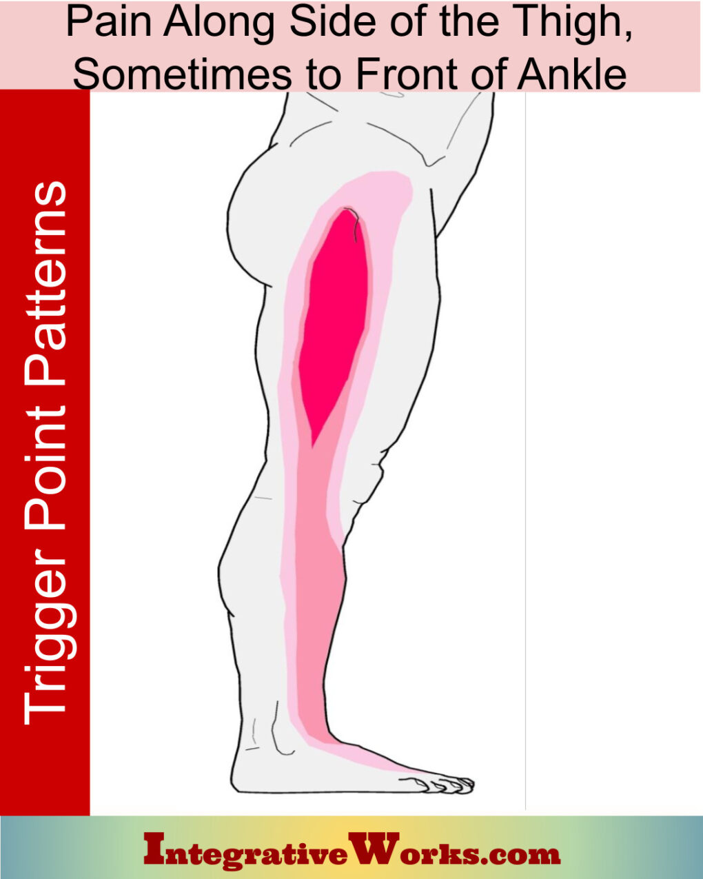
Pain Along Side of Thigh, Sometimes to Front of Ankle
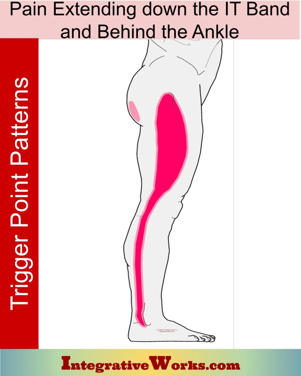
Pain Down the IT Band Extending Behind the Ankle
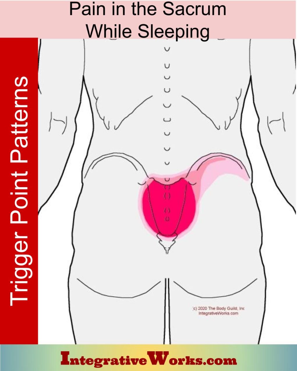
Pain in the Sacrum or Crest of the Hip When Sleeping on Side
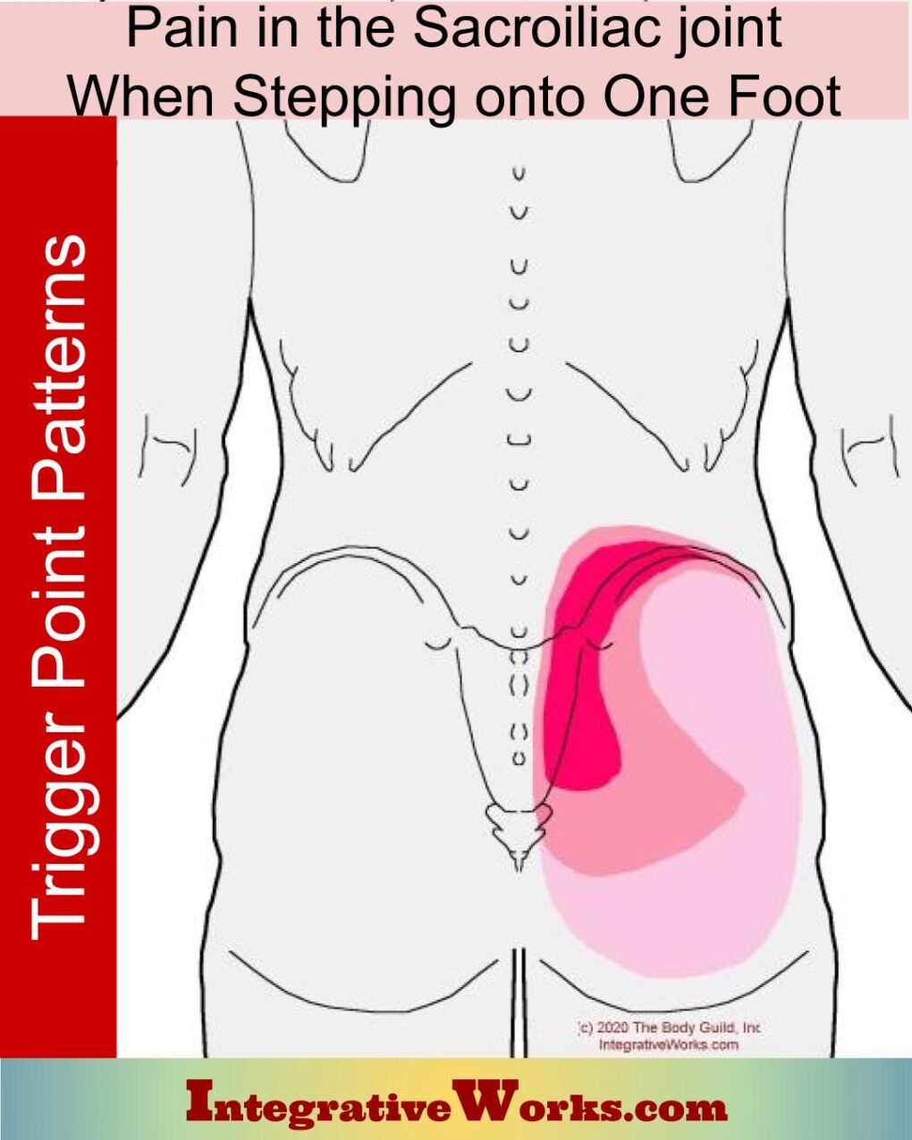
Pain Through the Hip Joint when Standing on One Foot
Self Care – Kneeling Stretch for Hip Flexors
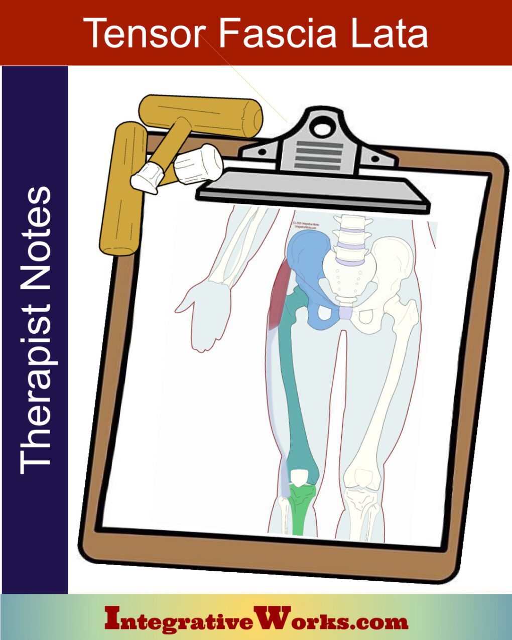
Tensor Fascia Lata – Massage Therapy Notes
Tensor Fascia Lata – Neuromuscular Massage Protocol
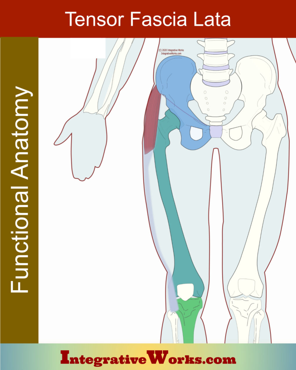
Tensor Fascia Lata (TFL) – Functional Anatomy
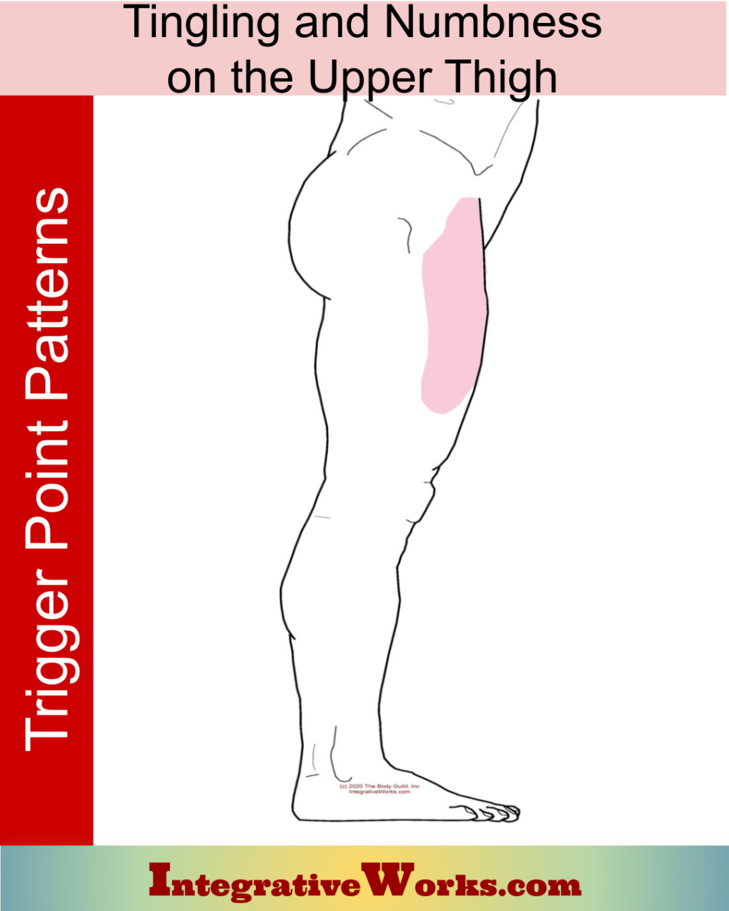
Tingling and Numbness in the Upper Thigh
Support Integrative Works to
stay independent
and produce great content.
You can subscribe to our community on Patreon. You will get links to free content and access to exclusive content not seen on this site. In addition, we will be posting anatomy illustrations, treatment notes, and sections from our manuals not found on this site. Thank you so much for being so supportive.
Cranio Cradle Cup
This mug has classic, colorful illustrations of the craniosacral system and vault hold #3. It makes a great gift and conversation piece.
Tony Preston has a practice in Atlanta, Georgia, where he sees clients. He has written materials and instructed classes since the mid-90s. This includes anatomy, trigger points, cranial, and neuromuscular.
Question? Comment? Typo?
integrativeworks@gmail.com
Interested in a session with Tony?
Call 404-226-1363
Follow us on Instagram

*This site is undergoing significant changes. We are reformatting and expanding the posts to make them easier to read. The result will also be more accessible and include more patterns with better self-care. Meanwhile, there may be formatting, content presentation, and readability inconsistencies. Until we get older posts updated, please excuse our mess.

