Overview
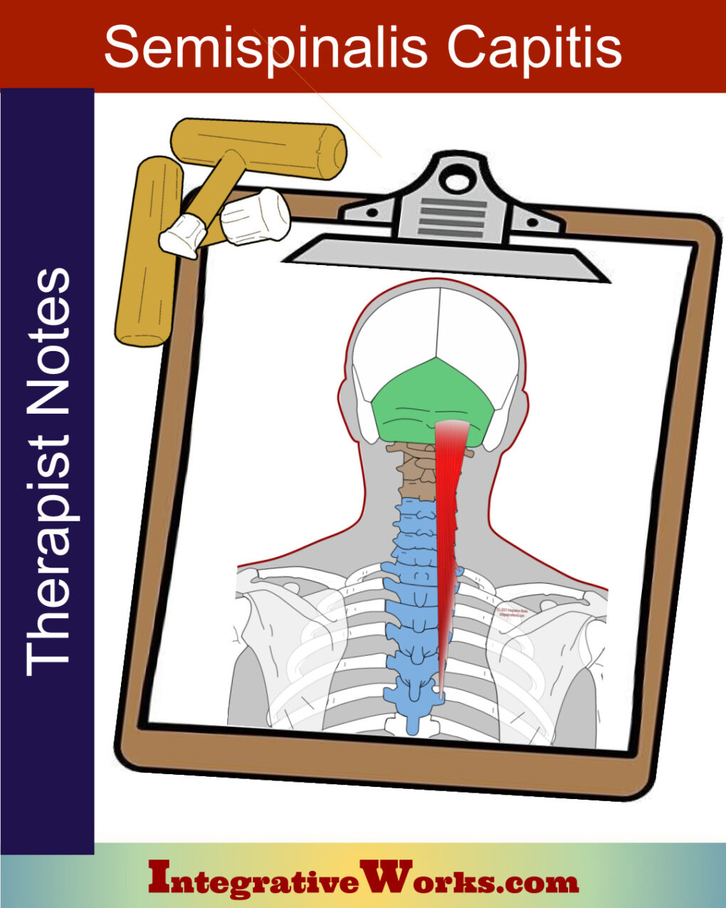
Semispinalis capitis anatomy is significant for managing pain. It is a large muscle that extends the head, perpetuates forward head posture, and squeezes the greater occipital nerve.
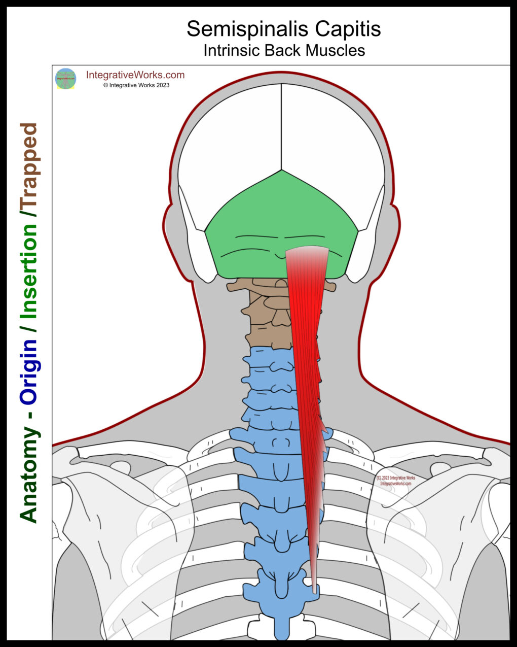
Origin
- articular processes of C4-C7
- transverse processes of T1-T6
Insertion
- between the superior and inferior nuchal ridges of the occipital ridge
Function
- extension and some rotation of the head
Nerve
- greater occipital nerve, posterior ramus of C2
- spinal nerve C3
Attachment Details
The functional anatomy of semispinalis capitis is significant regarding upper cervical pain. It attaches to the occiput and then usually skips the first three cervical vertebrae. After that, it has variable attachments to the vertebrae from C4 through T7.
Anomalies, Etc.
Dissection studies vary in their report of attachments on the thoracic vertebrae. They show that they differ in which vertebrae are attached, and some slips attach to the transverse or spinous processes.
Functional Considerations
Note how this muscle, which is usually thick and strong, supports a proper head position. Additionally, it skips the atlas and contributes to wedging it forward.
Greater Occipital Nerve Involvement
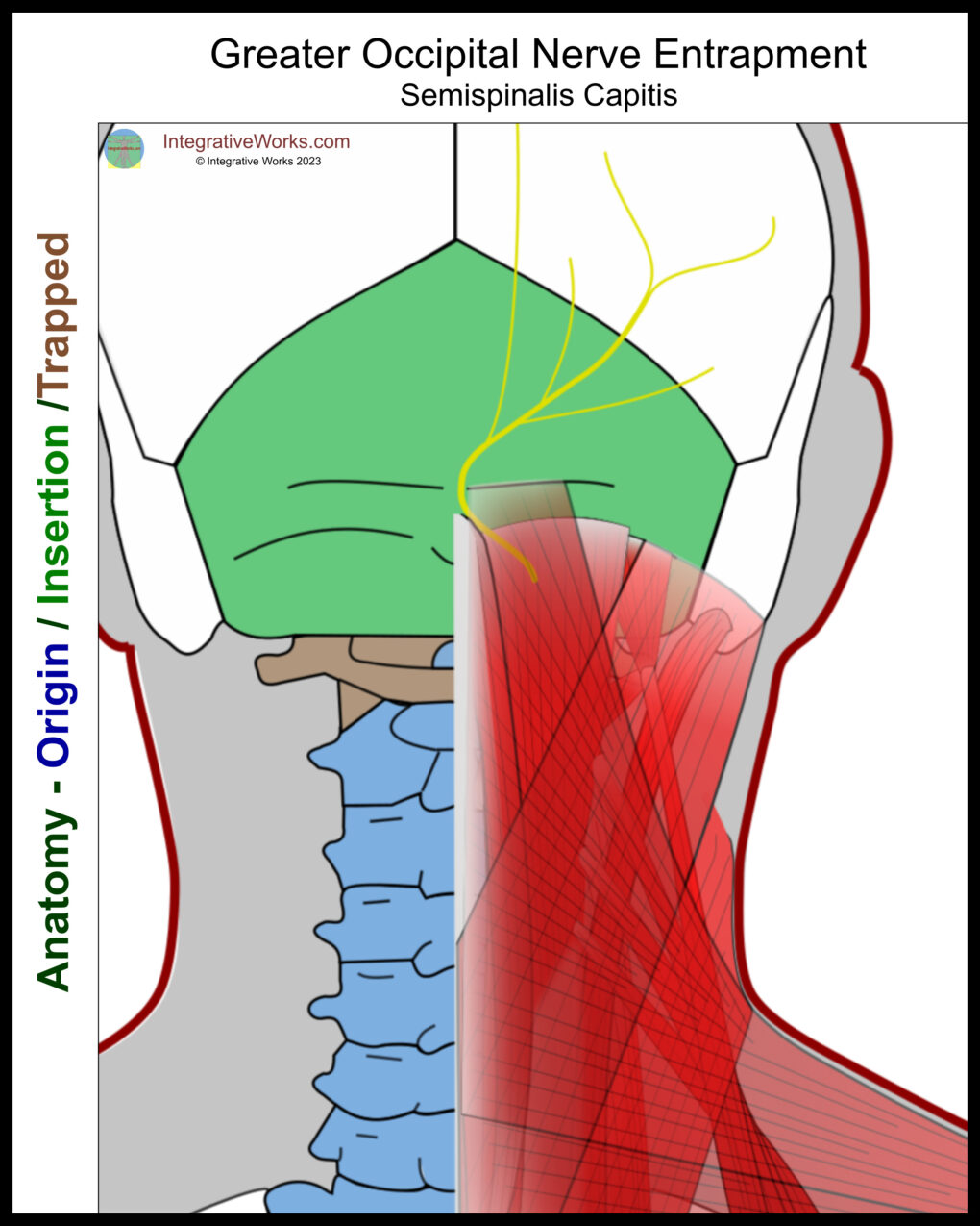
The greater occipital nerve (GON) is structurally vulnerable to tension in semispinalis capitis. This nerve branches off of the C2 dorsal ramus below the posterior arch of the atlas. Often, it has fibers from the C1 nerve root and occasionally from C3. The palpable tension of the C2 nerve root at its emergence is a reliable indicator of atlas displacement. It winds around the lower border of the obliquus capitis inferior. Then, it passes superficial to the suboccipital muscles. The nerve extends superiorly until it pierces semispinalis capitis and emerges deep to the trapezius. It passes through the attachment tendon on the superior nuchal line near the external occipital protuberance. It then passes under the galea aponeurosis.
The GON has variations in its path. Studies vary slightly but show that the GON pierces the semispinalis capitis about 90% of the time. However, it pierces the trapezius about 45% of the time. In rare instances, about 7% of the time, it pierces the obliquus capitis inferior.
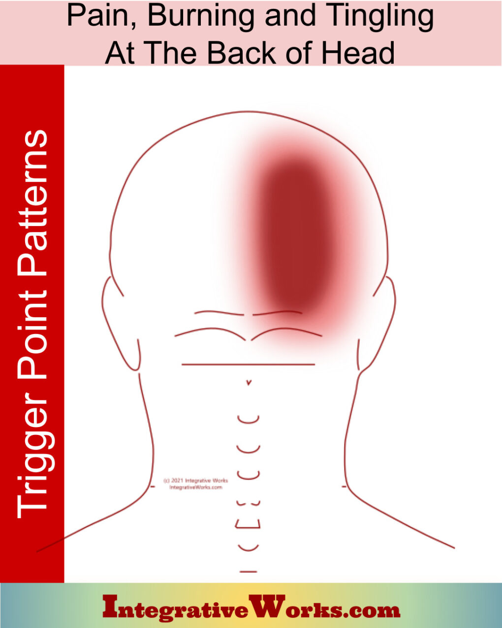
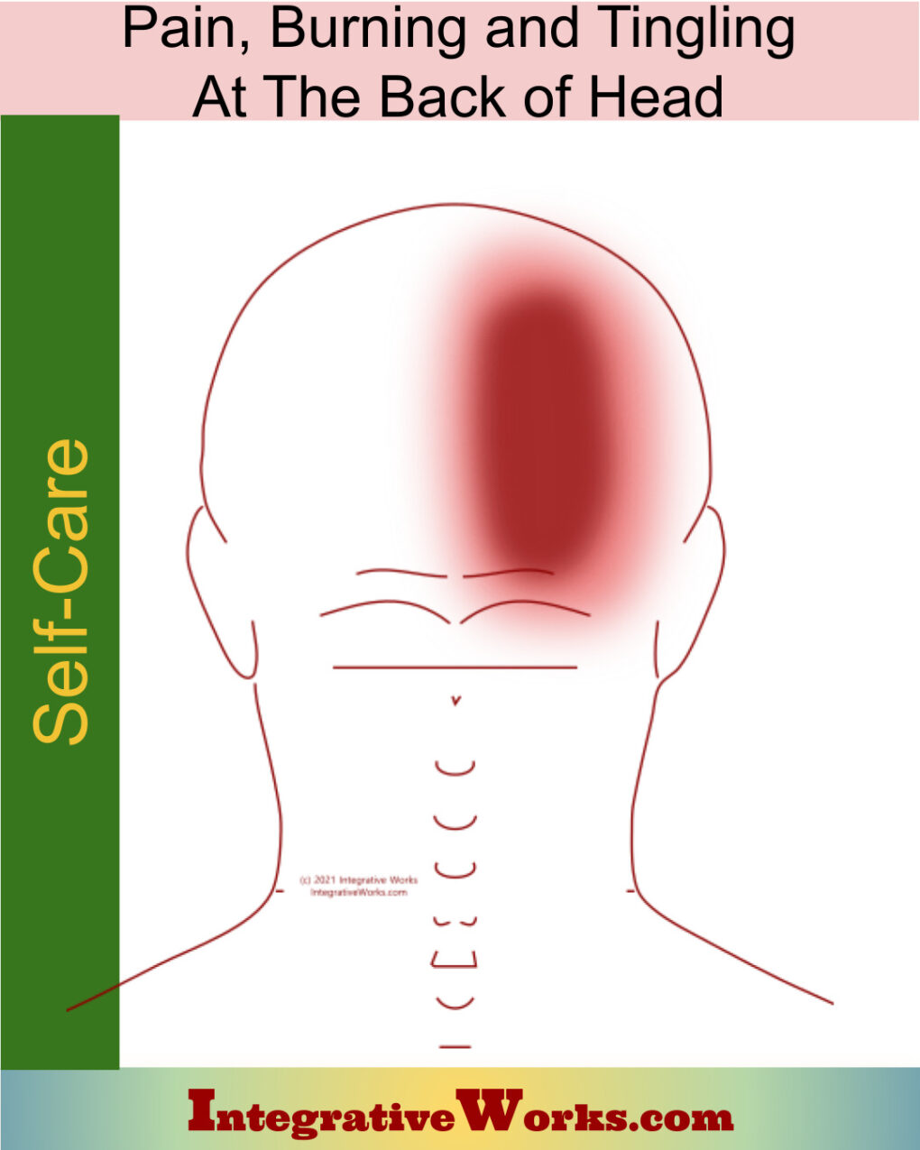
Tension in semispinalis capitis entraps the greater occipital nerve, creating posterior head pain and paraesthesia.
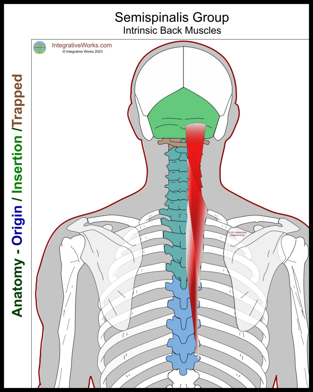
Semispinalis Muscle Group
The semispinalis muscle group includes:
Capitis attaches to the cranium base, but the other two are interspinous muscles. These muscles are deep to the extrinsic back muscles and superficial to other interspinous muscles. They provide another layer of paraspinal erectors to support the cervical muscles on the thoracic column.
Often, these muscles are considered to be another, deeper layer of the erector spinae. Comparatively, they have similar attachments and perform a similar function to the erectors just superficial to them. Likewise, they are a longer version of the multifidi muscles, which are deep to this semispinalis group.
All of the semispinalis muscles have statistically significant variations in their structure.
Related Posts
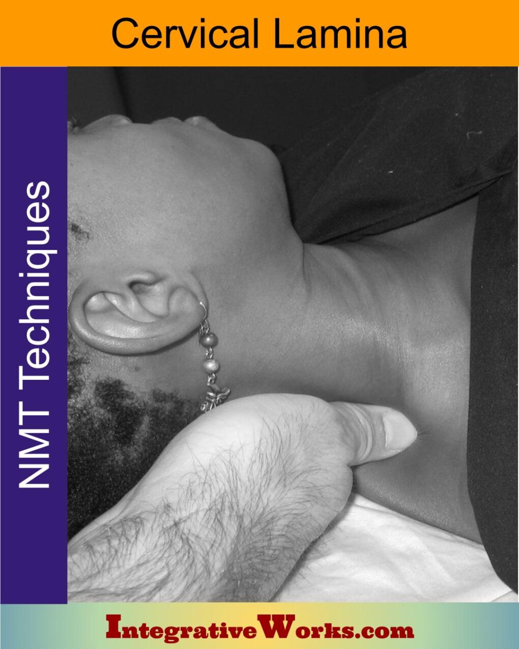
Cervical Lamina Supine – Neuromuscular Massage Protocol
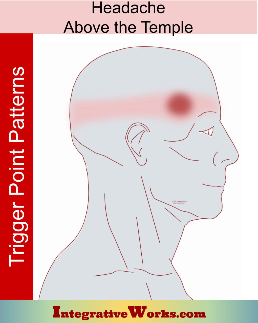
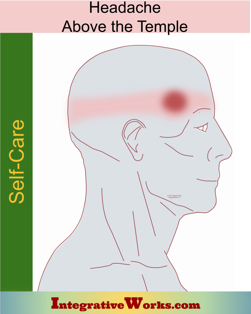
Headache Spot Just Above The Temple
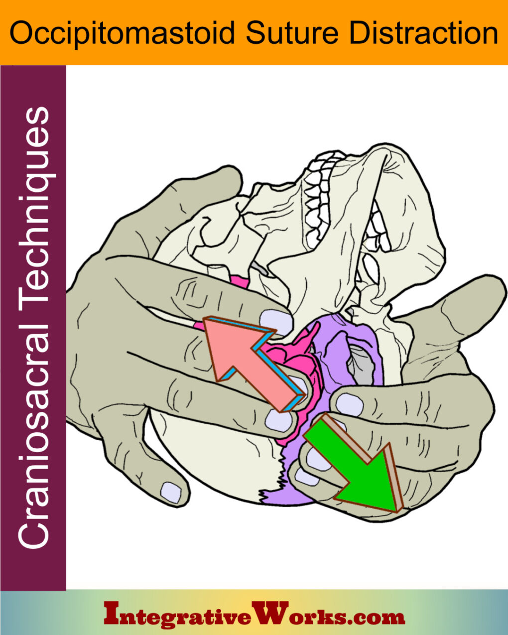
Occipitomastoid Suture Distraction- Craniosacral Techniques
Pain, Burning & Tingling on Back of Head
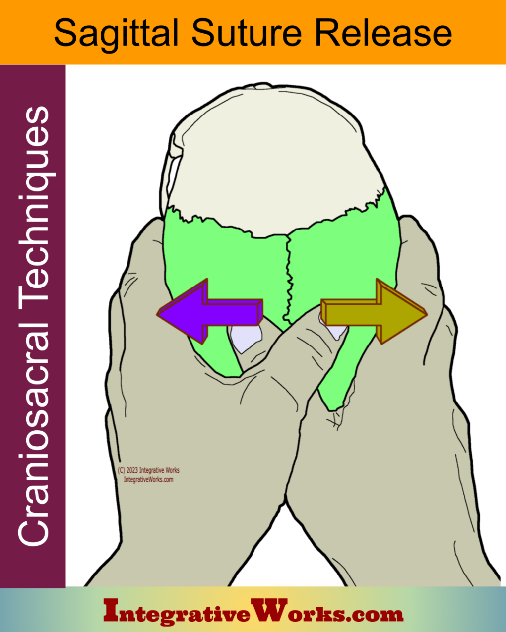
Sagittal Suture Release – Craniosacral Techniques
Self Care – Headache Spot Just Above The Temple
Self Care – Pain, burning and tingling in the back of the head
Semispinalis Capitis – Massage Therapy Notes
Sub-Occipitals Supine – Neuromuscular Massage Protocol
Support Integrative Works to
stay independent
and produce great content.
You can subscribe to our community on Patreon. You will get links to free content and access to exclusive content not seen on this site. In addition, we will be posting anatomy illustrations, treatment notes, and sections from our manuals not found on this site. Thank you so much for being so supportive.
Cranio Cradle Cup
This mug has classic, colorful illustrations of the craniosacral system and vault hold #3. It makes a great gift and conversation piece.
Tony Preston has a practice in Atlanta, Georgia, where he sees clients. He has written materials and instructed classes since the mid-90s. This includes anatomy, trigger points, cranial, and neuromuscular.
Question? Comment? Typo?
integrativeworks@gmail.com
Interested in a session with Tony?
Call 404-226-1363
Follow us on Instagram
*This site is undergoing significant changes. We are reformatting and expanding the posts to make them easier to read. The result will also be more accessible and include more patterns with better self-care. Meanwhile, there may be formatting, content presentation, and readability inconsistencies. Until we get older posts updated, please excuse our mess.

