Overview
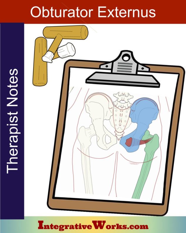
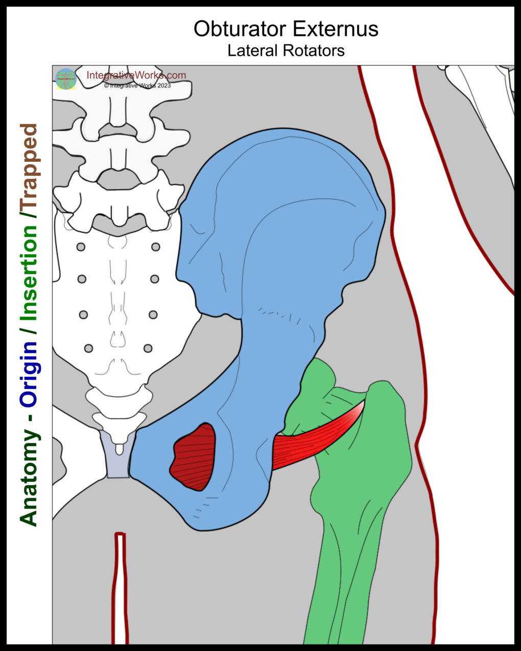
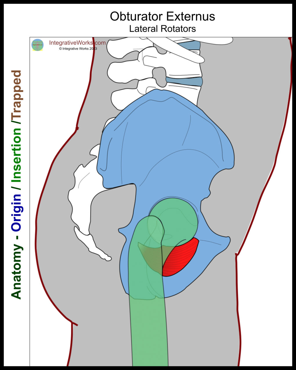
The obturator externus is a flat, triangular muscle that extends from the lower pelvis to the superior femur. The muscle has a superior belly and the main belly. Its name comes from its attachment to the external surface of the obturator foramen. Its counterpart, obturator internus, originates from the internal surface.
Origin
- medial lip of the obturator foramen
- rami of the ischium and pubis
- anterior/medial two-thirds of the external surface of the obturator membrane
Insertion
- trochanteric fossa near the superior aspect of the femoral neck
Function
- stabilization of the femoral head
- lateral rotation of the hip
- abduction of the hip, when flexed
Innervation
- posterior branch of the obturator nerve (L3-4)
Functional Considerations
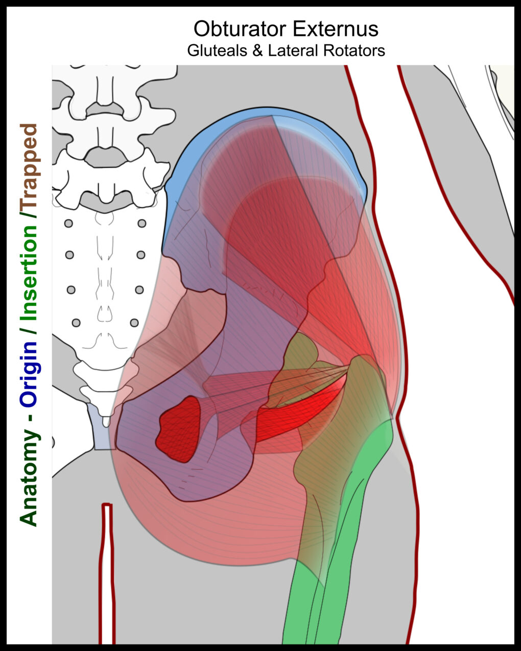
This muscle is positioned so that it assists in lateral rotation of the hip joint when standing. As the hip rotates into flexion and the insertion rotates posteriorly, it can assist in abduction of the hip joint.
When this muscle is tight, it entraps nerves that pass through the obturator and impacts bladder function.
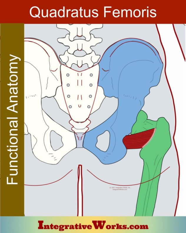
This muscle is the most anterior layer of the lateral rotators. Its origin is more accessible when the patient is supine than prone. Posteriorly, its insertion is more easily palpated after the superficial glutes, and the quadratus femoris release.
Anomalies, Etc.
This muscle is highly variable.
A muscle splits off the obturator externus in utero in about one-third of people. This muscle originates on the inferior pubic bone. Its insertion is variable but usually attaches to the aponeurosis of the adductor minimus. However, it may also attach along the femur along the pectineal line or lesser trochanter.
Without the superior belly, the main belly occurs in about twenty percent of cases.
Occasionally, a bursa separates the obturator externus from the femur.
Lateral Rotator or Adductor?
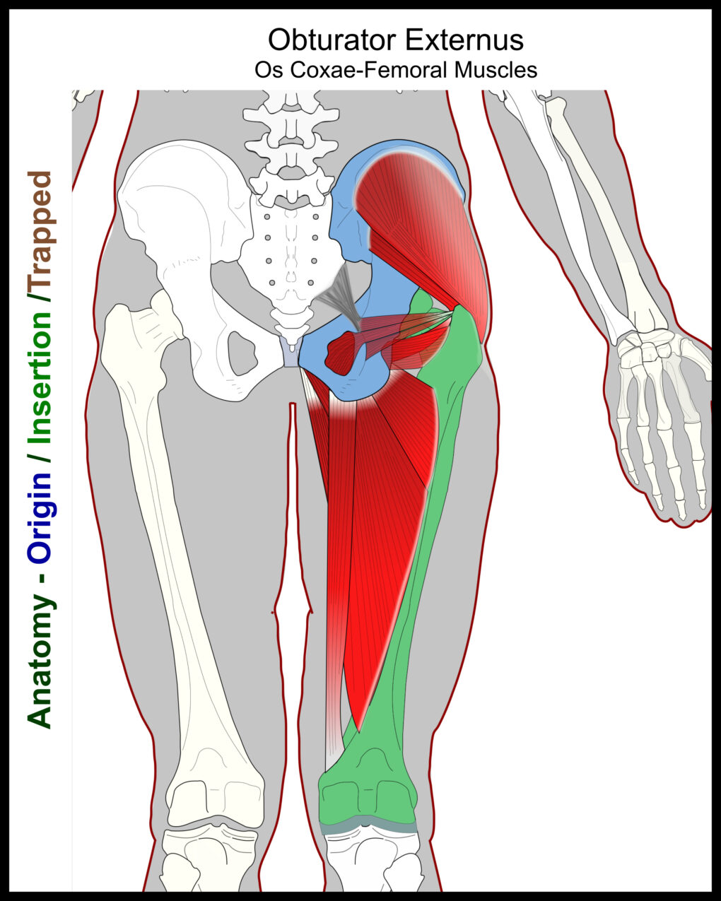
It is usually considered to be part of the external rotators of the thigh. Because of the anomalies that attach a bit lower on the thigh, it is sometimes considered to be part of the medial thigh group.
In other words, the origin on the anterior surface of the ischium with the insertion on the posterior femur makes it look like an adductor, especially when the femoral attachment is a bit lower.
Related Posts
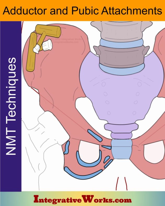
Adductor Pubic Attachments – Neuromuscular Massage Protocol
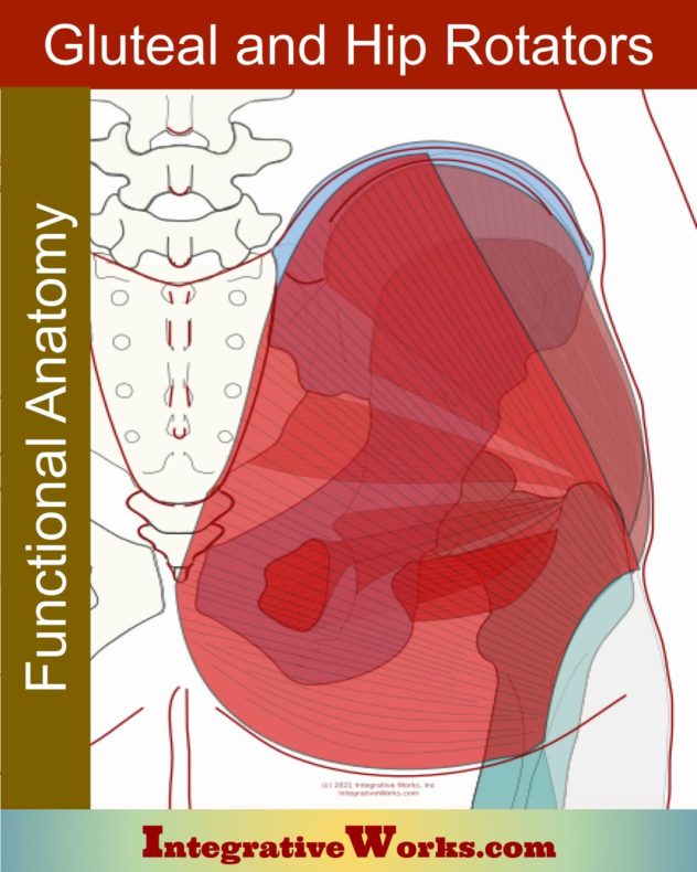
Gluteal and Lateral Hip Rotator Muscles – Functional Anatomy
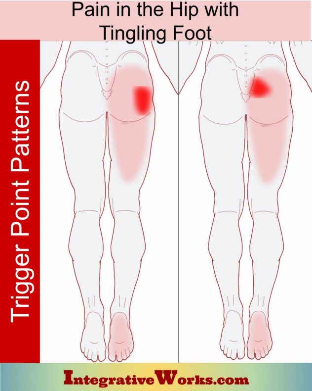
Hip Pain with Tingling Bottom of Foot
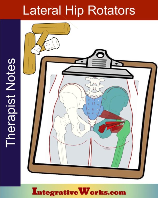
Lateral Hip Rotators – Massage Therapy Notes
Obturator Externus – Massage Notes
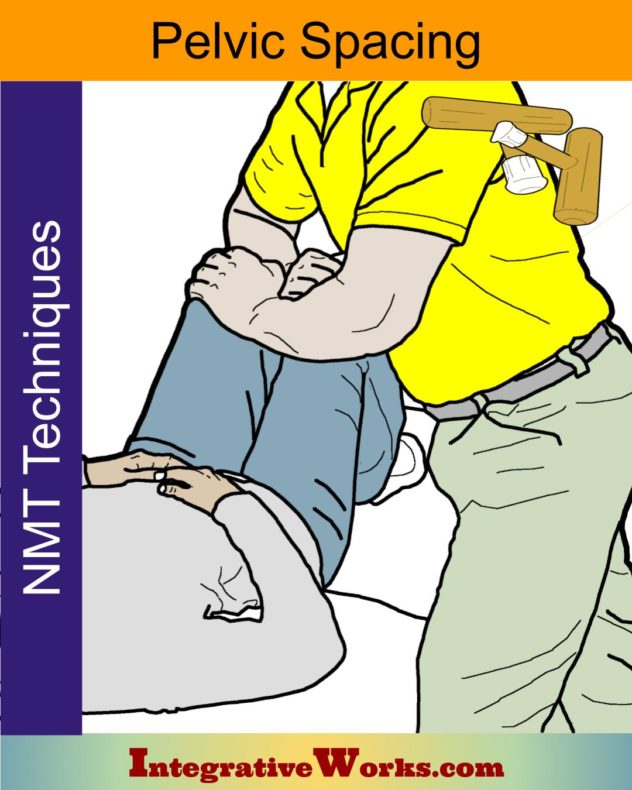
Pelvic Spacing – Techniques
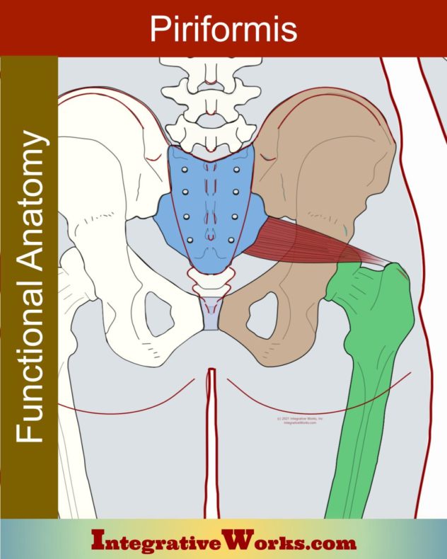
Piriformis – Functional Anatomy
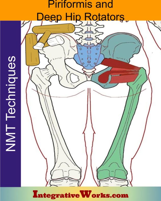
Piriformis & Deep Hip Rotators – Neuromuscular Massage Protocol
Quadratus Femoris – Functional Anatomy
Self Care – Releasing hip trigger points with a tennis ball
Self-Care – Hip Rotators
Self-Care – Side of Hip Pain from Childbirth or Cycling
Side of Hip Pain after Pregnancy or Cycling
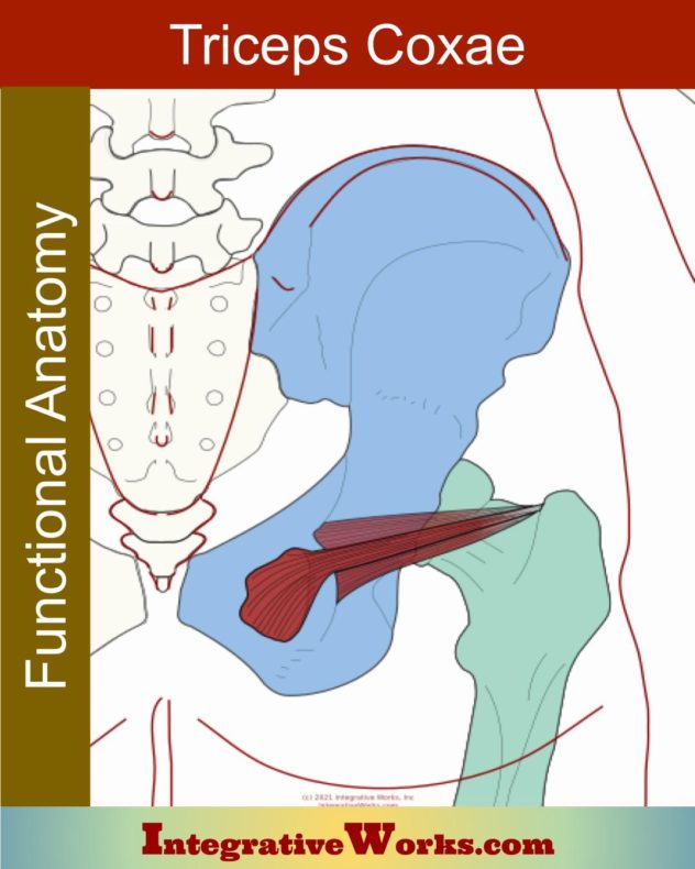
Triceps Coxae
Support Integrative Works to
stay independent
and produce great content.
You can subscribe to our community on Patreon. You will get links to free content and access to exclusive content not seen on this site. In addition, we will be posting anatomy illustrations, treatment notes, and sections from our manuals not found on this site. Thank you so much for being so supportive.
Cranio Cradle Cup
This mug has classic, colorful illustrations of the craniosacral system and vault hold #3. It makes a great gift and conversation piece.
Tony Preston has a practice in Atlanta, Georgia, where he sees clients. He has written materials and instructed classes since the mid-90s. This includes anatomy, trigger points, cranial, and neuromuscular.
Question? Comment? Typo?
integrativeworks@gmail.com
Follow us on Instagram

*This site is undergoing significant changes. We are reformatting and expanding the posts to make them easier to read. The result will also be more accessible and include more patterns with better self-care. Meanwhile, there may be formatting, content presentation, and readability inconsistencies. Until we get older posts updated, please excuse our mess.

