Overview
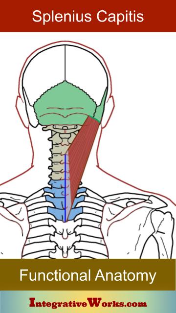
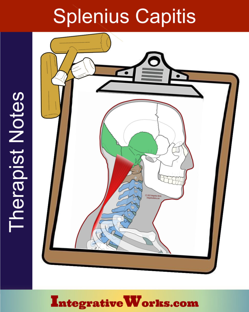
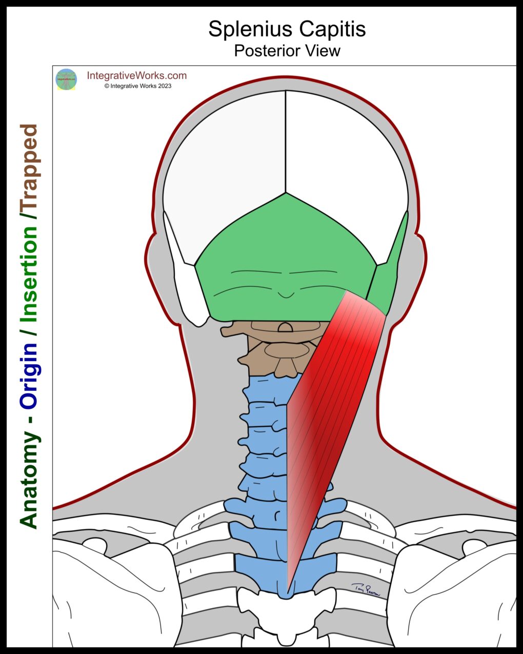
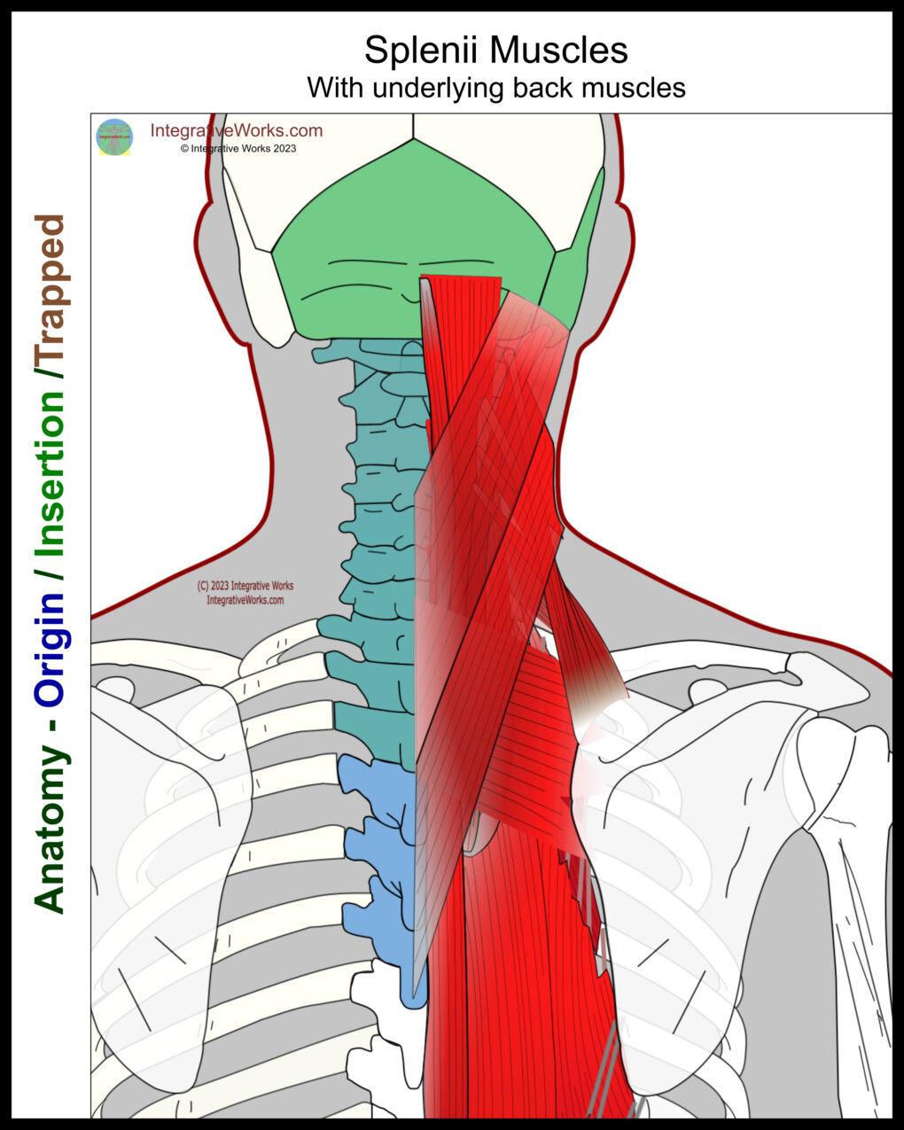
Splenius capitis is a broad, flat sheet that originates along the mid-line of the neck’s base. It wraps the lateral muscles and inserts onto the lateral base of the skull. Its relationship with the splenius cervicis complicates the anatomy.
Origin
- midline fascia over the ligamentum nuchae from C3 – C7
- the spinous processes of T1-T3
Insertion
- underneath the SCM on the superior nuchal line of the temporal bone and occipital bone
Function
- extension of the head
Innervation
- posterior rami of C3-4
Origin
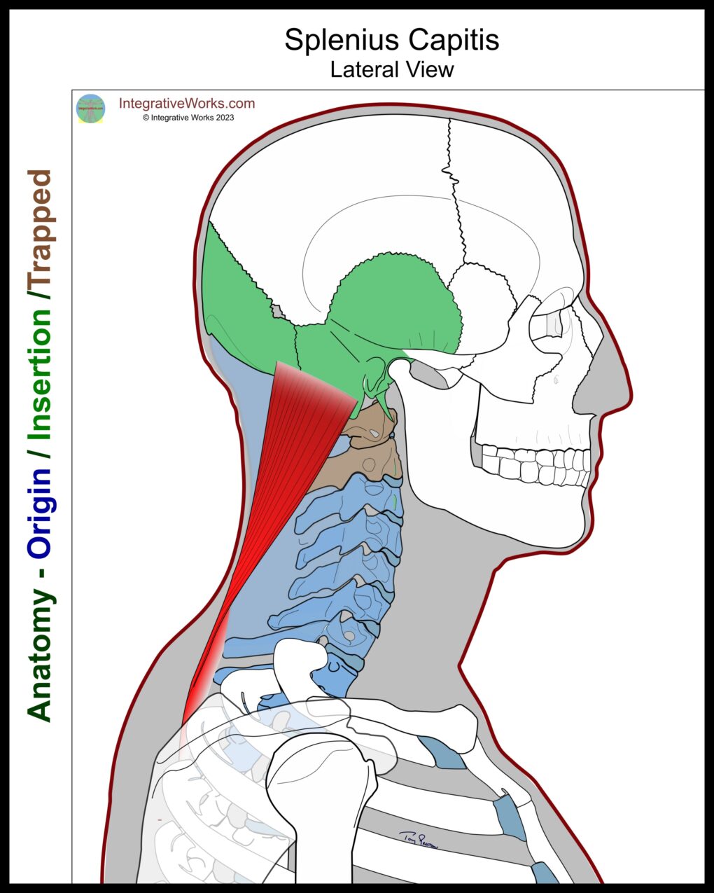
- midline fascia over the ligamentum nuchae from C3 – C7
- the spinous processes of T1-T3
Insertion
- underneath the SCM on the superior nuchal line of the temporal bone and occipital bone
Function
- extension of the head
Innervation
- posterior rami of C3-4
Downloadable on Etsy
Attachment Details
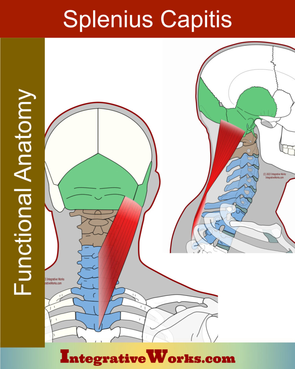
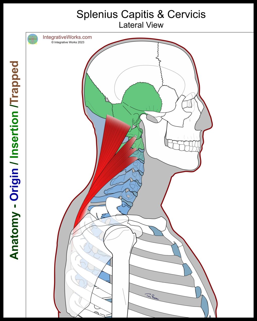
The splenii muscles have a long attachment along the center of the spine. Inferiorly, this sheet of muscle anchors from about C4 on the nuchal ligament to the spinous process of T6.
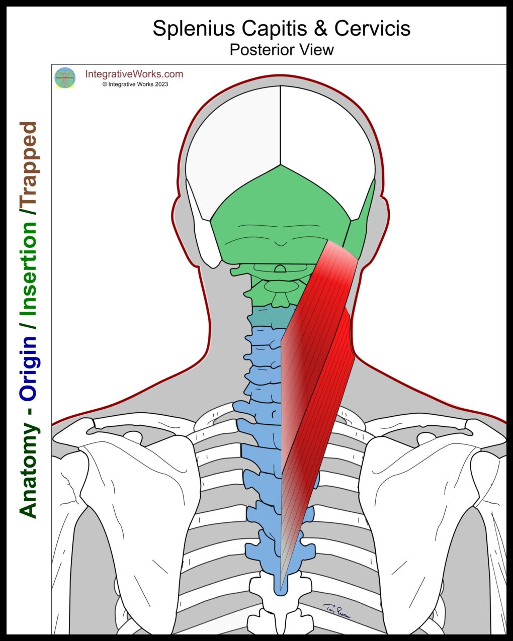
They form a broad, flat veneer of muscle that splits to become splenius capitis and splenius cervicis. This strap-like structure wraps around the lateral muscles of the neck. Along the way, the myofascial sheet divides into two sections as it extends superiorly. The splenius capitis attaches to the occipital and temporal bones while the splenius cervicis wraps around lateral neck musculature to attach to the spinous processes of C1-C3.
Downloadable on Etsy
Functional Considerations
As you can see from the illustration, the bones of origin (in blue) form a broad base from which the head and neck can be turned and extended.
A portion of the origin attaches through the nuchal ligament to the lower cervical vertebrae. There is stability along the lower cervical vertebrae with flexibility against contralateral musculature. This flexible attachment allows for play between the stable origin on the thoracic vertebrae and the bony attachments along the upper cervical vertebrae and cranial base.
Downloadable on Etsy
The Bandage
Splenii comes from a word meaning “bandage.” These muscles wrap around the deeper muscle of the lateral neck. Splenius capitis and cervicis form a “bandage” to retain those muscles.
Wikipedia entry for Splenius Capitis
Posts Related to Splenius Capitis
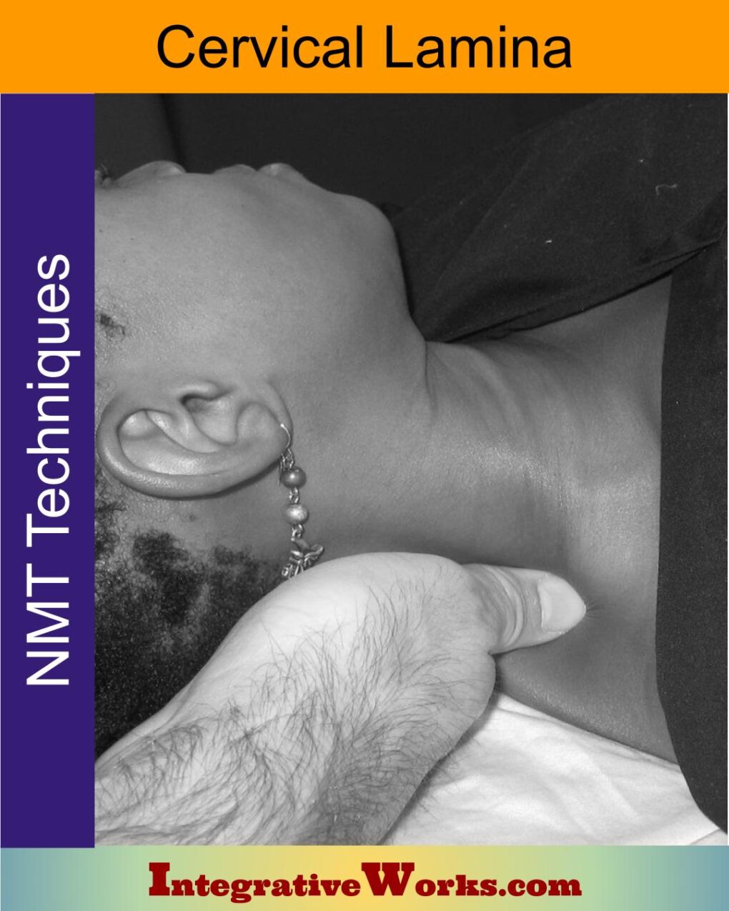
Cervical Lamina Supine – Neuromuscular Massage Protocol
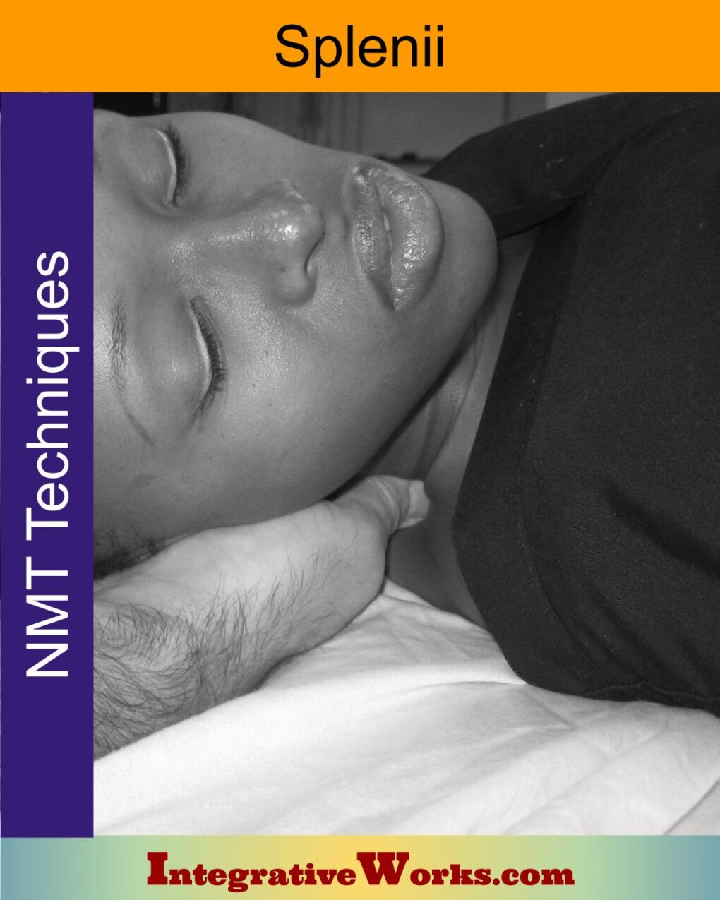
Cervical Splenii Supine – Neuromuscular Massage Protocol
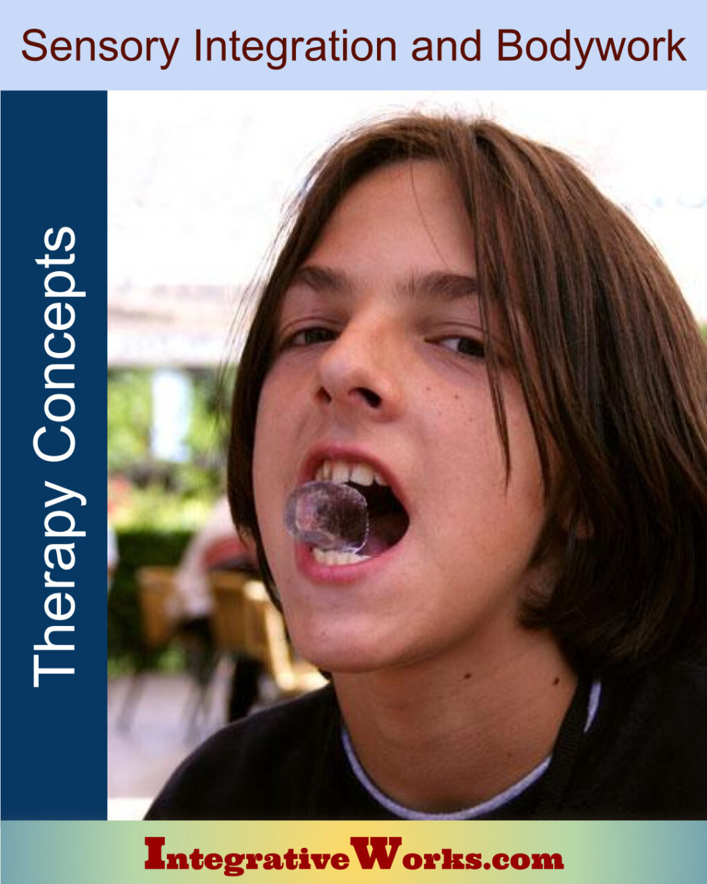
Connecting Sensory Integration and Bodywork
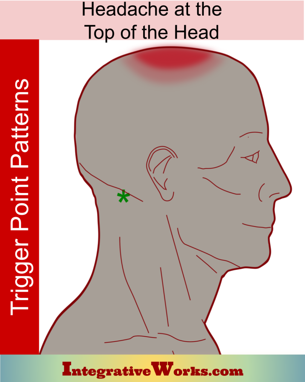
Headache on the Top of Your Head
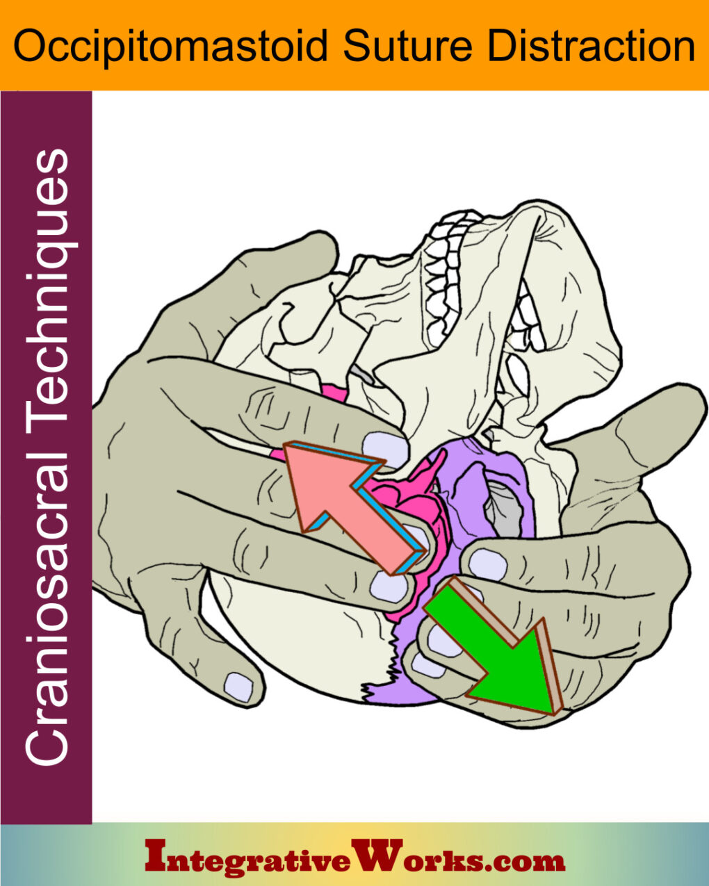
Occipitomastoid Suture Distraction- Craniosacral Techniques
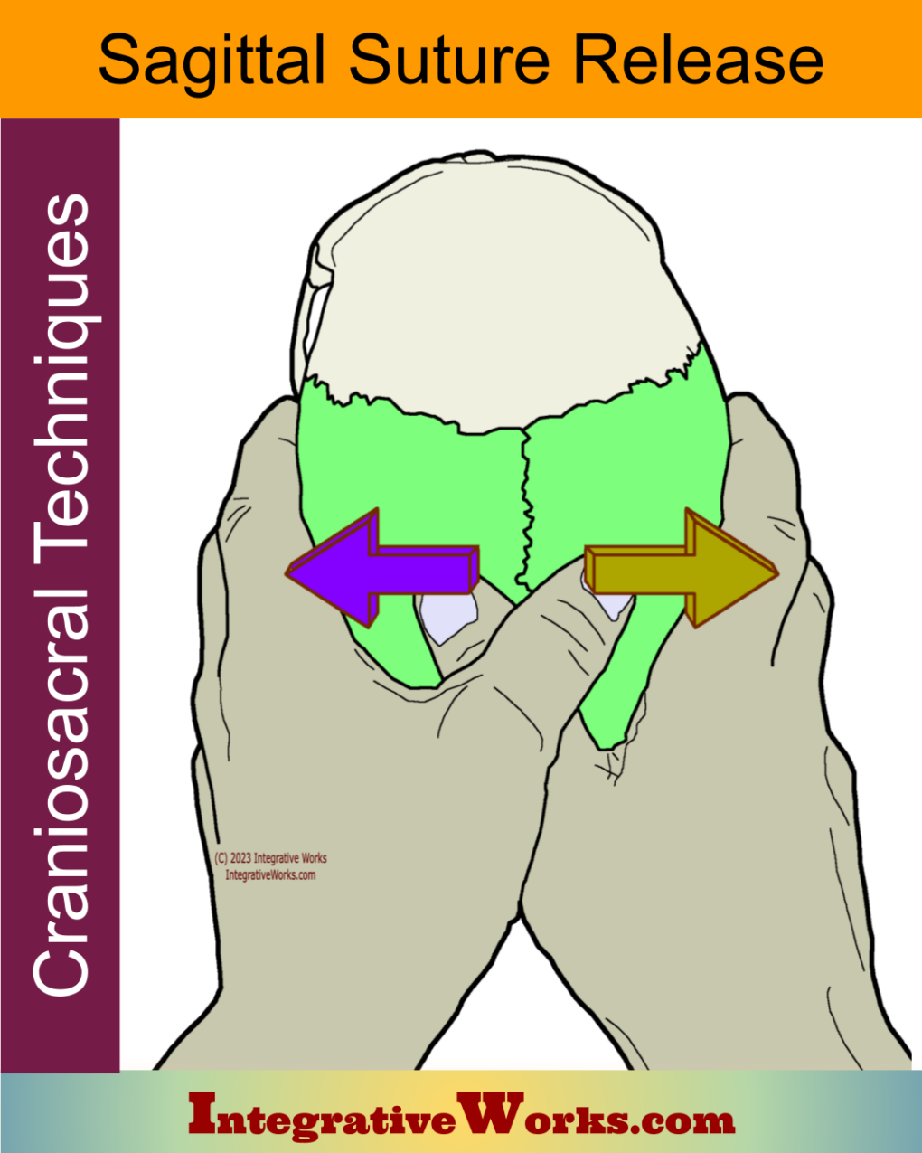
Sagittal Suture Release – Craniosacral Techniques
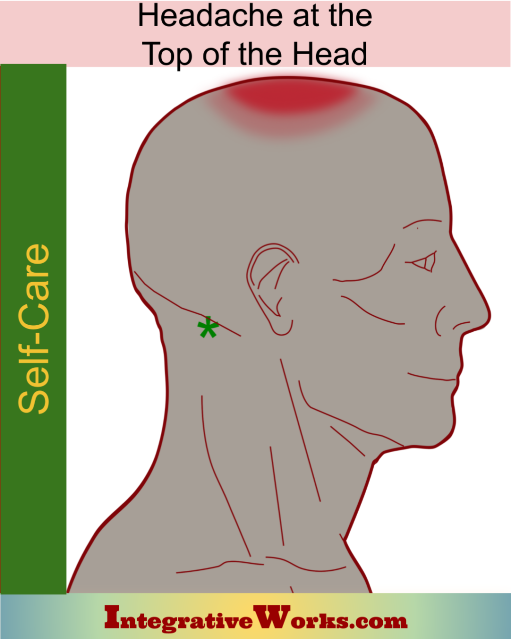
Self Care – Headache on the Top of Your Head
Splenius Capitis – Massage Therapy Notes
Splenius Capitis – Functional Anatomy
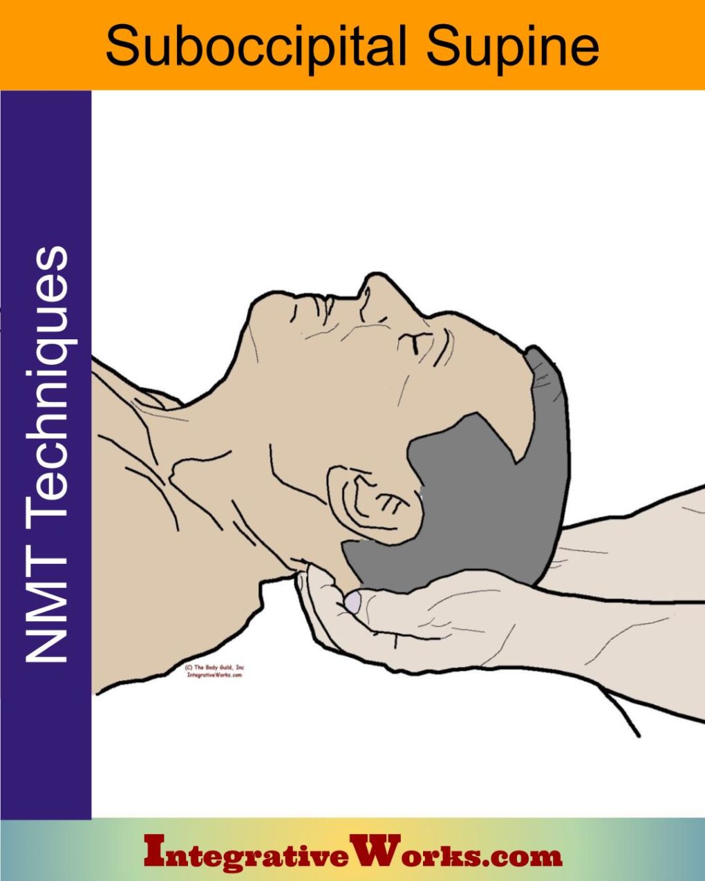
Sub-Occipitals Supine – Neuromuscular Massage Protocol
Support Integrative Works to
stay independent
and produce great content.
You can subscribe to our community on Patreon. You will get links to free content and access to exclusive content not seen on this site. In addition, we will be posting anatomy illustrations, treatment notes, and sections from our manuals not found on this site. Thank you so much for being so supportive.
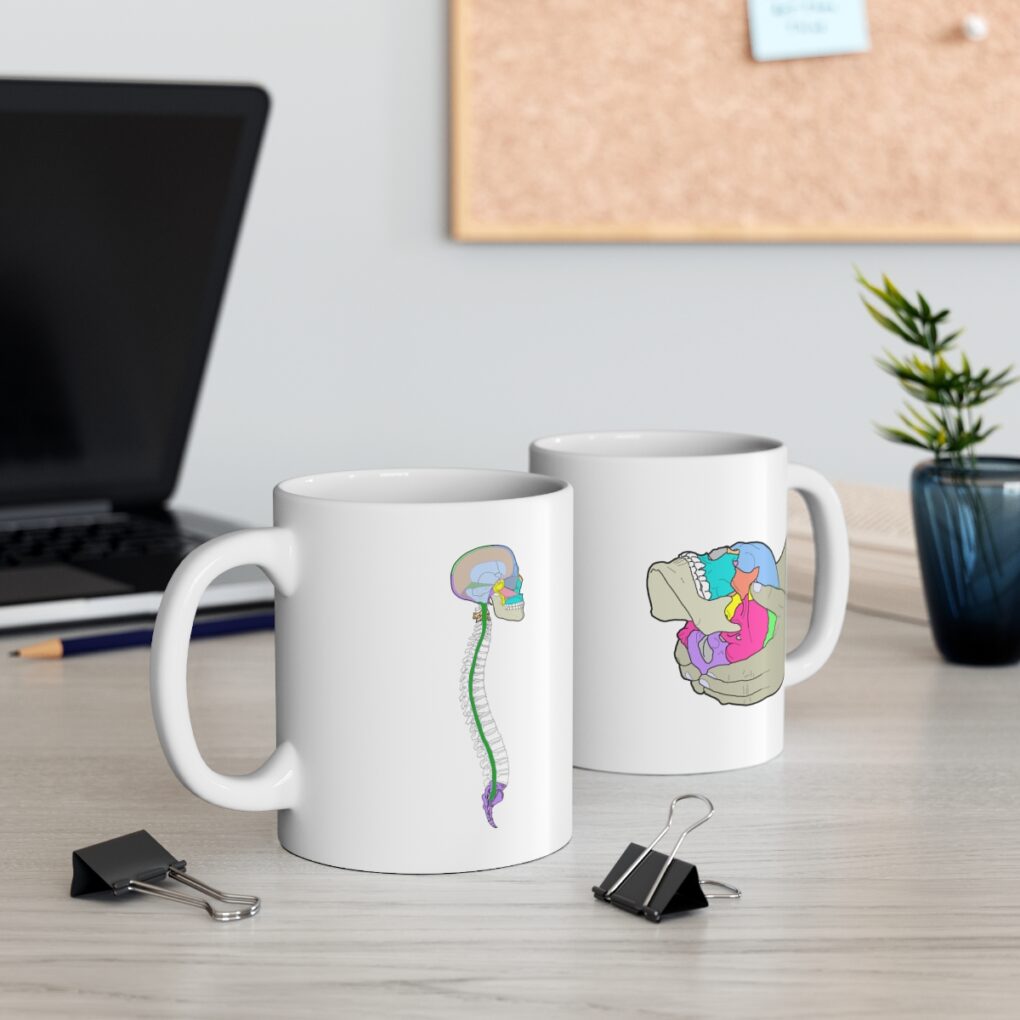
Cranio Cradle Cup
This mug has classic, colorful illustrations of the craniosacral system and vault hold #3. It makes a great gift and conversation piece.
Tony Preston has a practice in Atlanta, Georgia, where he sees clients. He has written materials and instructed classes since the mid-90s. This includes anatomy, trigger points, cranial, and neuromuscular.
Question? Comment? Typo?
integrativeworks@gmail.com
Interested in a session with Tony?
Call 404-226-1363
Follow us on Instagram
*This site is undergoing significant changes. We are reformatting and expanding the posts to make them easier to read. The result will also be more accessible and include more patterns with better self-care. Meanwhile, there may be formatting, content presentation, and readability inconsistencies. Until we get older posts updated, please excuse our mess.
