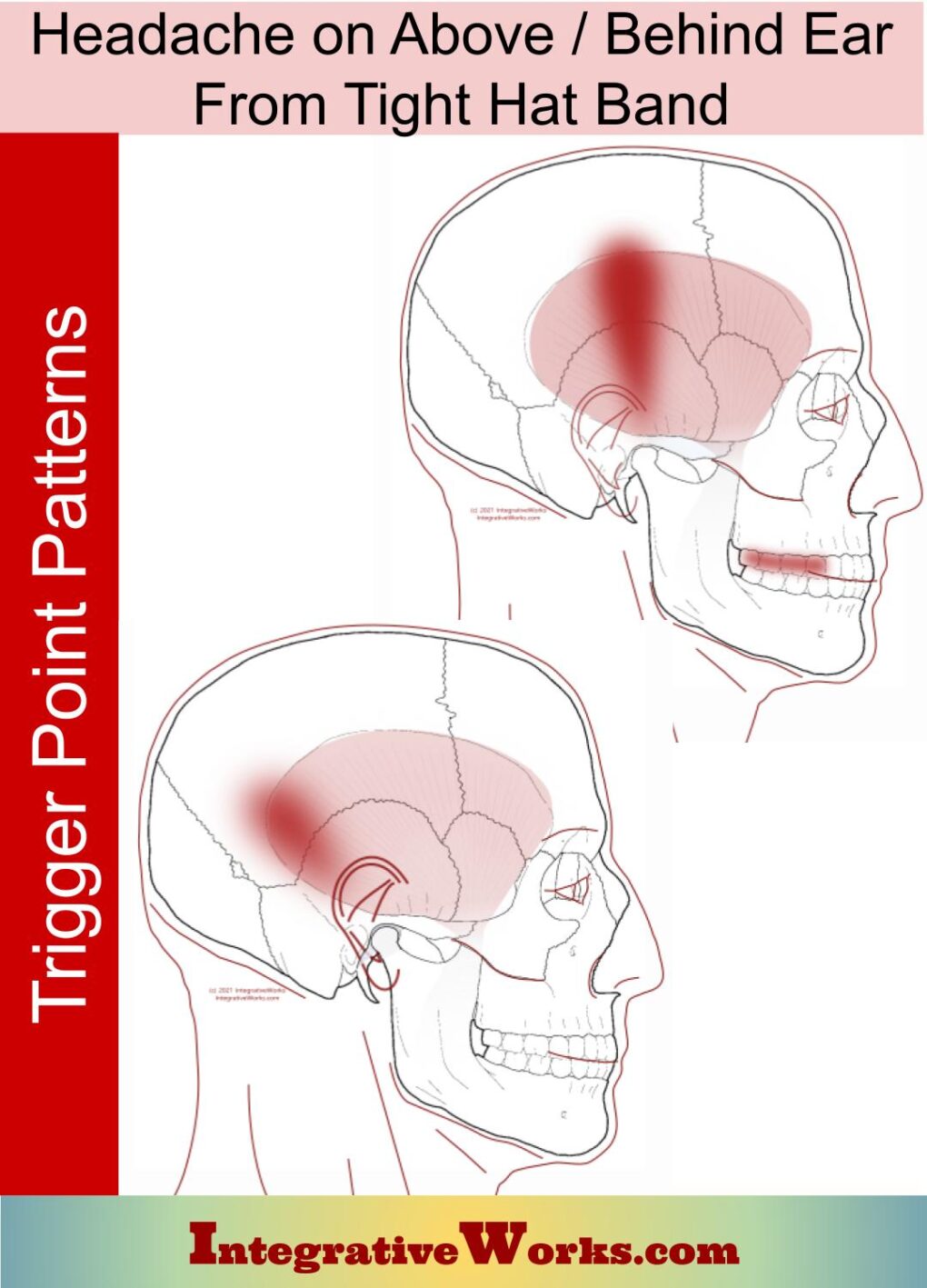
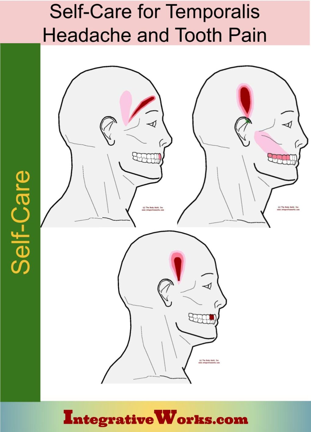
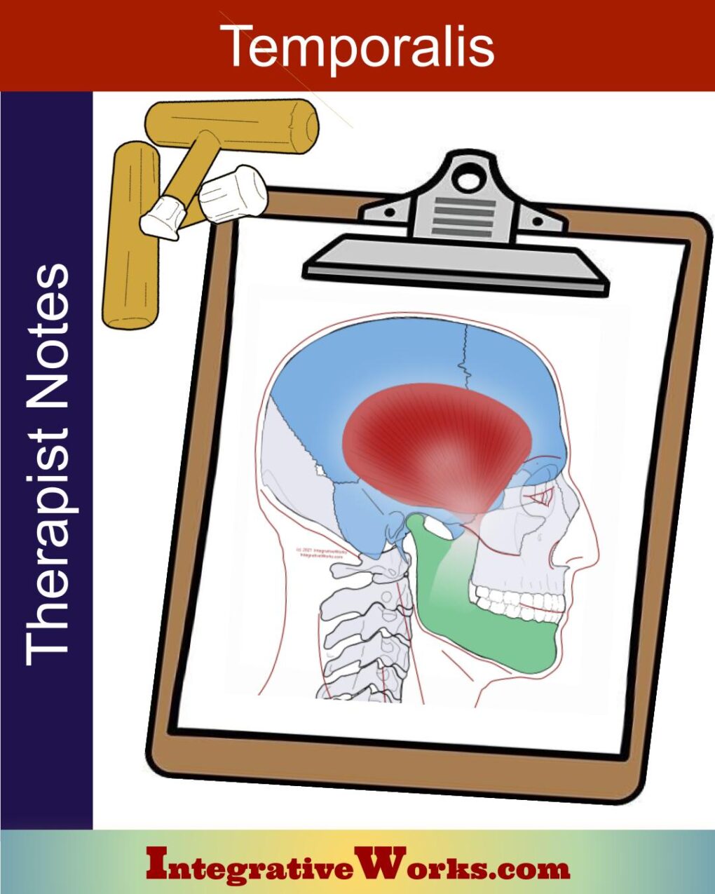
Overview
The temporalis muscle is a flat muscle on the lateral surface of the head. Its anatomy is a bit more complicated than expected when you look closer.
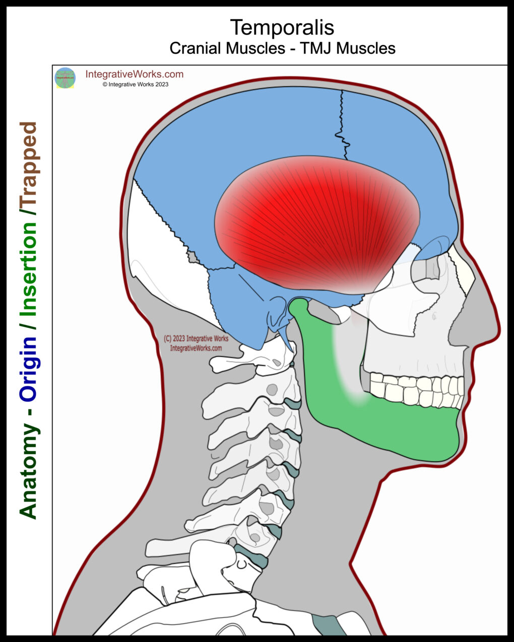
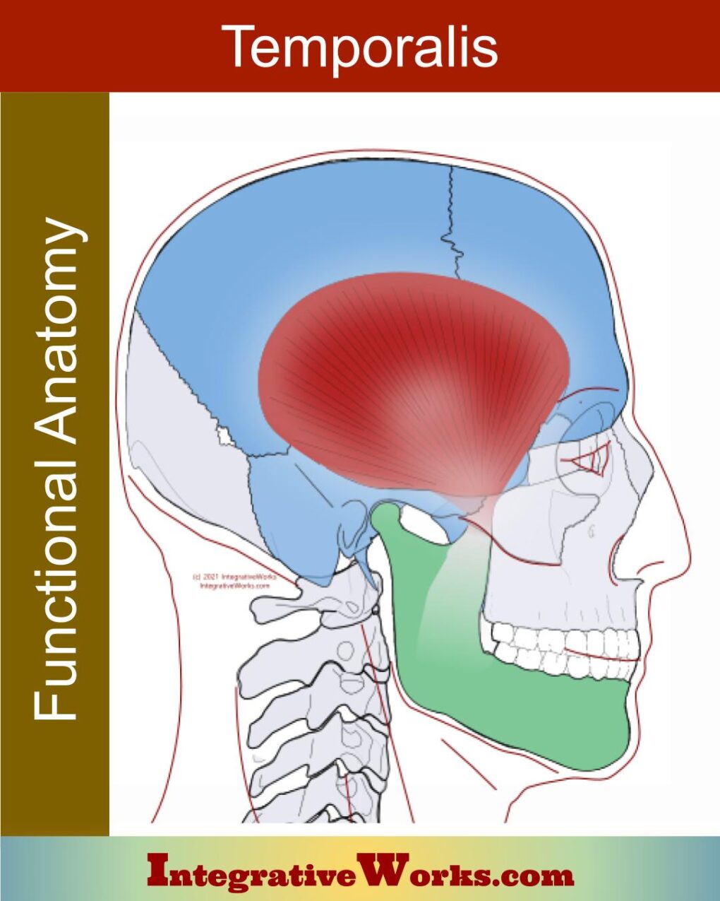
Origin
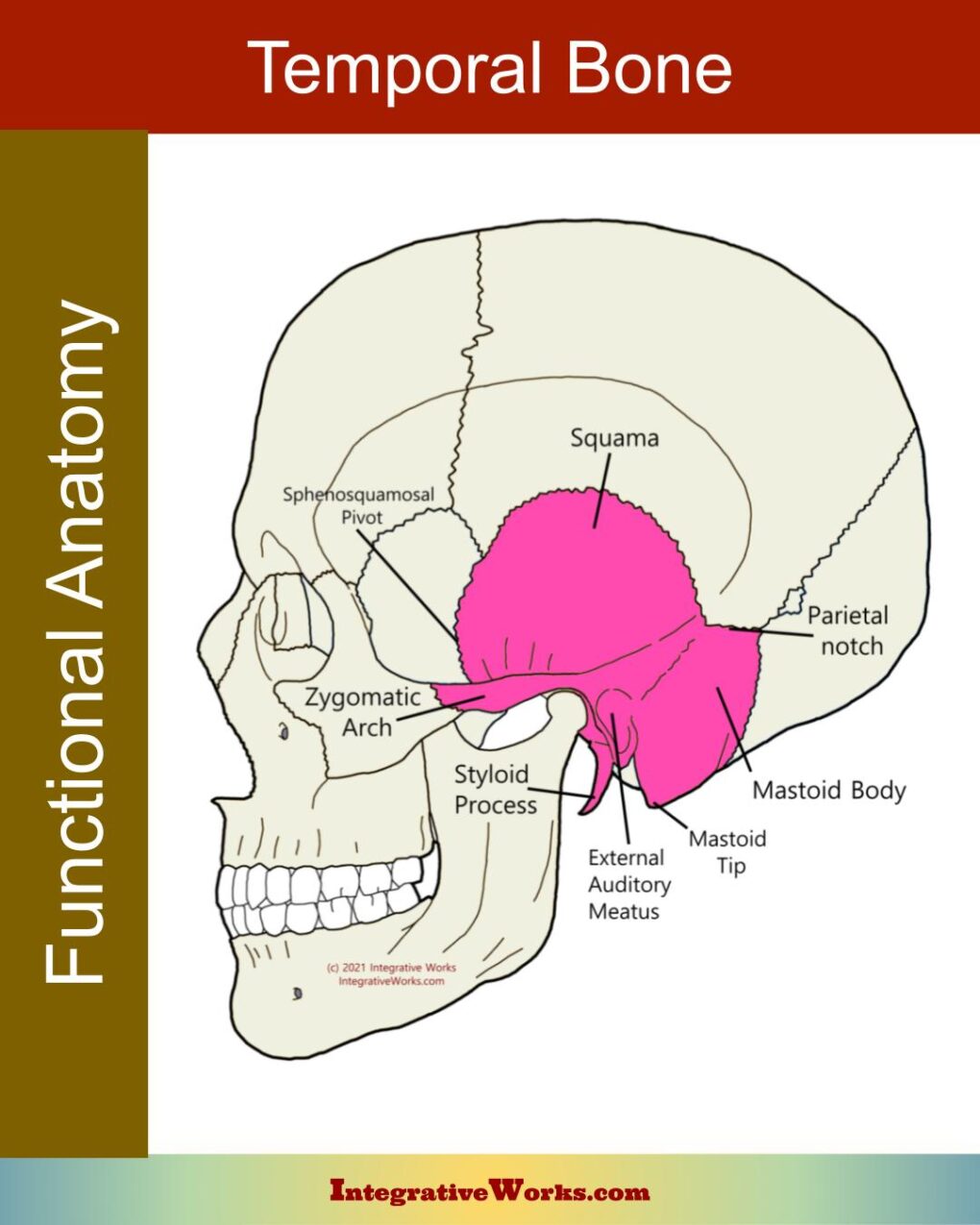
- temporal fossa of the parietal and frontal bone
- greater wing of the sphenoid
- medial surface of the zygomatic arch
- temporal fascia
Insertion
- coronoid process of the mandible
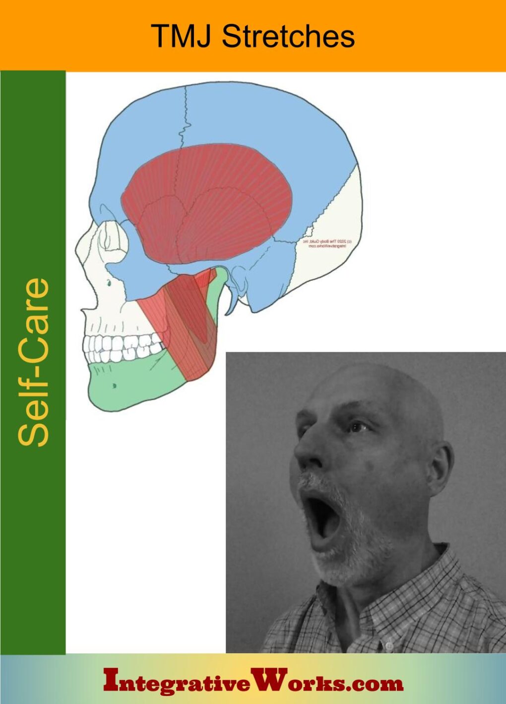
Function
- elevate the mandible
Innervation
- deep temporal branches of the mandibular nerve
Temporal Fascia
The temporal fascia covers the temporalis. Again, studies show varying results. Most texts discuss a multi-layered covering of the temporalis. Most of them seem to acknowledge a deep layer that adheres to the muscle. However, this study says that after 167 dissections, it doesn’t exist. Another study of 27 hemifaces states clearly that the superficial fascia distinctly separates from the deep fascia.
Consequently, this study concluded that harvesting the superficial fascia is safe for rhinoplasty. This study has an interesting discussion about a general structure that has a superficial and deep fascia. It also discusses how the layers split inconsistently. It discourages the terms “deep temporal fascia” and “superficial temporal fascia” they are confusing.
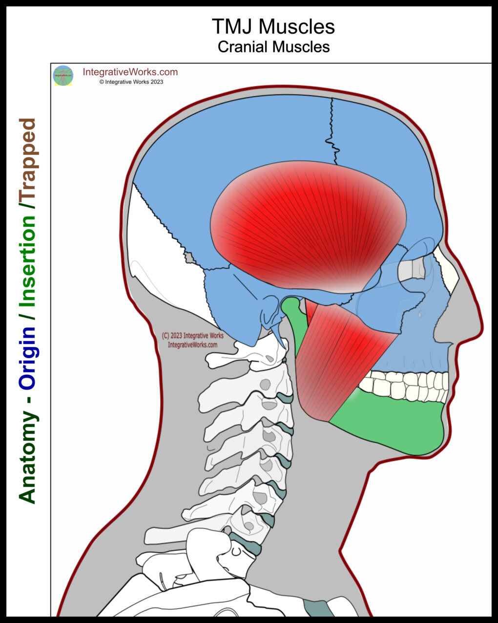
The temporalis anatomy, like infraspinatus and gluteus medius, reveals a triangular muscle with converging fibers. They all have a thick fascial covering. However, their attachments are unexpectedly complicated.
Attachment Details
To my surprise, most sites reported the origin of this muscle. They commonly state that it is the temporal fossa of the parietal bone. Any picture shows it differently. So, I went to the cadaver studies.
As expected, this study shows that it attaches to the frontal, parietal, and sphenoid bones. To my surprise, every dissection showed that it skips over the temporal squama. However, it attaches to the medial aspect of the zygomatic arch via the temporal fascia.
Insertion
The insertion is also more extensive than it appears at first glance. Commonly, illustrations show that it attaches firmly to the apex of the coracoid process. In fact, it usually extends down the medial surface to the anterior borders of the coronoid process and ramus. Its fibers often extend down and forward o the first molar.
Anomalies, Etc.
There seems to be a notion that this muscle has multiple sections that could identify as a different muscle. However, studies show that they have the same innervation and blood supply and should be considered part of the temporalis.
Related Posts
Support Integrative Works to
stay independent
and produce great content.
You can subscribe to our community on Patreon. You will get links to free content and access to exclusive content not seen on this site. In addition, we will be posting anatomy illustrations, treatment notes, and sections from our manuals not found on this site. Thank you so much for being so supportive.
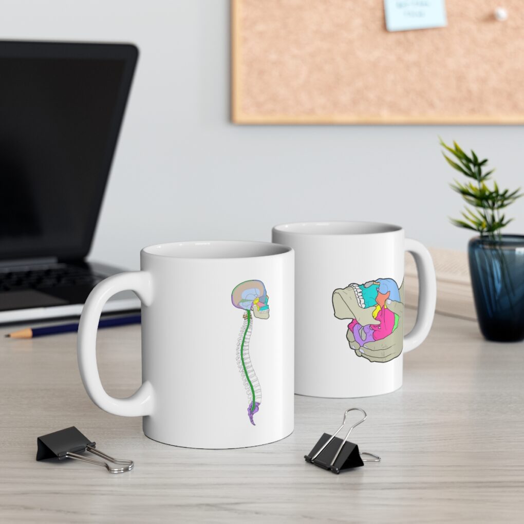
Cranio Cradle Cup
This mug has classic, colorful illustrations of the craniosacral system and vault hold #3. It makes a great gift and conversation piece.
Tony Preston has a practice in Atlanta, Georgia, where he sees clients. He has written materials and instructed classes since the mid-90s. This includes anatomy, trigger points, cranial, and neuromuscular.
Question? Comment? Typo?
integrativeworks@gmail.com
Interested in a session with Tony?
Call 404-226-1363
Follow us on Instagram

*This site is undergoing significant changes. We are reformatting and expanding the posts to make them easier to read. The result will also be more accessible and include more patterns with better self-care. Meanwhile, there may be formatting, content presentation, and readability inconsistencies. Until we get older posts updated, please excuse our mess.