Overview
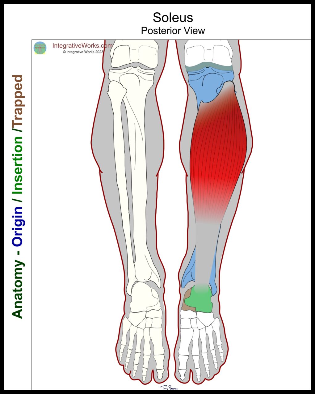
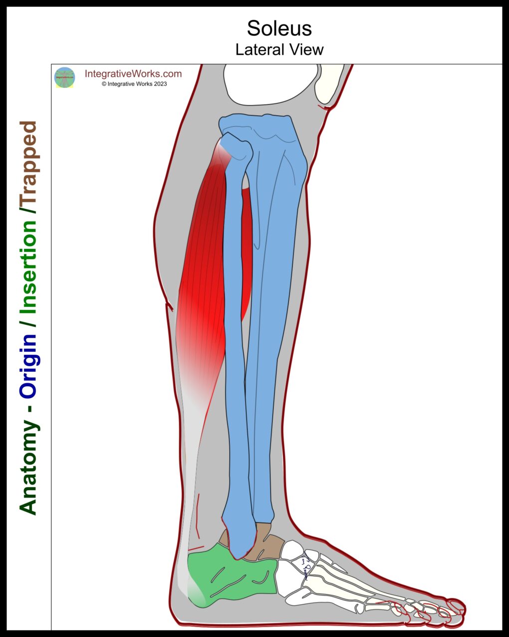
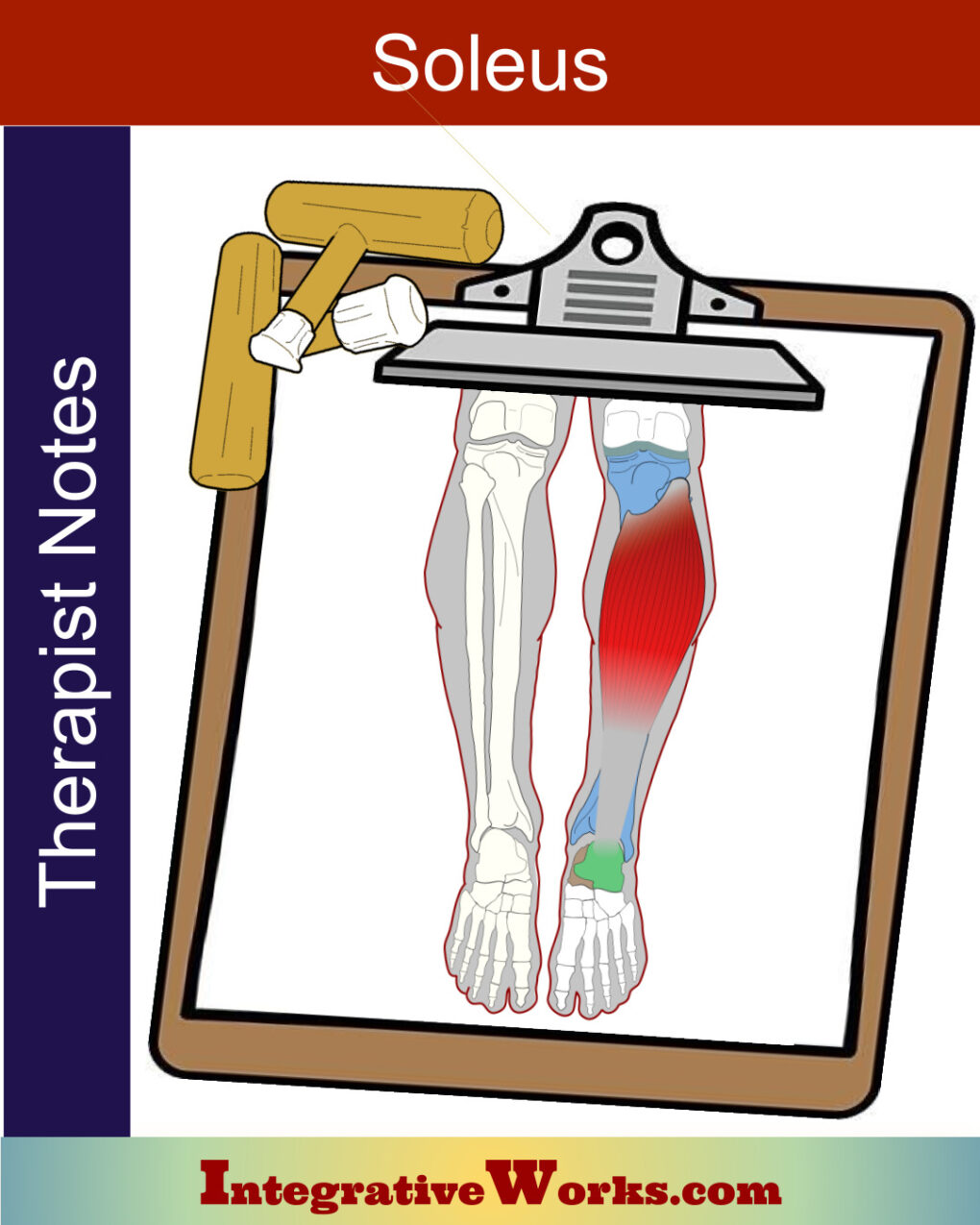
Here, you will find the anatomy of the soleus muscle. It is a flat muscle in the posterior low leg and part of the triceps surae.
Origin
- head of the fibula
- soleal line of proximal, medial tibia
Insertion
- Calcaneus
Function
- plantar flexion of the ankle
Nerve
- tibial nerve
Triceps Surae
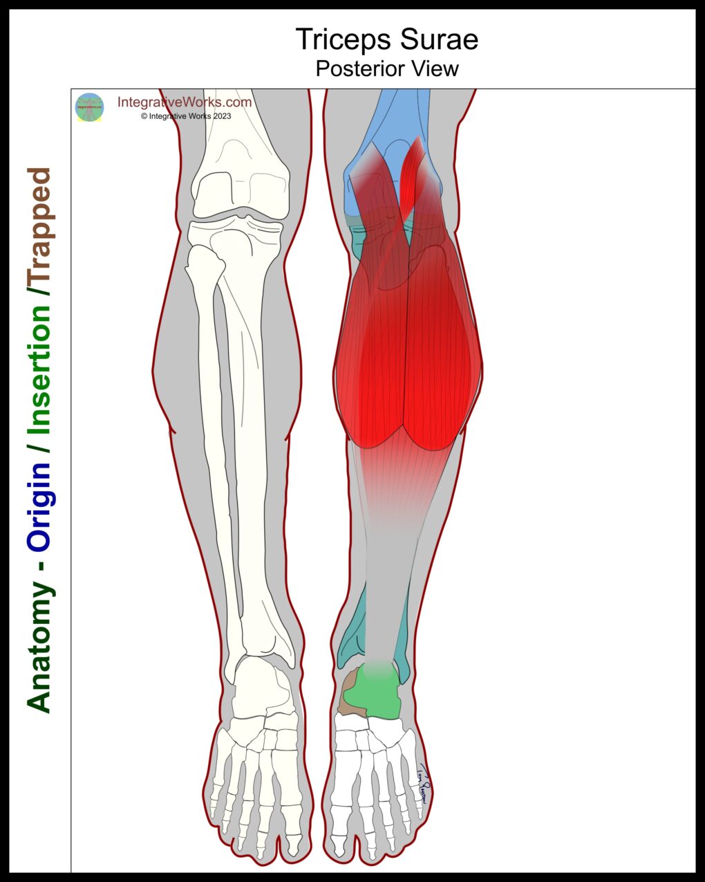
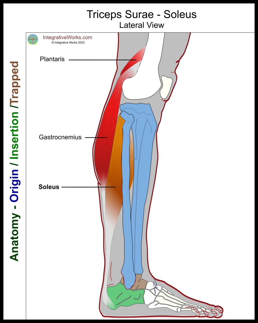
As the name implies, the triceps surae is a part structure of the calf. It consists of three muscles: the gastrocnemius, the soleus, and the plantaris.
The gastrocnemius is more superficial and originates on the distal femur. The bulbous shape of the gastrocnemius gives the upper calf its distinct form. On the other hand, the soleus, located beneath the gastrocnemius, originates on the proximal fibula and tibia. They attach to the calcaneus via the Achilles tendon.
The plantaris muscle is often considered a vestigial muscle absent in 7-20% of limbs. This muscle originates on the lateral, distal femur. It has a small belly in the popliteal fossa. The insertion tendon courses along the medial border of the soleus and Achilles tendon. Finally, it inserts onto the calcaneus with the Achilles tendon.
Functional Considerations
Most research agrees that the soleus is a postural muscle. Hence, it is more involved in maintaining a standing position than the gastrocnemius, which is more active in running, jumping, etc.
Its attachment to the fibular head connects it to the lateral hamstring. Consequently, it has a notable impact on the low back. Frequently, patients experience relief in their low back pain by stretching the soleus muscle.
Popliteal Artery, Vein
& Tibial Nerve
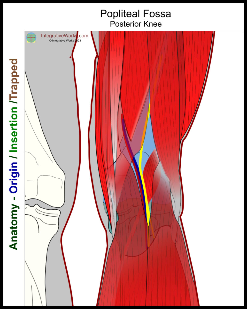
The popliteal artery and vein enter the popliteal fossa from the adductor hiatus in the adductor magnus muscle. At that point, they pass under the medial hamstrings. Next, they join the tibial nerve as it branches off the sciatic nerve. Then, they pass over the plantaris and popliteus muscles. Next, they pass under the heads of gastrocnemius. Finally, they pass under the lip of the soleus at the soleus canal.
Most commonly, circulation is compromised by tightness in the soleus canal or adductor hiatus.
Anomalies, Etc.
Rarely, Soleus has an accessory belly. Typically, it is in the distal portion. Upon initial evaluation, it is often mistaken for a tumor or other pathological growth.
Related Posts
Support Integrative Works to
stay independent
and produce great content.
You can subscribe to our community on Patreon. You will get links to free content and access to exclusive content not seen on this site. In addition, we will be posting anatomy illustrations, treatment notes, and sections from our manuals not found on this site. Thank you so much for being so supportive.
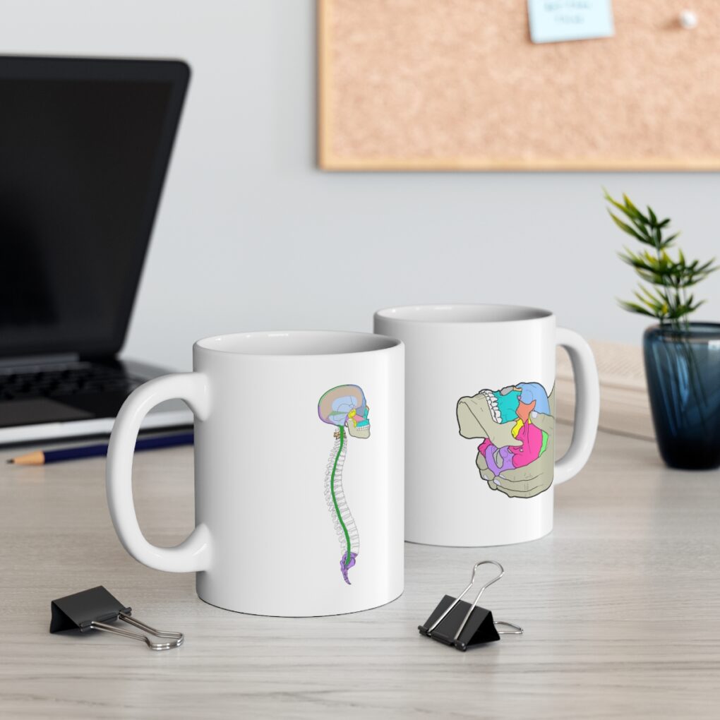
Cranio Cradle Cup
This mug has classic, colorful illustrations of the craniosacral system and vault hold #3. It makes a great gift and conversation piece.
Tony Preston has a practice in Atlanta, Georgia, where he sees clients. He has written materials and instructed classes since the mid-90s. This includes anatomy, trigger points, cranial, and neuromuscular.
Question? Comment? Typo?
integrativeworks@gmail.com
Interested in a session with Tony?
Call 404-226-1363
Follow us on Instagram

*This site is undergoing significant changes. We are reformatting and expanding the posts to make them easier to read. The result will also be more accessible and include more patterns with better self-care. Meanwhile, there may be formatting, content presentation, and readability inconsistencies. Until we get older posts updated, please excuse our mess.

