Here, you will find the anatomy of the pectoralis major, including functional considerations, variations and links to related posts.
Overview
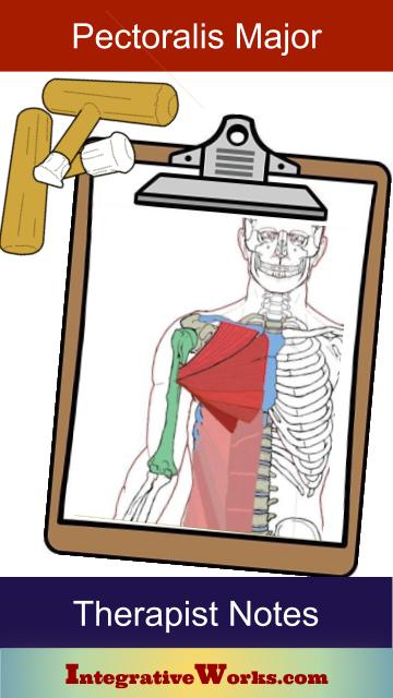
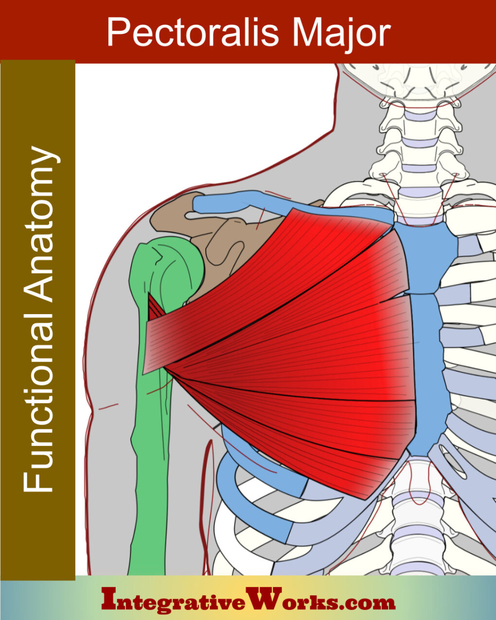
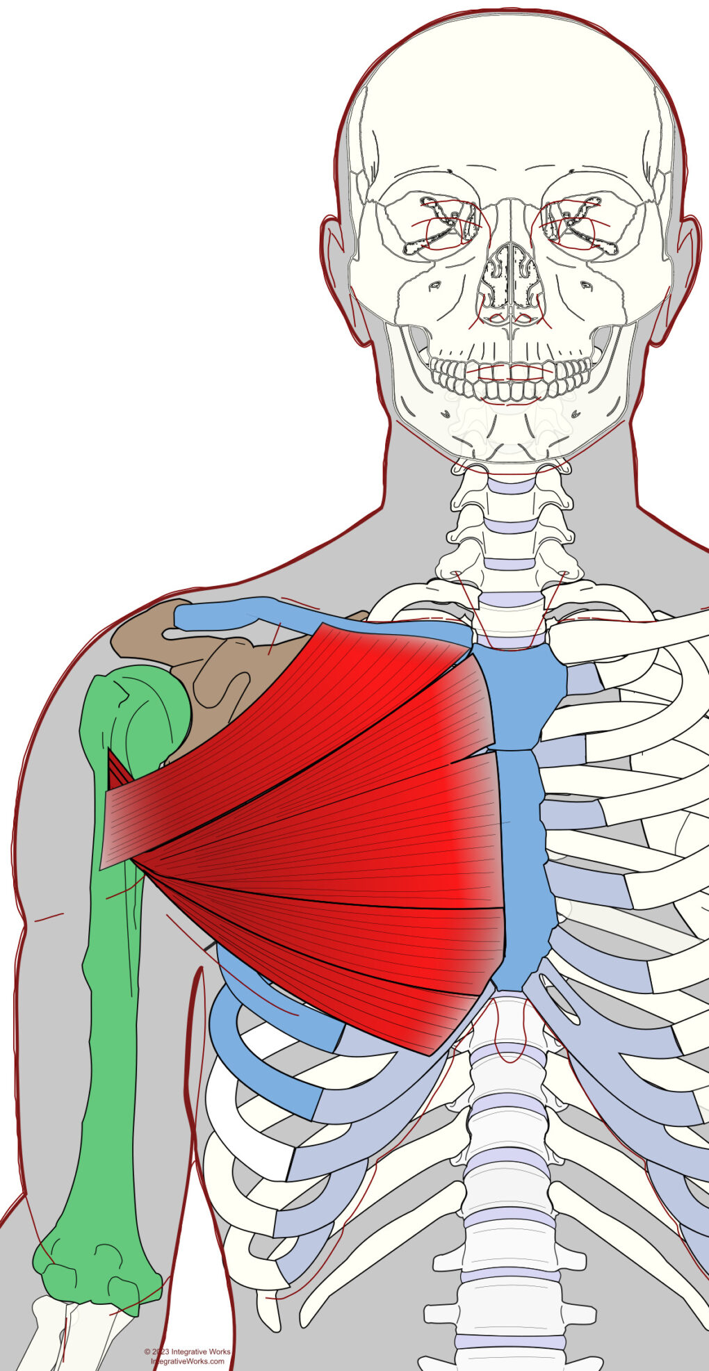
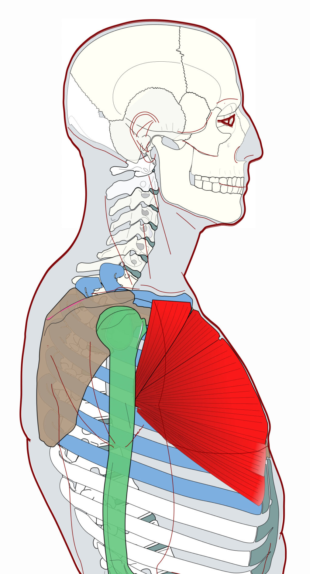
The Pectoralis major is a large, flat muscle on the anterior thorax. It originates on the anterior axial skeleton. Then, with a twist, it inserts on the proximal humerus.
Origin
- medial clavicle
- sternum
- costal cartilages of ribs 1-6
- abdominal aponeurosis
Insertion
- lateral lip of the bicipital groove
Function
- flexion of humerus
- adduction of humerus
- medial rotation of humerus
Nerve
- medial pectoral nerve (C8, T1)
- lateral pectoral nerve (C5, C6, C7)
Functional Considerations
The extensive mobility of the glenohumeral joint dramatically expands the list of functions of the pectoralis major. Let’s look at that through a few exercises. First, the bench press motion activates “horizontal flexion” as the humerus travels medially at shoulder level. Next, dips activate flexion as the humerus is brought anteriorly. Cable flyes create horizontal flexion with adduction as the humerus travels medially and inferiorly. Pullovers extend the humerus as, from overhead, it travels inferiorly. With latissimus dorsi and subscapularis, it also medially rotates the humerus.
Anomalies, Etc.
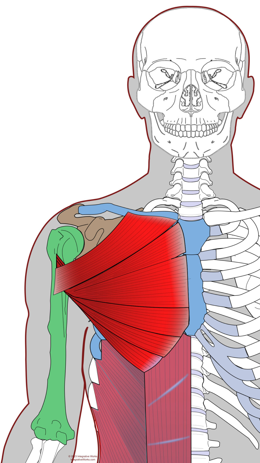
Dissection studies show that the pectoralis major varies dramatically. There are commonly 2-5 laminates, sometimes 6 or 7, and sometimes the entire pectoralis is missing (Poland Syndrome). Also, the overlap of sections varies among studies. The variation illustrated here is common and I used it because, well, it was fun to illustrate and shows how differently laminates vary in their pull on the rib cage.
It is commonly taught that it only has a clavicular and sternal section, However, this is not the case in the majority of cases. So, this example has costal and abdominal sections, which ties the humerus directly into the fascia of the abdominals. Eventually, this inserts on the pubic bone and shows an interesting step in the evolutionary progression.
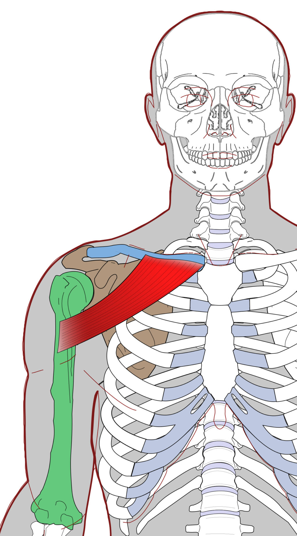
Clavicular section
Origin: medial half of the clavicle
Insertion: lateral lip of bicipital groove, starting at the greater tubercle
Function: flexion, horizontal flexion, and adduction of the glenohumeral joint
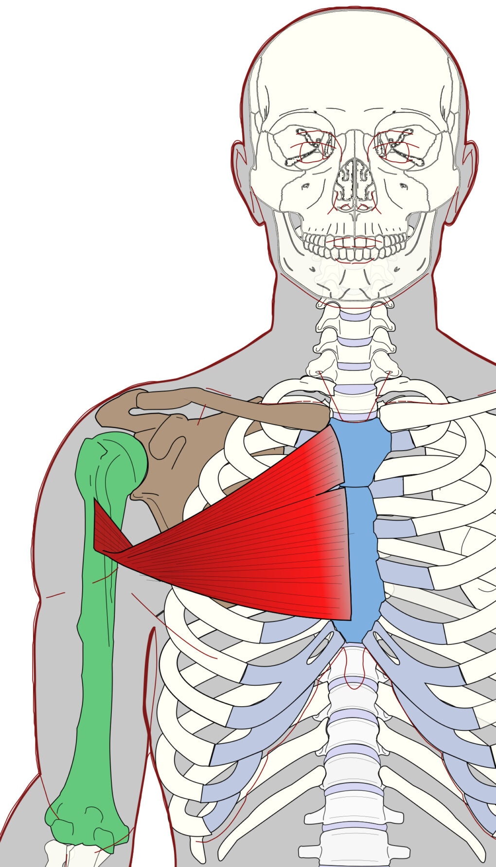
Sternal section
Origin: manubrium and upper 3/4 of sternum, at times, the superficial fascia of external oblique
Insertion: lateral lip of bicipital groove, starting at the greater tubercle
Function: horizontal flexion and adduction of the glenohumeral joint, some internal rotation of the humerus, protraction of the scapula
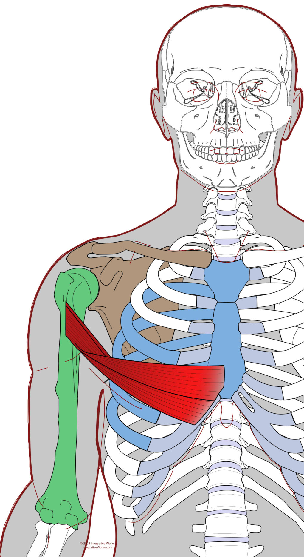
Costal sections
Often, there are two costal sections. The ventral portion lies on the superficial surface of the sternal section at the medial attachment. The dorsal section attaches to the costal cartilages inferior to the sternal section. These sections twist around the sternal and clavicular sections to attach beneath them on the bicipital groove. This twist, like the twist in latissimus dorsi, straightens when the arm is overhead.
Origin: costal cartilages of ribs. This may vary and attach as superior as rib 2 and as inferiorly as rib 8. The ventral section attaches to the lower sternum, superficial to the sternal section. Also, this may attach to the superficial fascia of the external oblique.
Insertion: bicipital groove deep to the sternal and clavicular attachments.
Function: protraction and depression of the scapula, adduction, depression, and horizontal flexion of the humerus
Abdominal section
This section, which is often absent, ties the humerus into the fascia overlapping the abdominals, creating a line of muscle from the upper extremity, across the torso, to the pubic bone.
Origin: superficial fascia of the rectus abdominus and/or external oblique.
Insertion: bicipital groove deep to the sternal and clavicular attachments.
Function: protraction and depression of the scapula, adduction, depression and horizontal flexion of the humerus
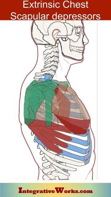
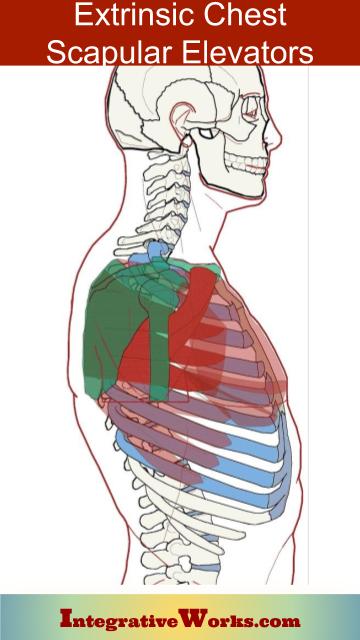
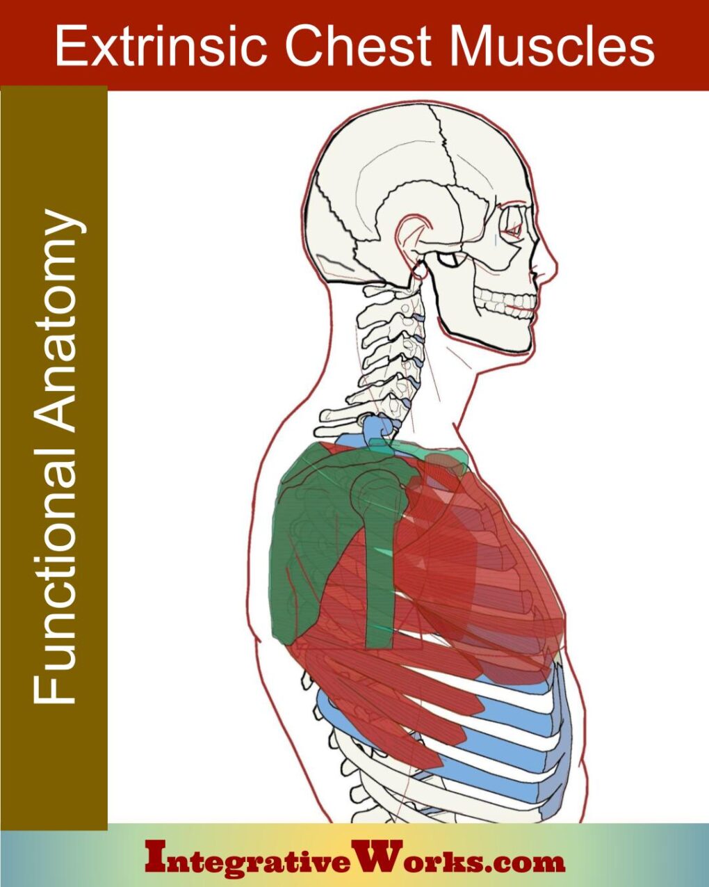
Extrinsic chest muscles do more than protract the scapula. They also balance the extrinsic back muscles in elevation, depression, and rotation of the scapula.
Related Posts
Support Integrative Works to
stay independent
and produce great content.
You can subscribe to our community on Patreon. You will get links to free content and access to exclusive content not seen on this site. In addition, we will be posting anatomy illustrations, treatment notes, and sections from our manuals not found on this site. Thank you so much for being so supportive.
Cranio Cradle Cup
This mug has classic, colorful illustrations of the craniosacral system and vault hold #3. It makes a great gift and conversation piece.
Tony Preston has a practice in Atlanta, Georgia, where he sees clients. He has written materials and instructed classes since the mid-90s. This includes anatomy, trigger points, cranial, and neuromuscular.
Question? Comment? Typo?
integrativeworks@gmail.com
Interested in a session with Tony?
Call 404-226-1363
Follow us on Instagram

*This site is undergoing significant changes. We are reformatting and expanding the posts to make them easier to read. The result will also be more accessible and include more patterns with better self-care. Meanwhile, there may be formatting, content presentation, and readability inconsistencies. Until we get older posts updated, please excuse our mess.

