Overview of anatomy

Longissimus is part of the erector spinae group, which also includes iliocostalis and spinalis. Like the others, longissimus has three sections. However, one of the sections has two parts, so four sections. As a result, often, the explanation of anatomy is simplified. That’s understandable, as it is both complex and variable.
All nine sections of the erector spinae gather into a fascial compartment that extends along the spine.
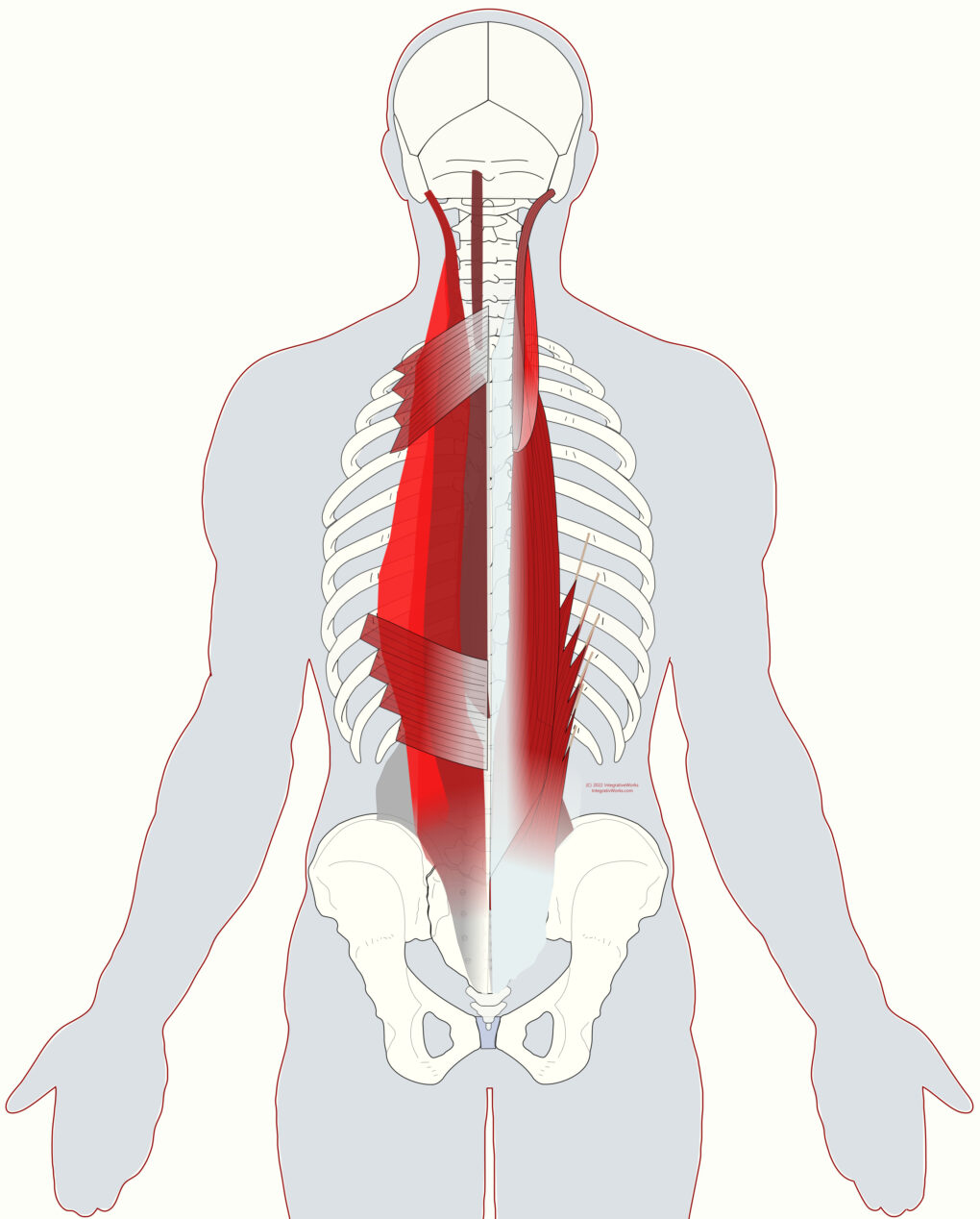
vs. a more detailed longissimus
Variations in Studies and Illustrating
Often, spinal erectors, including longissimus, are illustrated as bundles. I’ve created one of those classic looks on the right. I’ve included the serratus posterior muscles in the illustration, as they bundle the erectors together.
However, I’ve not seen that arrangement on the right in actual cadavers. I often find this kind of duplication in illustrations. More famous illustrators, especially Netter, set often not questioned standards. His illustration of the Iliotibial band is another example. I digress.
Problems in Research
I have to admit that the research on illustrating this was frustrating and exhausting for several reasons. First, many of the studies on the web are about livestock. Secondly, the studies are seldom clear in their description and images of the attachments. Thirdly, they don’t separate the sections in the images as clearly. Many of them have broad, sweeping sections that are misleading. Finally, at times, they use the same cadaver images in different studies with differences in descriptions of attachments. This study finds discrepancies among texts as well.
In this case, a muscle section originates on the ilium’s crest and attaches to the lumbar transverse processes. Some studies include this muscle with iliocostalis and in other studies with longissimus. However, it is typically listed in longissimus, so I’ve included it here.
A Different Perspective
I humbly present this format to provoke some thought. As the studies vary, I went with studies that best matched the cadavers I’ve seen.
Erectors, The Big Picture
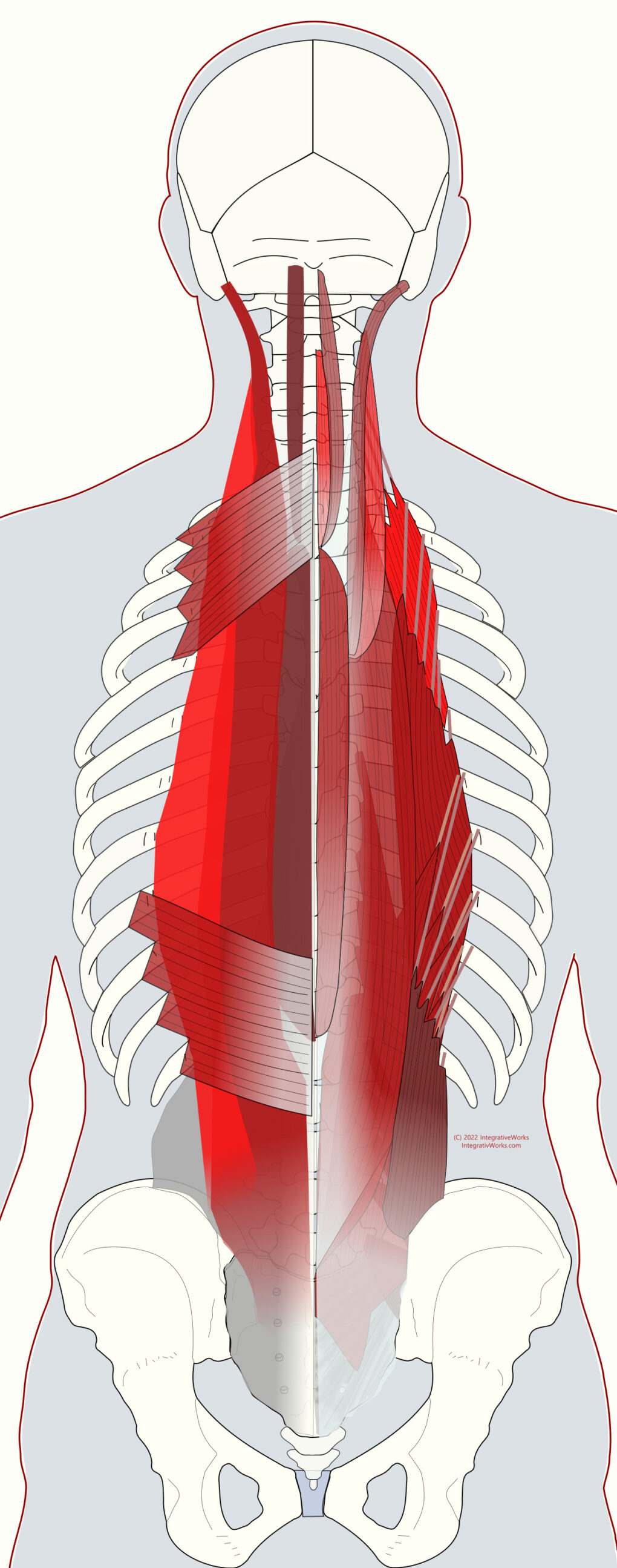
Erector spinae is regarded as three groupings of muscle: iliocostalis, longissimus, and spinalis. Commonly, each grouping then has three sections. Iliocostalis has lumborum, thoracis and cervicis. Longissimus has thoracis, colli (cervicis), and capitis. Spinalis also has thoracis, colli, and capitis.
On the Left
On the left, it shows a typical grouping of the erector spinae:
- Iliocostalis is bright red.
- Longissimus is darker red.
- Spinalis is dark brown.
- Serratus posterior superior, and inferior bundle the muscles together.
On the Right
Iliocostalis, longissimus, and spinalis are color-coded by their sections on the right.
- Lumborum sections are shown in dark brown.
- Thoracis sections are in dark red.
- Cervicis/Colli sections are in bright red.
- Capitis sections are in a lighter brown.
- The longissimus lumborum section is under a couple of layers and, therefore, not shown in this view.
Longissimus, The Overview
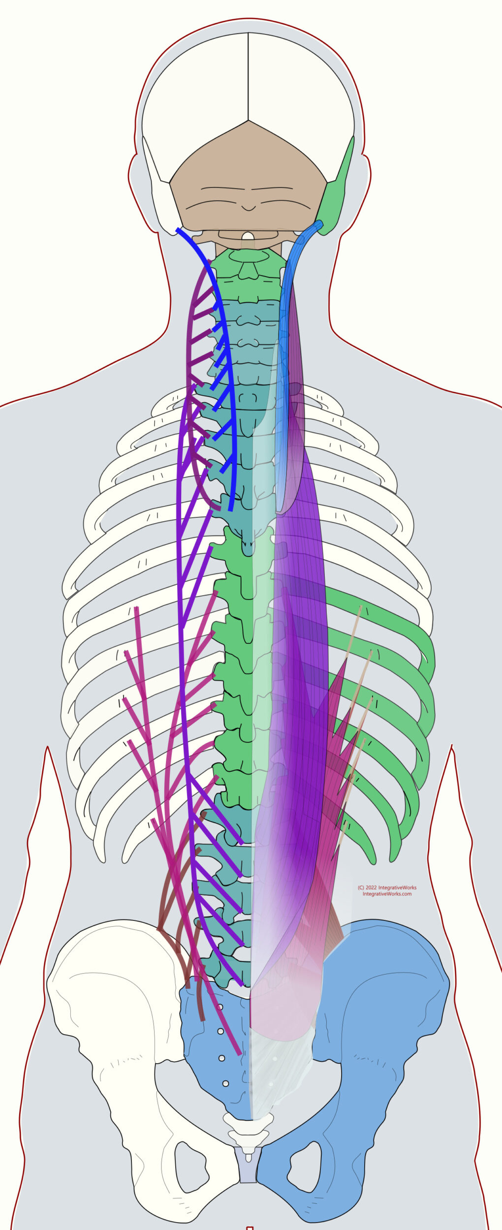
Longissimus is the longest group of erectors, extending from the sacrum to the cranium.
It has three or four sections, depending on how you count them. You can see from this illustration that this erector group creates a complex arrangement. Like the lumbar vertebrae, some bones serve as the insertion of one section and the origin for others. Cadaver studies vary in their descriptions of attachments, making this more complicated.
Most texts speak of three sections.
- Thoracis – This section connects the pelvis to the thoracic vertebrae and ribs. It is shown here as the lower three sections that originate from the pelvis and lumbar vertebrae. This section extends along the lumbar vertebrae and ribs.
Anatomy texts typically separate the deepest, most inferior section of the thoracis from the others. It is referred to as longissimus thoracis-lumborum or longissimus lumborum. - Cervicis – This section connects the thoracic vertebrae to the mid-cervical vertebrae.
- Capitis – This section originates from the lower cervical and upper thoracic vertebrae. Then, it inserts on the temporal bone.
Let’s go over each of these in more detail.
Longissimus Lumborum
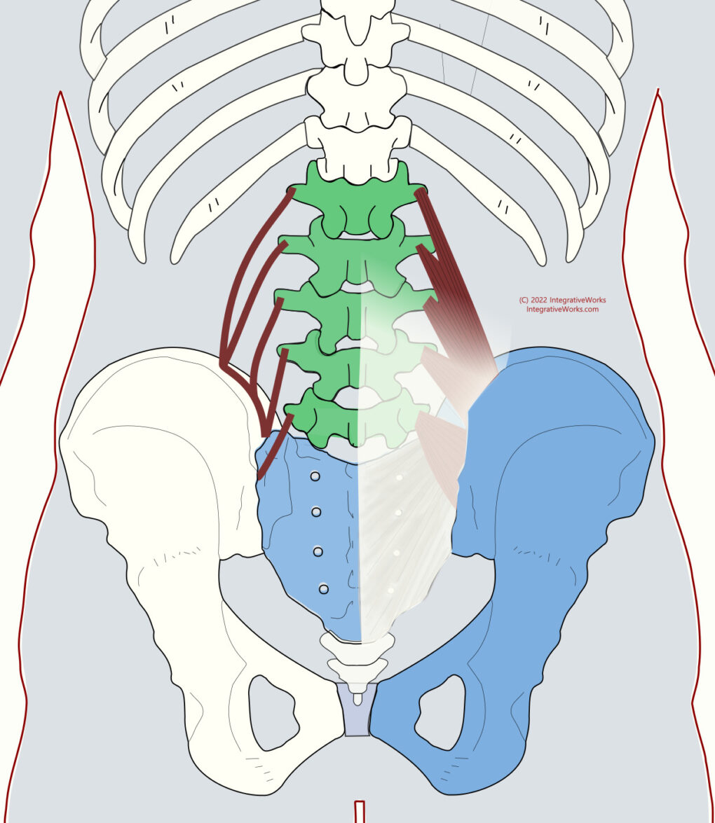
This section of longissimus is described differently from one text to another.
Most texts include this muscle as part of the longissimus thoracis. Accordingly, it is sometimes called the longissimus thoracis lumborum. A few studies describe a muscle with the same attachments as part of iliocostalis.
It has a superior section that connects the top 4 lumbar vertebrae to the crest of the ilium. Also, an inferior section ties the 5th lumbar to the ventral, medial surface of the ilium.
Origin
- superior section – the medial crest of the ilium via the lumbar intramuscular aponeurosis
- inferior section – posterior sacroiliac ligament on the ventral, medial surface of the ilium.
Insertion
- superior section – transverse processes of L1-L4
- inferior section – transverse process of L5
Function
- Extend the spine
- Laterally flex the spine
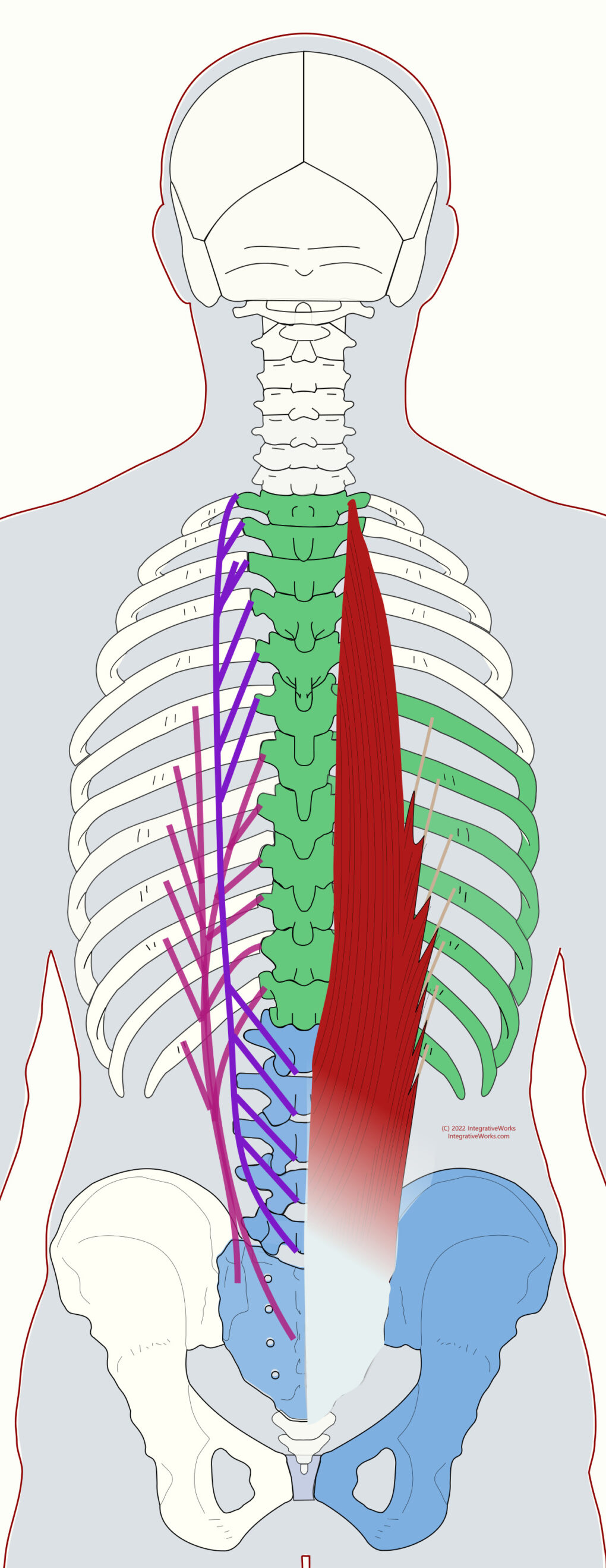
A variation where the deeper section attaches to the sacrum, lower seven ribs and lower seven vertebrae. The superficial section attaches to the lumbar vertebrae and upper thoracic vertebrae.
Longissimus Thoracis
The descriptions of this section of the muscle are highly variable. Incidentally, I’ve found more studies that vary than studies that agree. However, there is a pattern to these studies that makes sense. So, let’s start with the basics of how this muscle attaches.
Origin
- Median crest of the sacrum via the lumbar aponeurosis
- Lumbar transverse and accessory processes
Insertion
- Transverse processes of thoracic vertebra
- Ribs between the tubercle and transverse process
Function
- Extend the spine
- Laterally flex the spine
Nerve
- lateral branches of posterior/dorsal rami of cervical spinal nerves
Variations
Cadaver studies and anatomical references vary a great deal on this muscle. However, there is a pattern.
Texts vary in the number of sections and where they attach. They are especially variable in the upper attachment of longissimus thoracis. Some texts indicated that longissimus attaches to all thoracic vertebrae and ribs. On the other hand, other texts claim that it attaches to all thoracic vertebrae but not all the ribs. Still, other texts say that it only attaches to the ribs up to the 5th rib, And others say to the 3rd rib. In addition, some texts say that its lateral attachments are on the angle of the rib. However, most say that it attaches between the angle and the tubercle of the rib. Texts also vary in the number of fascicles or sections.
However, even though they may disagree on how many sections, most texts agree that the sections/fascicles with a more superior origin insert more superiorly and are more superficial. For example, the sections that originate in the upper lumbar vertebrae are more superficial and insert on the upper thoracic vertebrae (and ribs). In comparison, the sections that originate in the sacrum are deeper and attach to the lower thoracic vertebrae and ribs.
Longissimus Cervicis
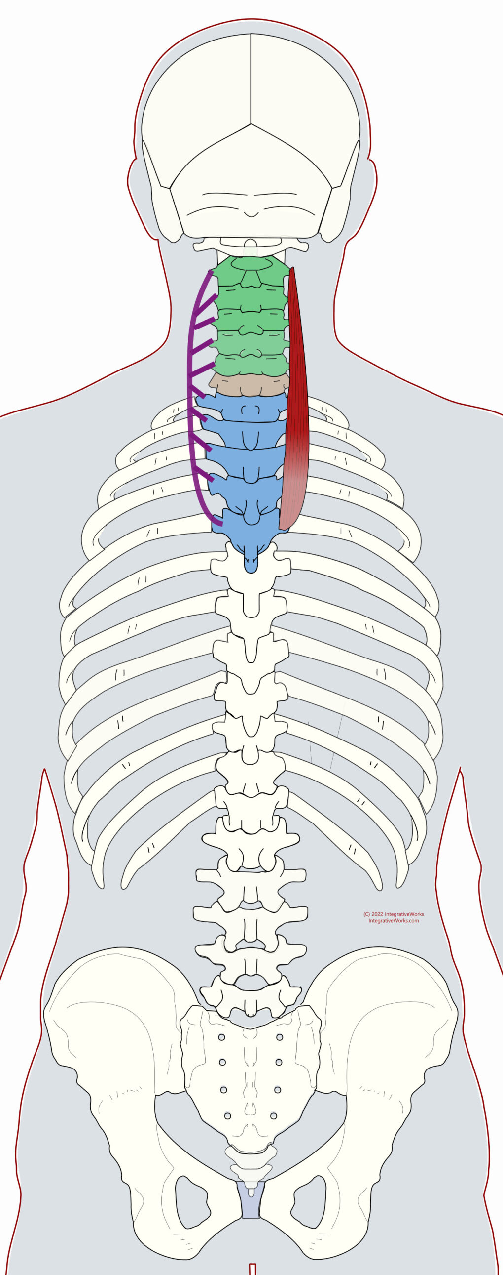
Longissimus cervicis is a small muscle that attaches to the upper thoracic and middle cervical vertebrae.
Origin
- transverse processes of T1-T5
Insertion
- transverse processes of C2-C6
Function
- extend the neck
- laterally flex the neck
Nerve
- lateral branches of posterior/dorsal rami of cervical spinal nerves
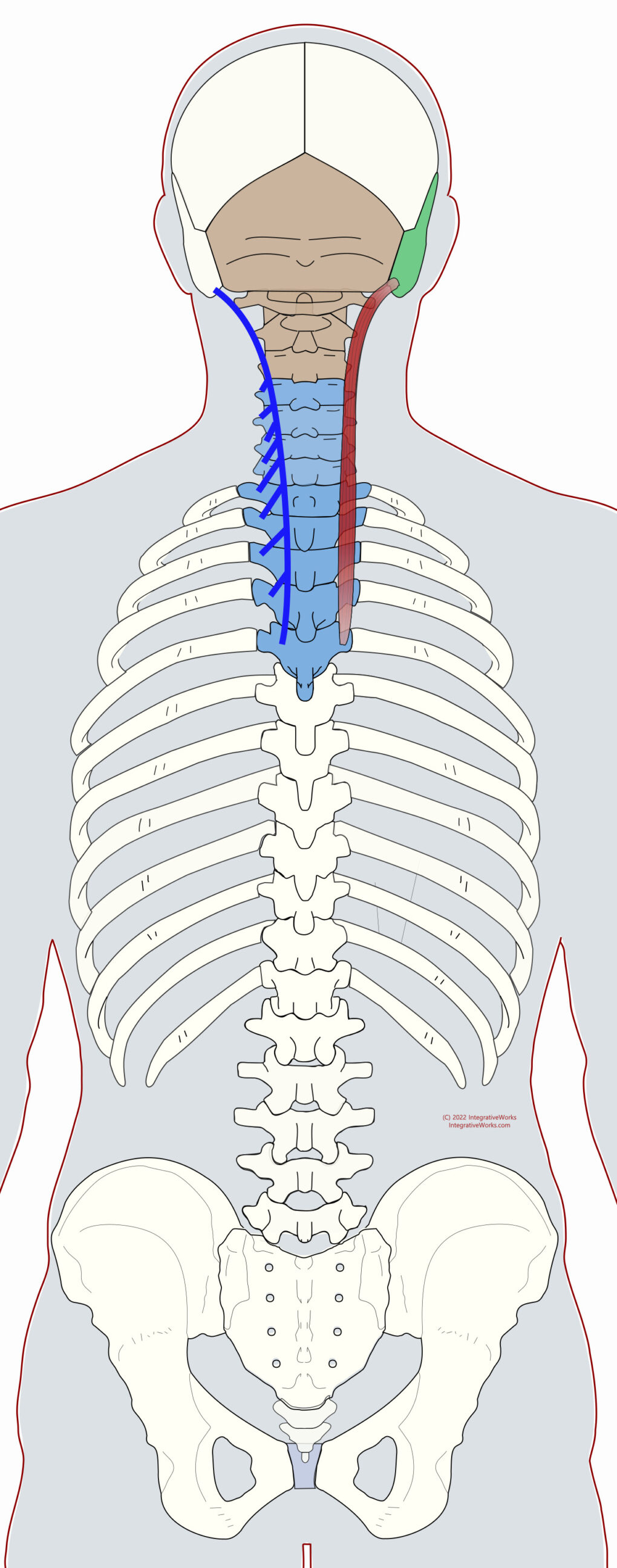
Longissimus Capitis
Longissimus cervicis is a small muscle that attaches to the upper thoracic and middle cervical vertebrae to the cranium.
Origin
- transverse processes of C3-T5
Insertion
- mastoid process of the temporal bone
Function
- extend the neck
- laterally flex the neck
Nerve
- lateral branches of posterior/dorsal rami of cervical spinal nerves
Anomalies, Etc.
The studies on this muscle show notable variances. This study looks at the differences in the direction of the fibers in women.
Posts related to Longissimus
Buttock Pain with Tightness into the Back
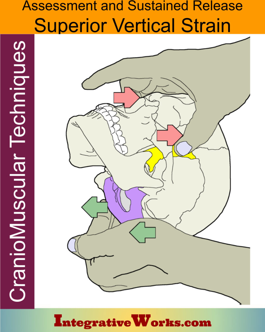
CranioMuscular – Superior Vertical Strain
Erector Spinae – Neuromuscular Massage Protocol
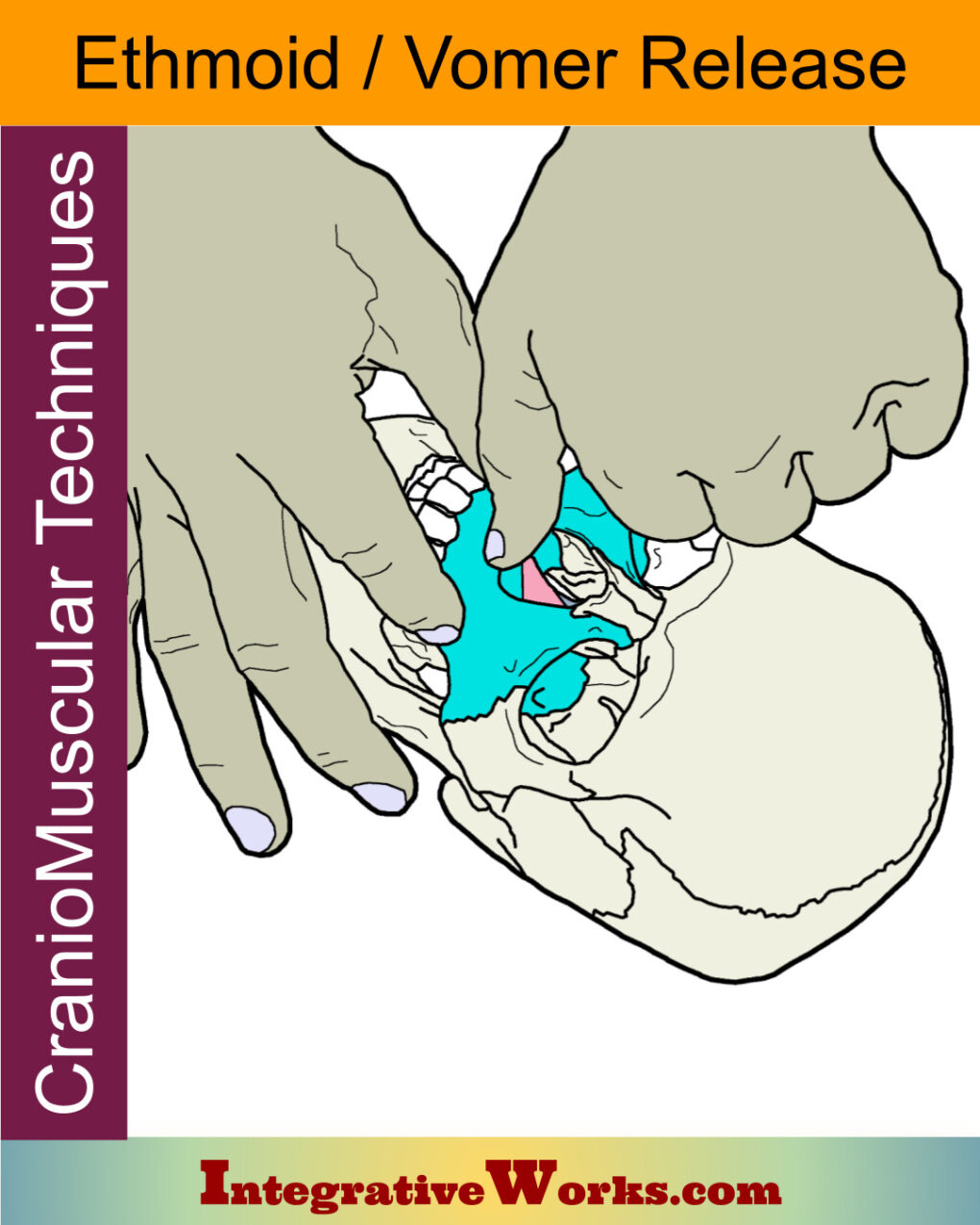
Ethmoid / Vomer Distraction
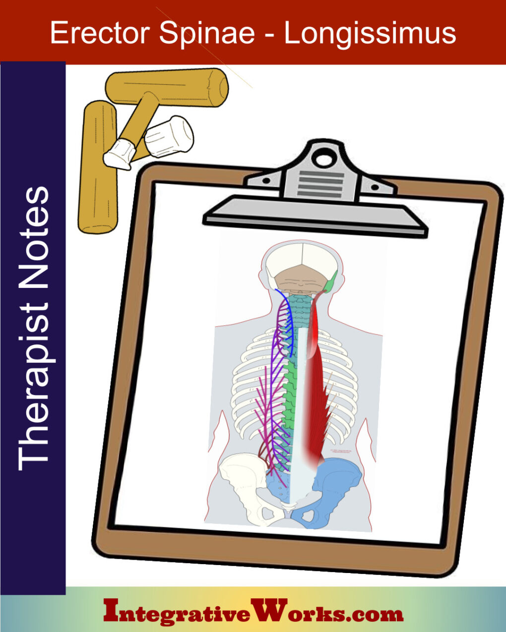
Longissimus – Massage Therapy Notes
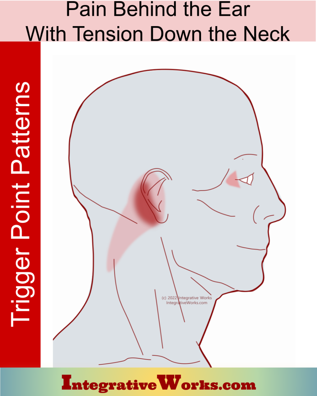
Pain Behind the Ear with Tension Down the Neck
Sciatica, Degenerative Discs and Trigger Points
Self-Care – Buttock Pain with Tightness in the Back
Self-Care – Pain Behind the Ear with Tension Down the Neck
Self-Care – Stiff Low Back Pain to the Crest of the Hip
Stiff Low Back Pain to the Crest of the Hip
Thoracolumbar Release with AO-45
Support Integrative Works to
stay independent
and produce great content.
You can subscribe to our community on Patreon. You will get links to free content and access to exclusive content not seen on this site. In addition, we will be posting anatomy illustrations, treatment notes, and sections from our manuals not found on this site. Thank you so much for being so supportive.
Cranio Cradle Cup
This mug has classic, colorful illustrations of the craniosacral system and vault hold #3. It makes a great gift and conversation piece.
Tony Preston has a practice in Atlanta, Georgia, where he sees clients. He has written materials and instructed classes since the mid-90s. This includes anatomy, trigger points, cranial, and neuromuscular.
Question? Comment? Typo?
integrativeworks@gmail.com
Interested in a session with Tony?
Call 404-226-1363
Follow us on Instagram

*This site is undergoing significant changes. We are reformatting and expanding the posts to make them easier to read. The result will also be more accessible and include more patterns with better self-care. Meanwhile, there may be formatting, content presentation, and readability inconsistencies. Until we get older posts updated, please excuse our mess.


