Overview
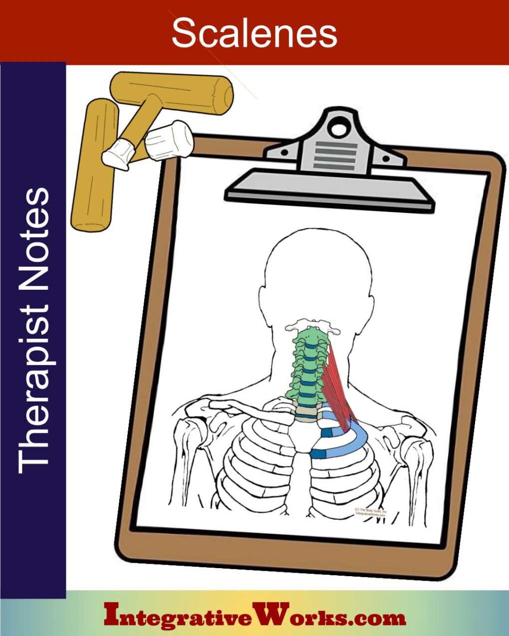
Scalene anatomy is more variable than most muscles. They vary in their attachments and size in 40-71% of the reviewed studies. Scalenes are 3 (sometimes 4) muscles on either side of the neck.
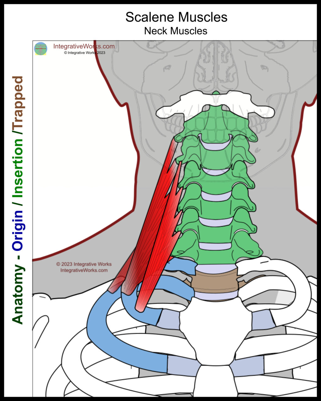
This is one of those muscles that makes it hard to say which end is the origin and which end is the insertion. Sometimes, we look at the lower attachments as the origin because we flex the neck. Contrarily, we may see the upper attachments as the origin of assisted breathing. This post uses the lower attachments as the origin for consistency in color coding and discussion.
Origin
- Ribs 1-2
- (fascia of the pleural cavity)
Insertion
- C2-C7
Function
- (forward) Flex the neck
- Laterally flex the neck
- Rotation of the neck
- Assisted breathing
Nerve
- Posterior rami of C3-C8
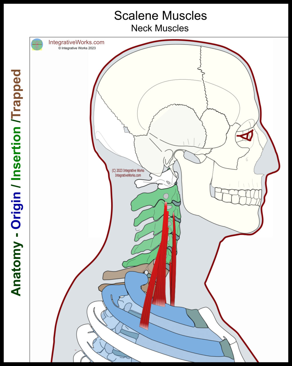
Overall Structure
The three bellies are named for their position; scalenus anterior, scalenus medius, and Scalenus posterior. Inconsistently, there is an anomalous belly is called scalenus minimus. But, as a group, scalenes are lateral cervical muscles that connect the cervical vertebrae to the upper thoracic cage. They are long muscles with fibers of uneven length.
- Scalenus anterior lies almost entirely under the clavicular head of the sternocleidomastoid in the pedicle groove. It passes anterior to the cervical nerves and subclavian artery but behind the subclavian vein.
- Scalenus posterior lies mainly under the anterior border of the trapezius. It passes posterior to the cervical nerves and the subclavian artery.
- Scalenus medius lies in the posterior cervical triangle between the other two scalenes.
- Scalenus minimus is between the scalenus medius and the brachial plexus.
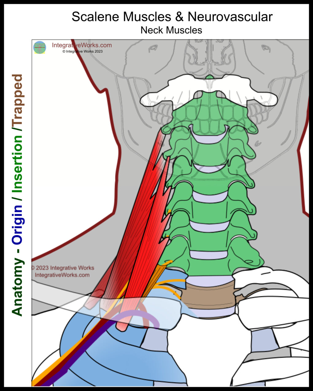
Neurovascular Structures
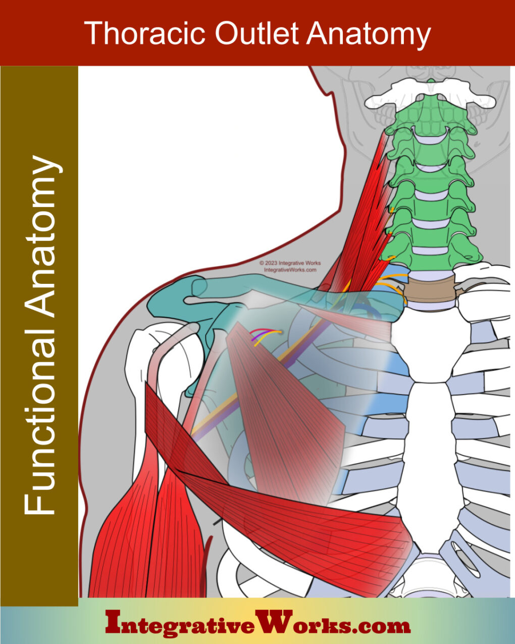
The scalenes entrap the neurovascular that extends across the first rib. The attachments on the first rib are quite variable. Some dissections reveal a smaller attachment of the scalenus anterior. However, others reveal a crescent-shaped attachment. Additionally, there is the highly variable presence of scalenus minimus.
The structures of the first rib from posterior to anterior:
- scalene, posterior
- scalene, middle
- brachial plexus
- scalene, minimus (when present)
- subclavian artery
- scalene, anterior
- subclavian vein
- clavicle
Anatomy
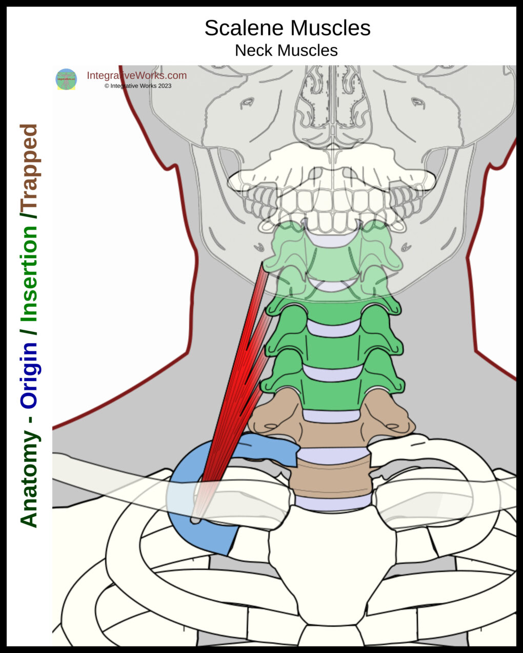
Scalenus Anterior
Origin
- 1st Rib
Insertion
- anterior tubercles of C3-C6
Scalenus anterior originates from a flat tendon on the first rib just under the clavicular SCM, medial to the thoracic bundle. It inserts along the anterior tubercles on the transverse process of C3-C6. The upper attachment on C3 is usually a more prominent tubercle than the other cervical vertebrae and is easily distinguished with palpation.
Additionally, there are reports of it extending to attach to the second rib.
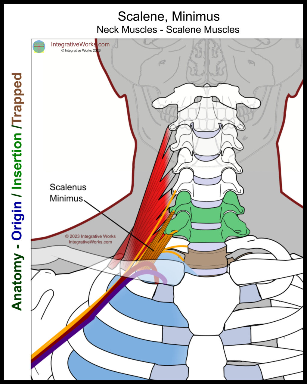
Scalenus, minimus
Origin
- suprapleural fascia
Insertion
- C7, (C6)
Dissection information on the scalenus minimus is highly variable. It is reported from 7-71% of the time and varies in its attachments.
Scalenus minimus is the most variable of the scalene muscles. It originates from the fascia of the thoracic pleura and sometimes the first rib. Usually, the minimus inserts on the posterior tubercle of C7, and, sometimes C6. It passes posterior to the subclavian artery.
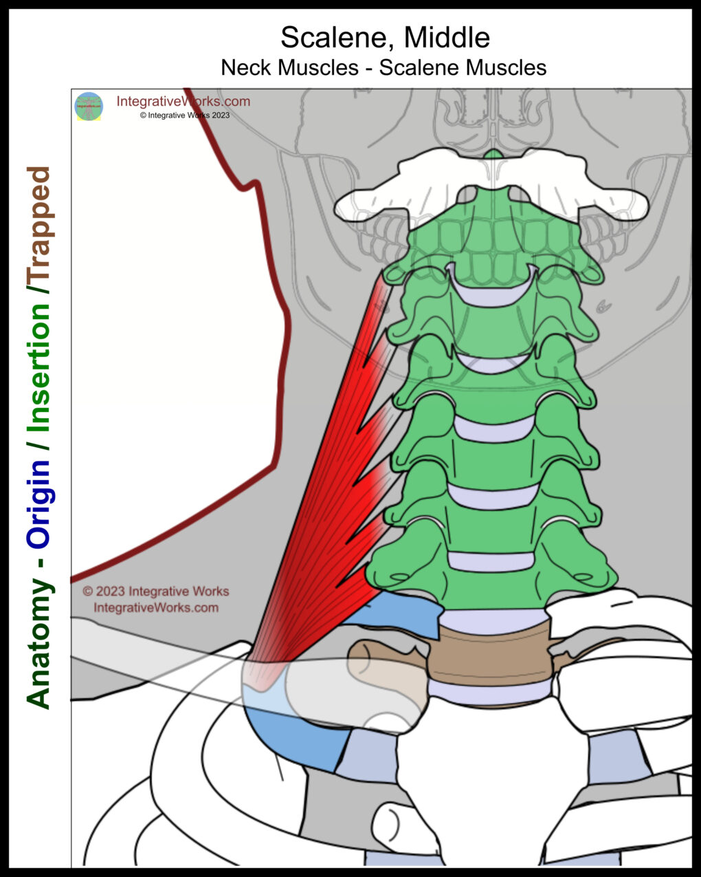
Scalenus medius
Origin
- 1st Rib
Insertion
- posterior tubercle of C2-C7
Scalenus medius originates from a flat tendon on the 1st rib lateral to the thoracic neurovascular bundle. It inserts on the posterior tubercles of the transverse processes of C2-C7. This traps C7-T1 between origin and insertion.
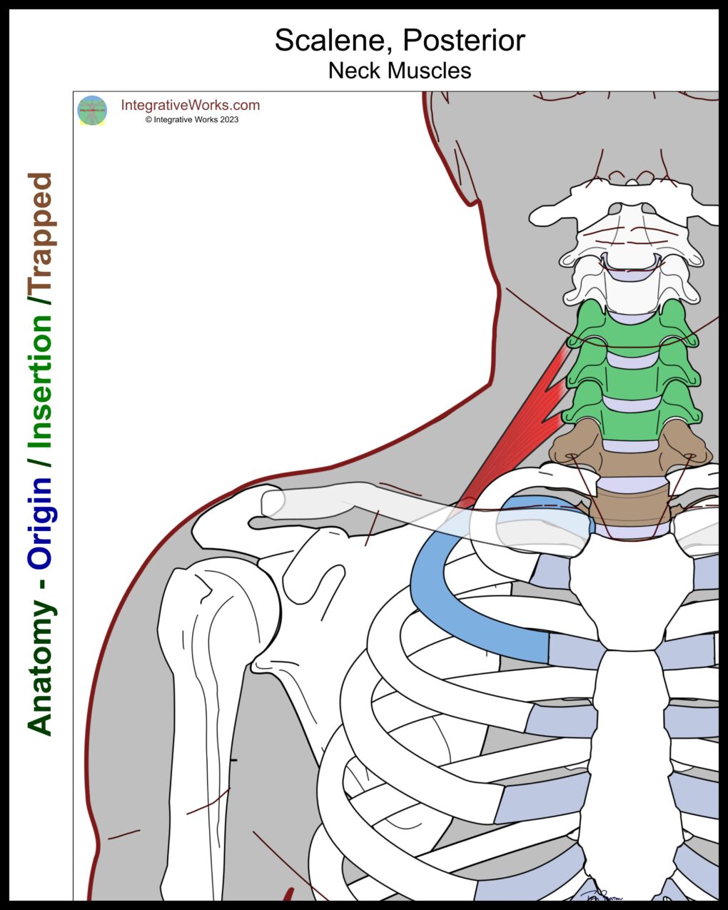
Scalenus posterior
Origin
- Lateral aspect of 2nd Rib
Insertion
- posterior tubercle C3-C6
The Scalenus posterior originates from a flat tendon on the lateral aspect of the 2nd rib. It inserts on the posterior tubercles of the C4-C6 or C7.
This traps T1 and T2 between the bones of origin and insertion.
The Scalenus posterior has very similar attachments to iliocostalis cervicis and serratus posterior superior. They pull on the same structures and synergize closely. They are essential to resolving forward head posture and lateral flexion of the lower cervicals.
Functional Considerations
Scalenes draw the cervical vertebrae toward the upper ribs. The implications of this can be complicated and argued at length.
- Contraction creates lateral flexion of the neck when the ribs are fixed.
- Contraction lifts the thoracic cage when the neck is fixed.
Lifting Ribs to Breathe
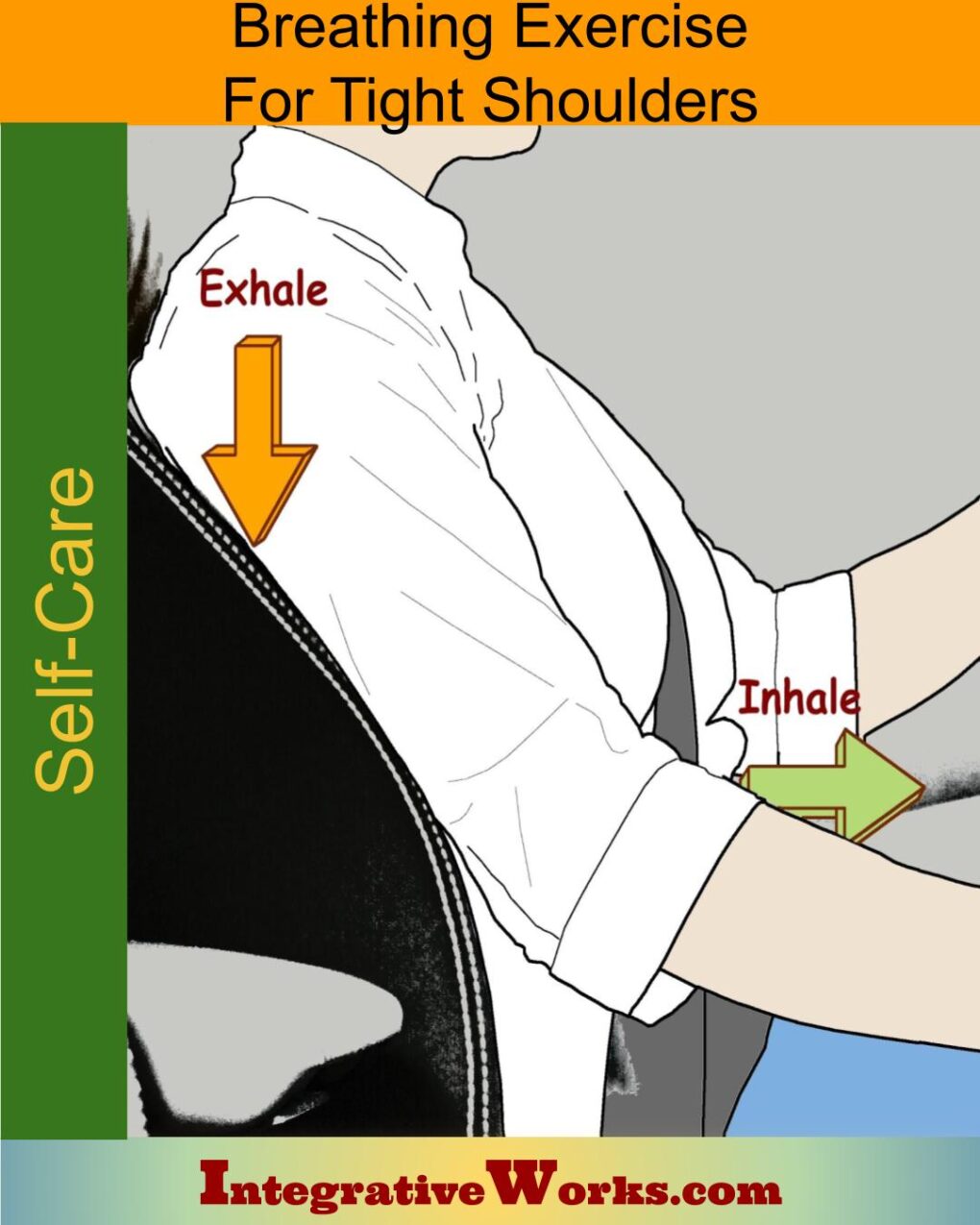
During inhalation, especially when the trunk is flexed, scalenes lift the upper ribs from a fixed vertebral column to create volume in the chest cavity. Depending on position and trigger point activity, the scalene muscles synergize with sternocleidomastoid, iliocostalis cervicis, and serratus posterior superior. In addition, Trapezius and levator scapula support the pectoral girdle so that the serratus anterior and pectoralis minor can also assist.
Scalenus anterior is a poor neck flexor but becomes chronically shortened in Forward-Head Posture. This is complemented on the back of the upper neck by the short extensors of the head. Those include the sub-occipitals, splenius capitis, and semispinalis capitis. After the more superficial phasic muscles are released, the deep tonic muscles still support Forward-Head Posture.
The Scalenus posterior has very similar attachments to iliocostalis cervicis and serratus posterior superior. They pull on the same structures and synergize closely. They are essential in resolving forward head posture and lateral flexion of the lower cervical vertebrae.
Lateral Flexion and Rotation
When the ribs are stabilized and the vertebrae are allowed to move, they laterally flex the neck. They synergize with the sternocleidomastoid, lower sections of the levator scapula, and upper trapezius.
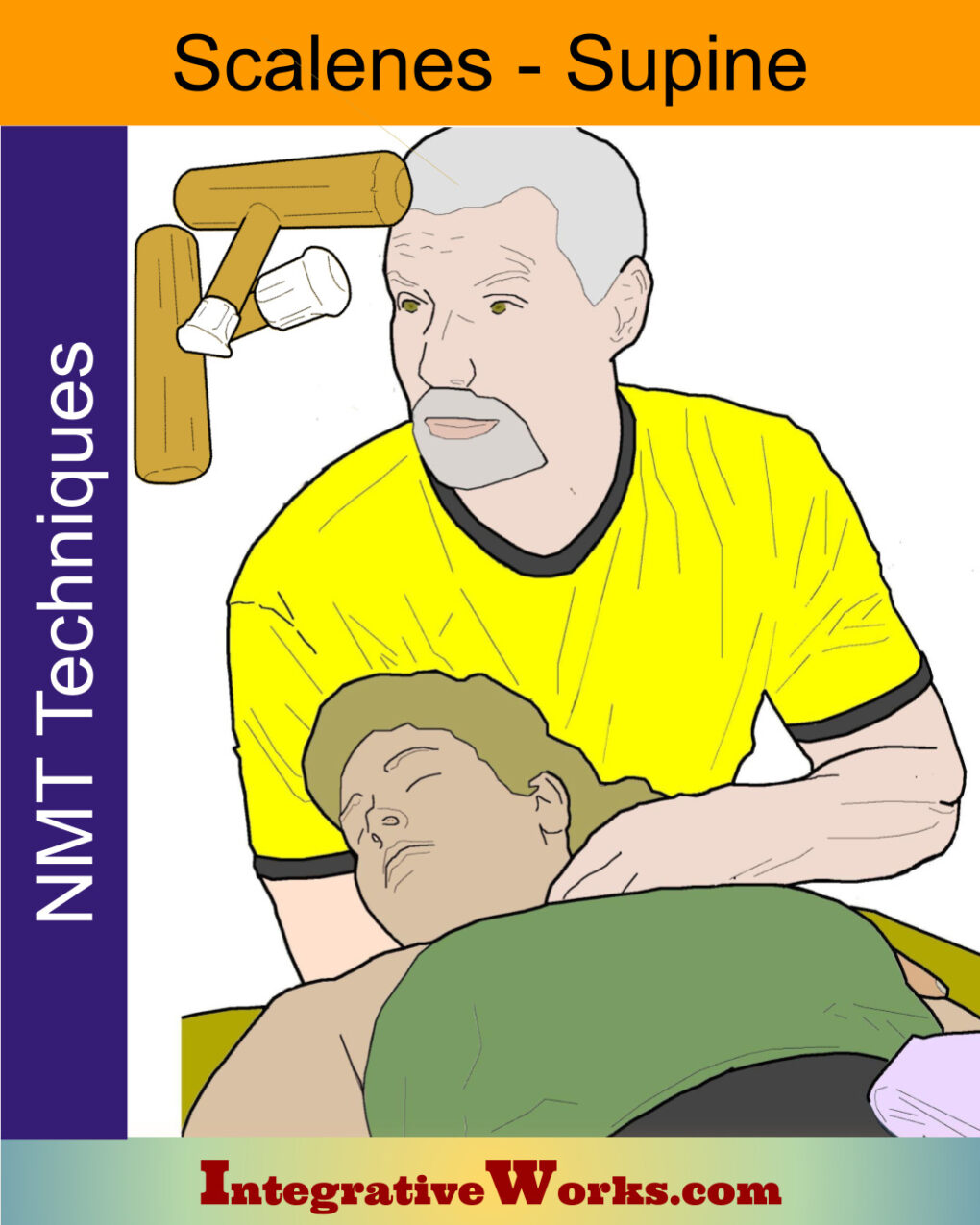
Anatomy books are confusing and contradictory about the role that scalenes play in the rotation of the vertebrae. Furthermore, no electro-myographical studies were found to support these claims. That said, scalene referral patterns can be more easily elicited when the head is rotated ipsilaterally while laterally flexing toward the opposite shoulder.
Helpful Collections
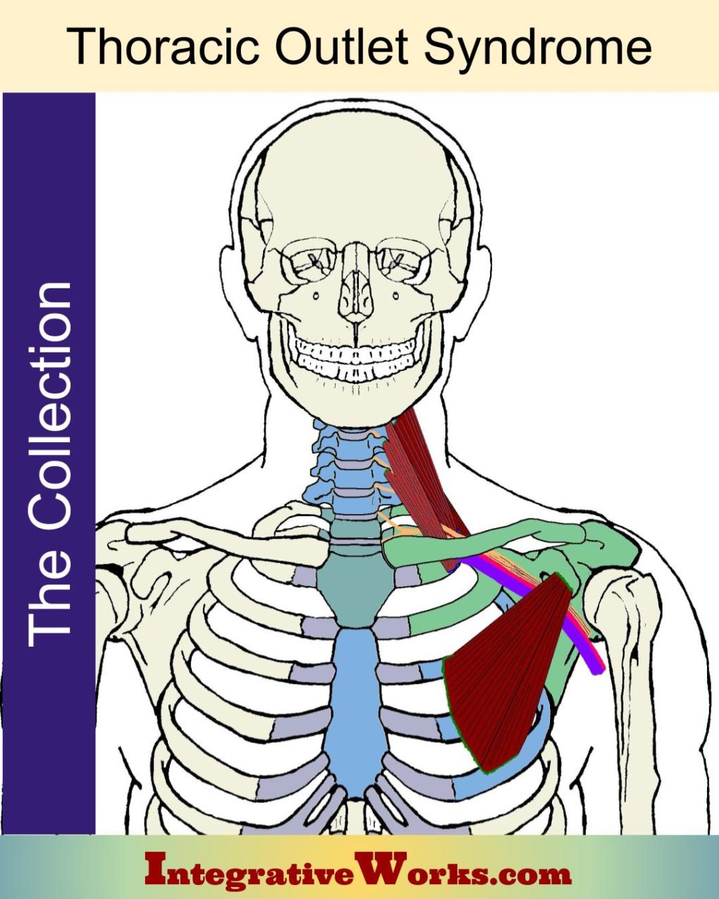
Thoracic Outlet Syndrome
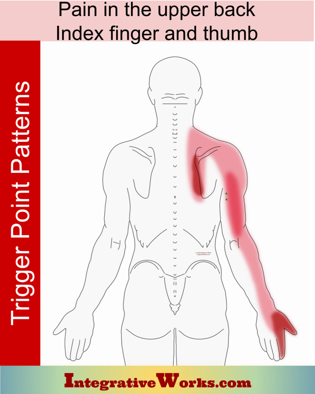
Scalenes are a vital part of thoracic outlet syndrome. They entrap nerves and blood vessels as they pass into the upper extremity. Also, they create patterns of pain, restriction, and swelling in the arm.
This collection contains posts about the thoracic outlet, including:
- involved muscles
- pain patterns
- exercises
- anatomy and physiology
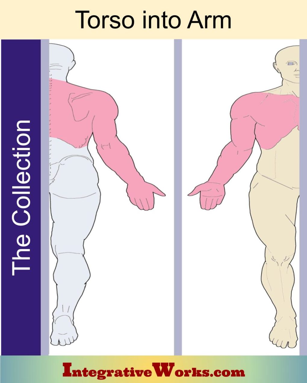
Torso Into Arm
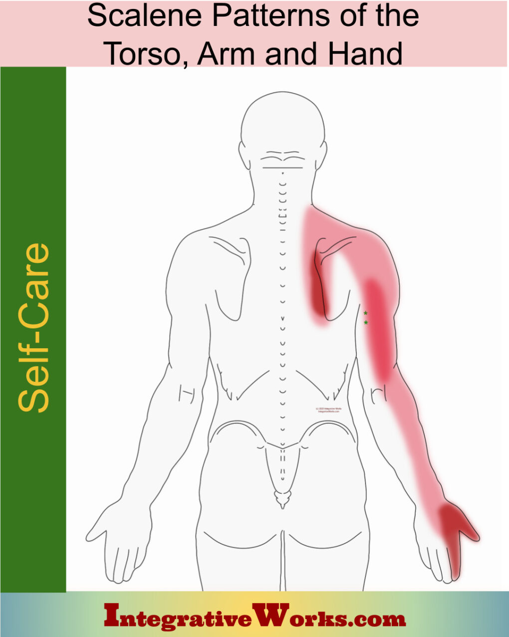
Scalene muscles are often suspected when pain occurs in the arm and hand. However, this is both useful and confusing. Many referral patterns overlap scalene referral patterns. It leads the novice therapist to treat scalenes too often.
This Collection contains posts about trigger points that refer from the torso into the arm. It can be a helpful, quick reference for comparing patterns and sorting out the real problem.
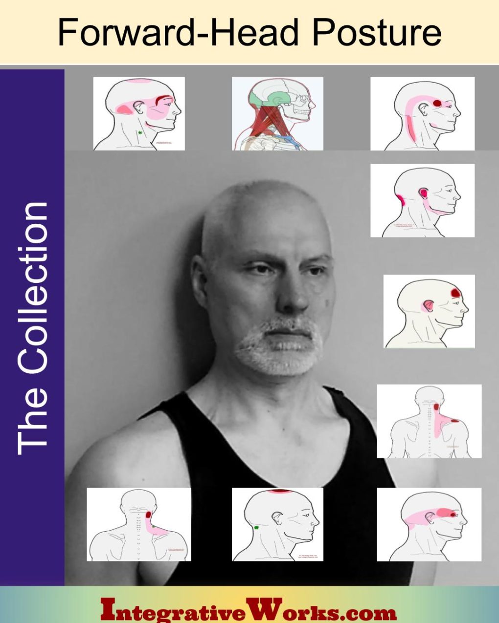
Address the Underlying Postural Problems
This muscle contributes to Forward-Head posture. Chronically, it becomes short and strong. Then, once the head has become imbalanced over the trunk, this muscle is supported to become shorter and stronger.
If you have Forward-Head Posture, review this collection, especially the self-care exercise Tuck, Tilt, Turn, and Lift.
Posts related to Scalenes
Do You Find Trigger Point Illustrations Confusing?
Pain in the Torso and Arm: Signs, Pain Patterns, Self-care, Therapy Notes…
Pain in the upper back, index finger, and thumb
Scalenes – Massage Therapy Notes
Scalenes Supine – Neuromuscular Massage Protocol
Self Care – Breathing exercise for upper back pain
Self Care – Breathing Exercises to Reduce Shoulder Tension
Self Care – Scalene Pain of Upper Back, Arm & Hand
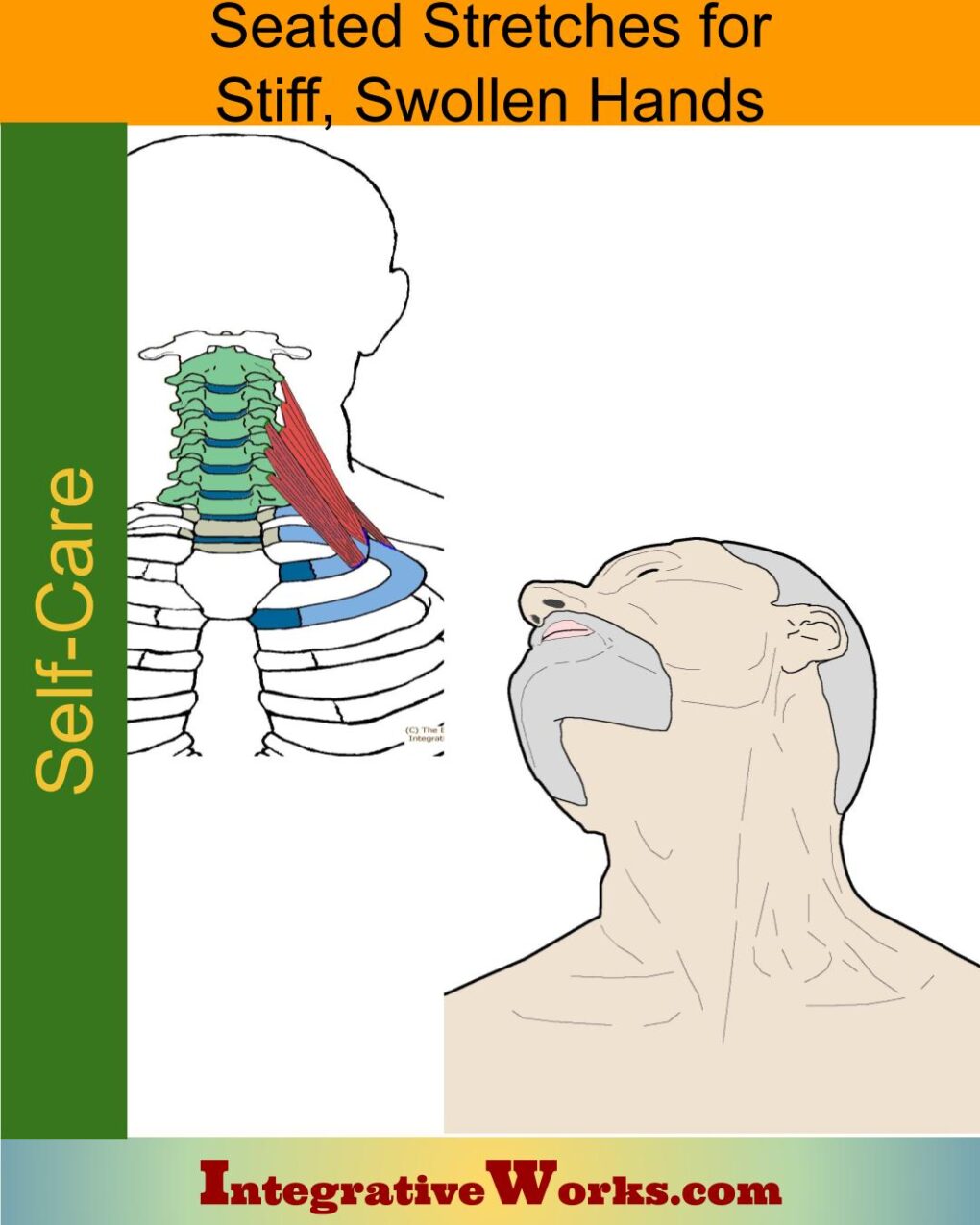
Self Care – Seated Stretches for Stiff, Swollen Hands
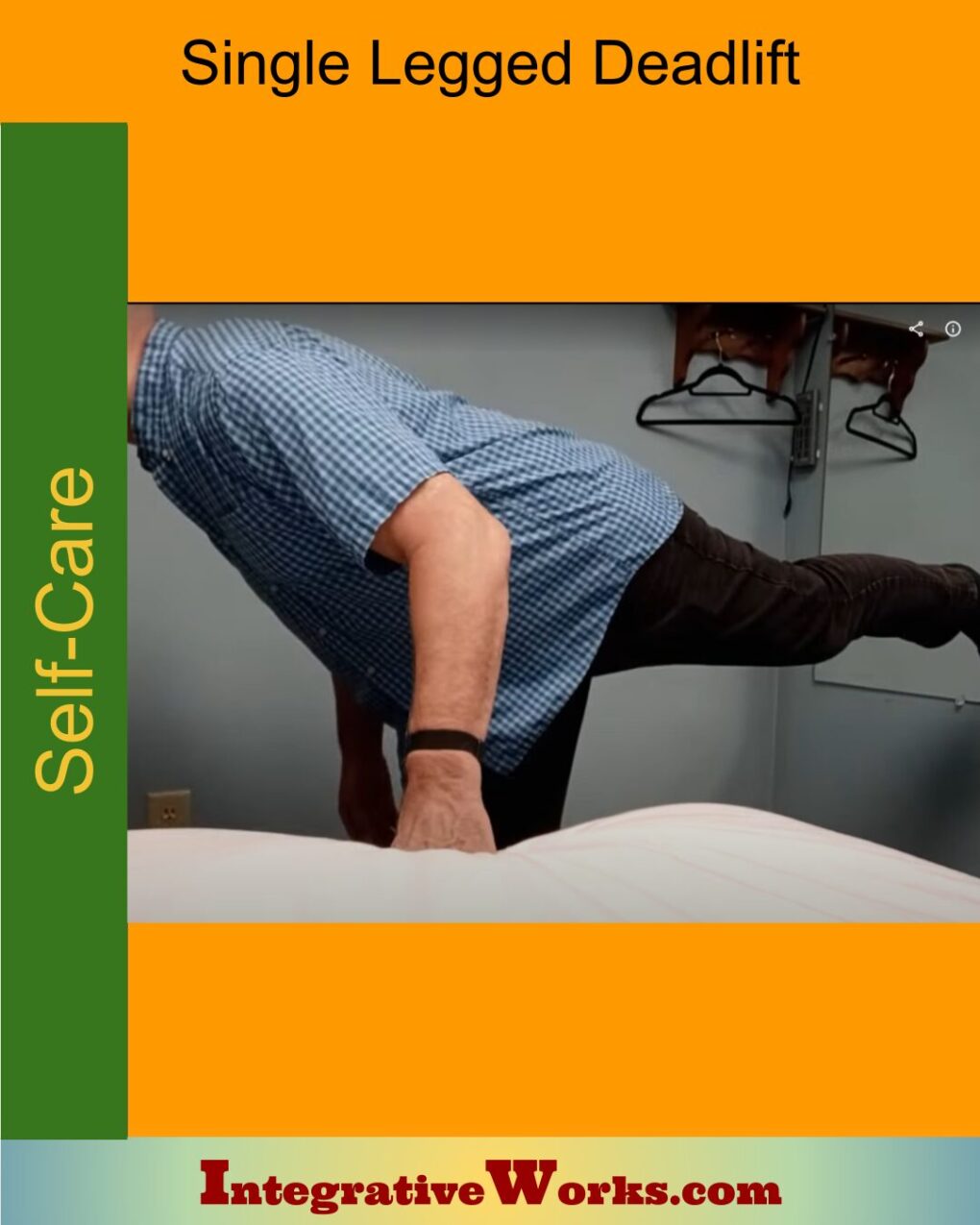
Self Care – Single-Legged Deadlifts
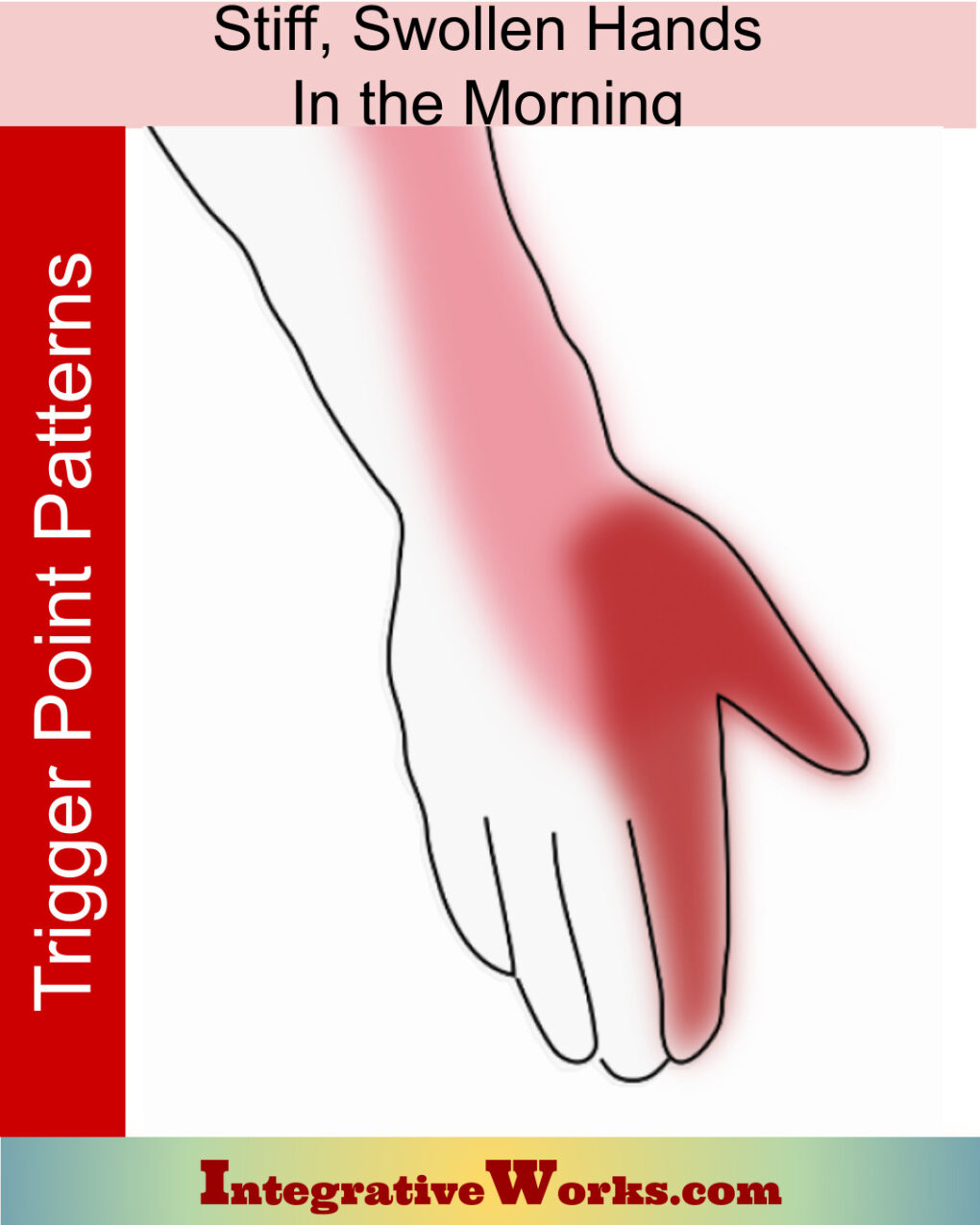
Stiff, Swollen Hands in the Morning
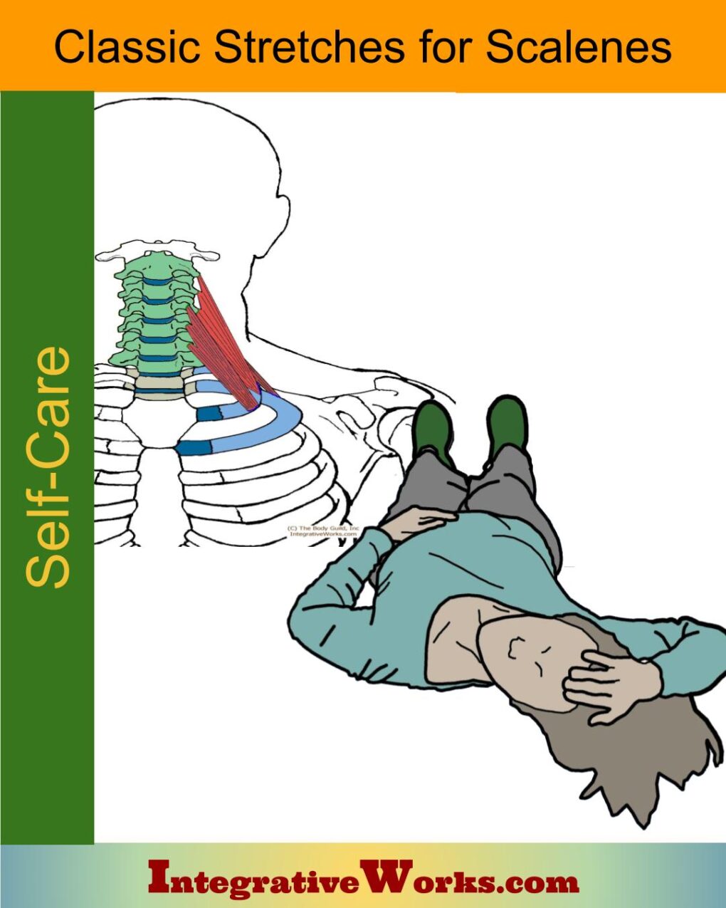
Stretching for scalene muscles
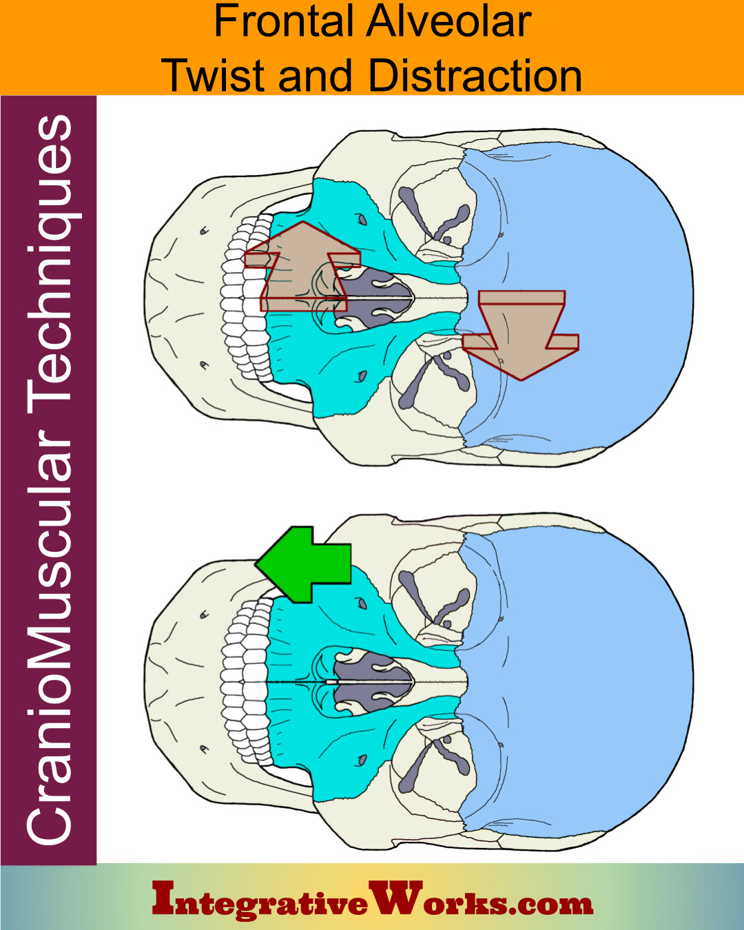
Structural Twist and Distraction of Frontal-Maxilla
Thoracic Outlet – Functional Anatomy
Wikipedia entry for scalene muscles
Support Integrative Works to
stay independent
and produce great content.
You can subscribe to our community on Patreon. You will get links to free content and access to exclusive content not seen on this site. In addition, we will be posting anatomy illustrations, treatment notes, and sections from our manuals not found on this site. Thank you so much for being so supportive.
Tony Preston has a practice in Atlanta, Georgia, where he sees clients. He has written materials and instructed classes since the mid-90s. This includes anatomy, trigger points, cranial, and neuromuscular.
Question? Comment? Typo?
integrativeworks@gmail.com
Interested in a session with Tony?
Call 404-226-1363
Follow us on Instagram

*This site is undergoing significant changes. We are reformatting and expanding the posts to make them easier to read. The result will also be more accessible and include more patterns with better self-care. Meanwhile, there may be formatting, content presentation, and readability inconsistencies. Until we get older posts updated, please excuse our mess.

