This post is temporarily available to the public.
Support us & see more at IntegrativeWorks Patreon.
Overview
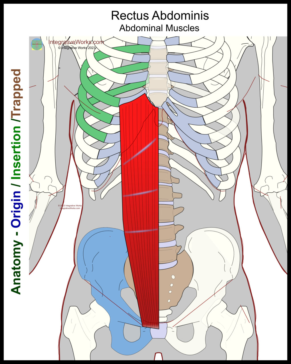
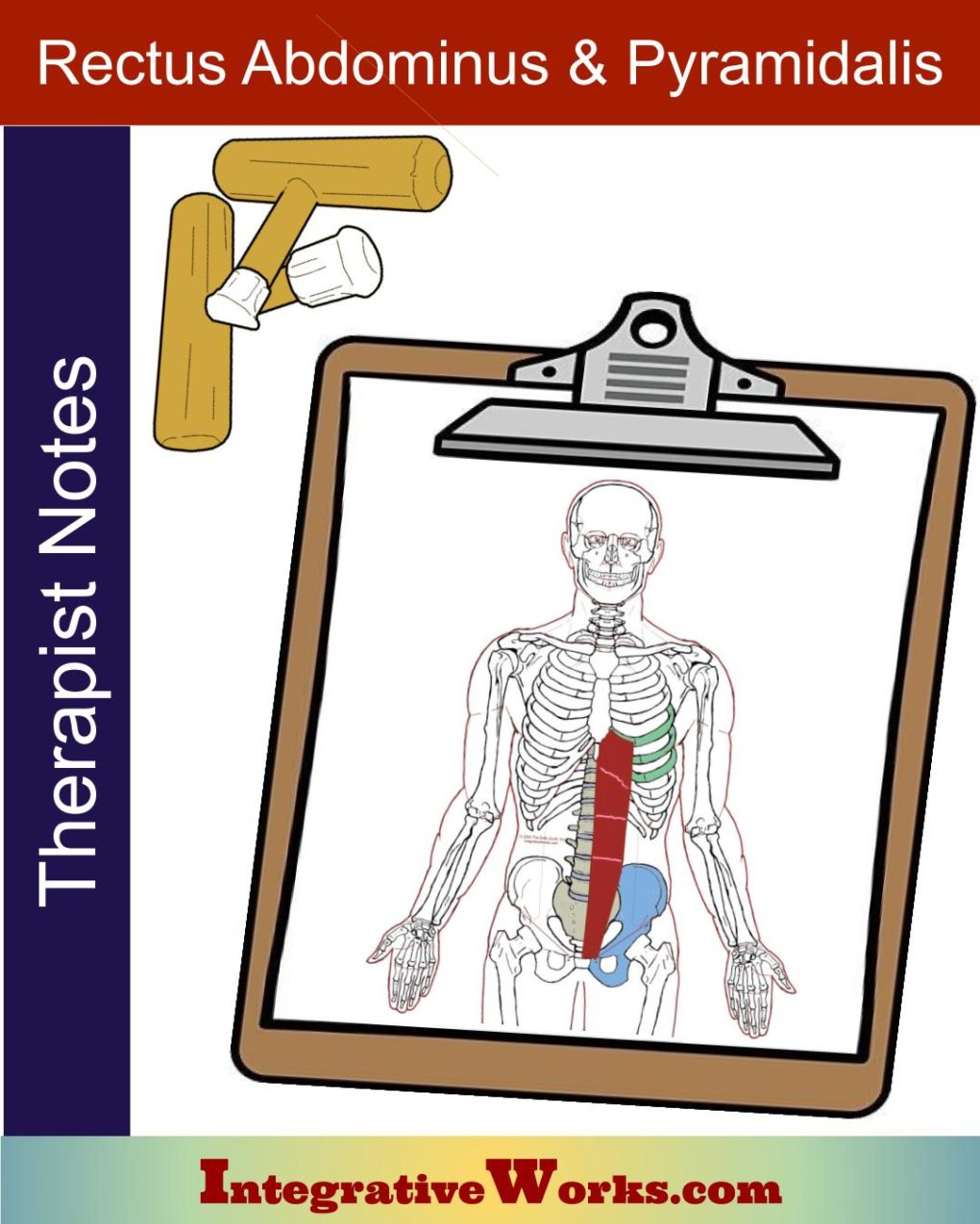
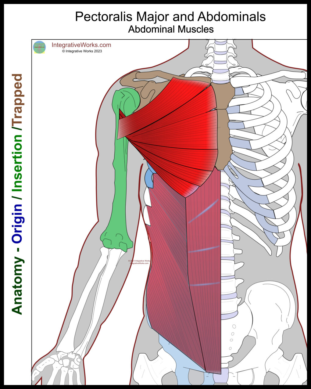
Rectus abdominis seems simple but is notably variable. It is a broad, flat muscle on the anterior abdomen with fibers that connect the pubic bone to the arch of the ribs. It is one of those muscles that are hard to label with an “origin” or “insertion,” but we’ll do that for illustrative purposes.
Origin
- pubic crest of pubic bone
- pubic symphysis
Insertion
- costal cartilages of ribs 5-7
- xiphoid process (when present)
Function
- flexion of the trunk
- Compression of the abdominal viscera
Innervation
- the thoracoabdominal nerves from (T7-T11)
- subcostal nerve (T12)
Rectus Sheath
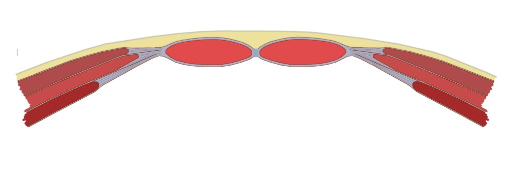
The rectus sheath is a tough fibrous aponeurosis that forms a compartment for the rectus abdominis. The rectus sheath can be divided into upper and lower portions divided by the arcuate line. The arcuate line is a semi-circular border found posterior to the rectus abdominis between the umbilicus and pubic symphysis.
Upper and Lower Rectus Sheath
- Above the arcuate line – The superficial portion is formed by the aponeurosis of the external oblique and a portion of the aponeurosis of the internal oblique. The posterior part is formed by the aponeurosis of the transverse abdominis and a portion of the aponeurosis of the internal oblique.
- Below the Arcuate line – The aponeuroses of the transverse abdominis, internal, and external oblique pass anteriorly to the rectus abdominis. Only the transverse fascia passes posteriorly.
The level of the arcuate line varies in its position. In some cases, it is above the umbilicus. In most cases, it is just below the umbilicus. In other cases, it is near the pubic symphysis.
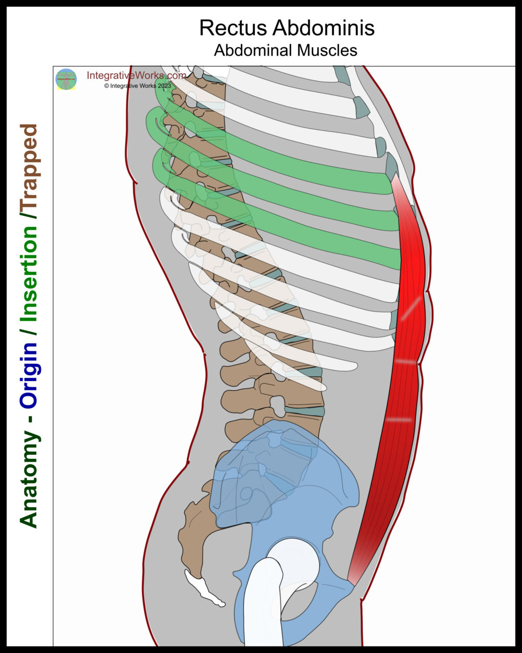
The rectus abdominis is wider, laterally near the ribs, and thicker anterior-to-posterior near the pubic bone.
Functional Considerations
This muscle draws the hips and ribs together, which flexes the trunk. During the sit-up, it is active in all phases but most active when the lower thoracic and lumbar spine are flexing between the time that the scapulae come off the ground and the iliac crest comes off the ground.
When the spinal erectors stabilize the trunk, it compresses the distended abdomen. This assists in activities like defecation or labored breathing. In addition, this muscle stabilizes the trunk when moving the head and/or legs. When supine, the rectus abdominis engages to lift the head. It engages when the legs are raised. It is active when carrying a backpack or pulling down and forward with the arms.
Rectus Abdominis vs. Iliopsoas
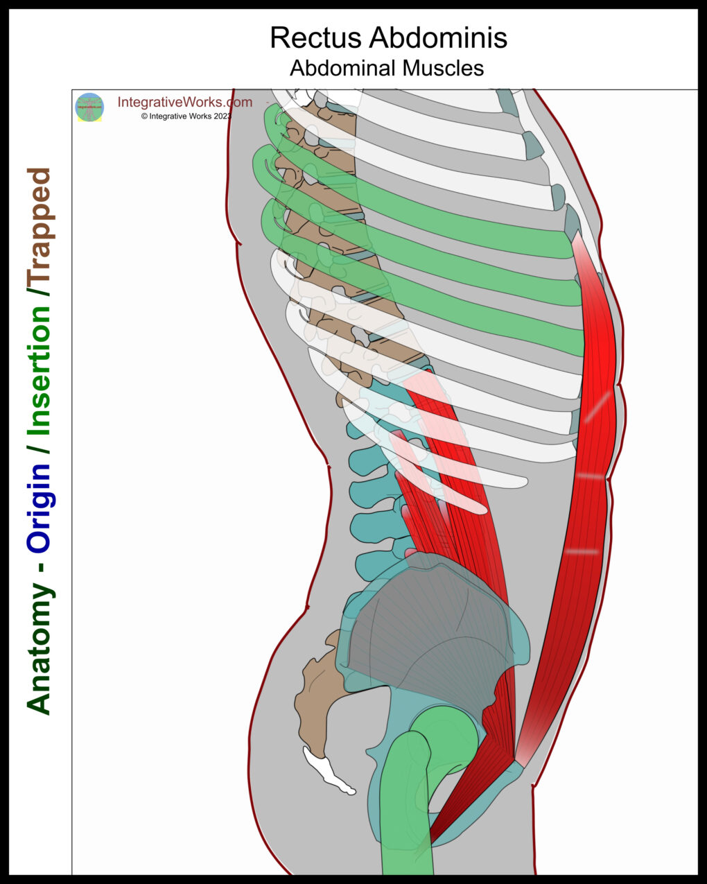
Rectus abdominis and iliopsoas have an antagonistic relationship.
- As the rectus femoris contracts, it flexes the lumbar spine. In a standing position, it can also posteriorly rotate the pelvis.
- The psoas major tightens as the low back flattens to restore the lumbar curve. Additionally, the iliacus anteriorly rotates the pelvis in the standing position.
This fundamental relationship is complicated by other factors, such as the tension of obliques and visceral fat.
Anomalies, Etc.
The muscle is divided into sections by 3, sometimes 4, rarely only 2, tendon-like bands across the muscle fibers. The top band is usually just under the xiphoid, the bottom one is near the umbilicus, and the third is usually between those. Here is one study on that variability.
The construction of the arcuate line is also variable. In fact, this study of 24 cadavers states, “The shape and position of the arcuate line were neither symmetrical nor constant, and neither was the arrangement of the nerve supply to the rectus abdominis muscle or to the overlying skin.”
Posts related to Rectus Abdominis
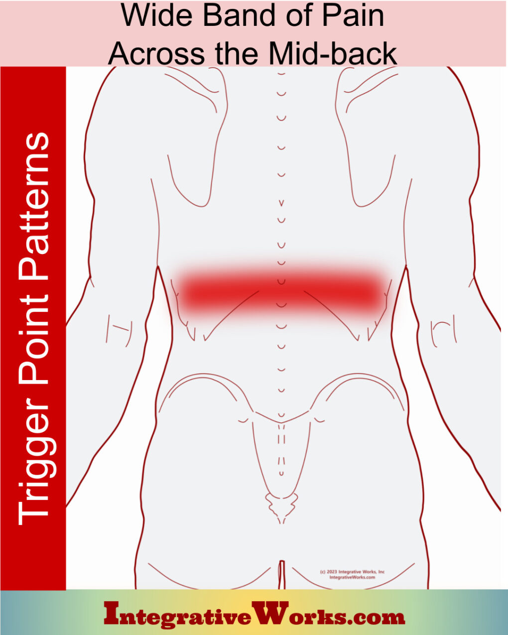
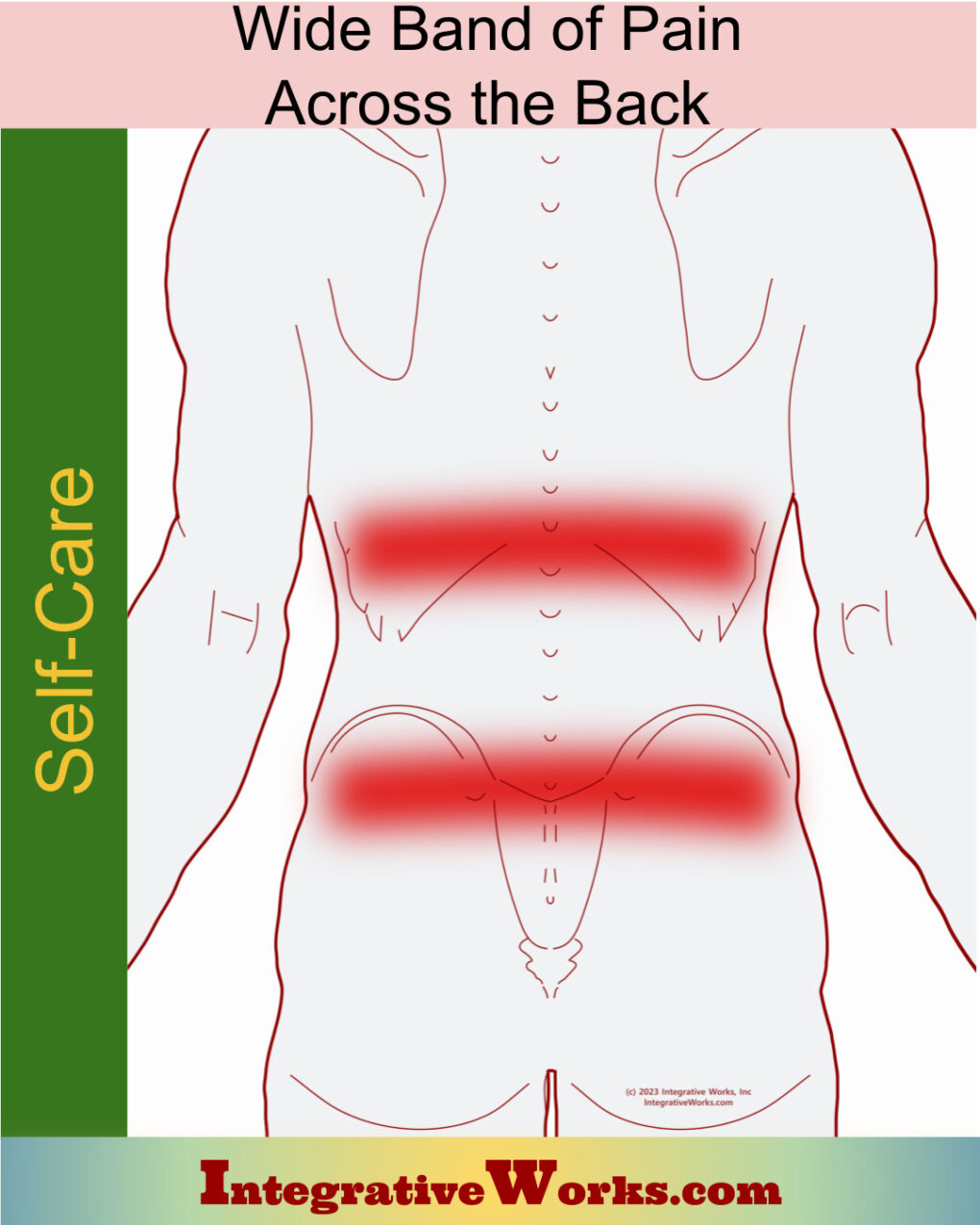
Band of Pain Across the Mid-back
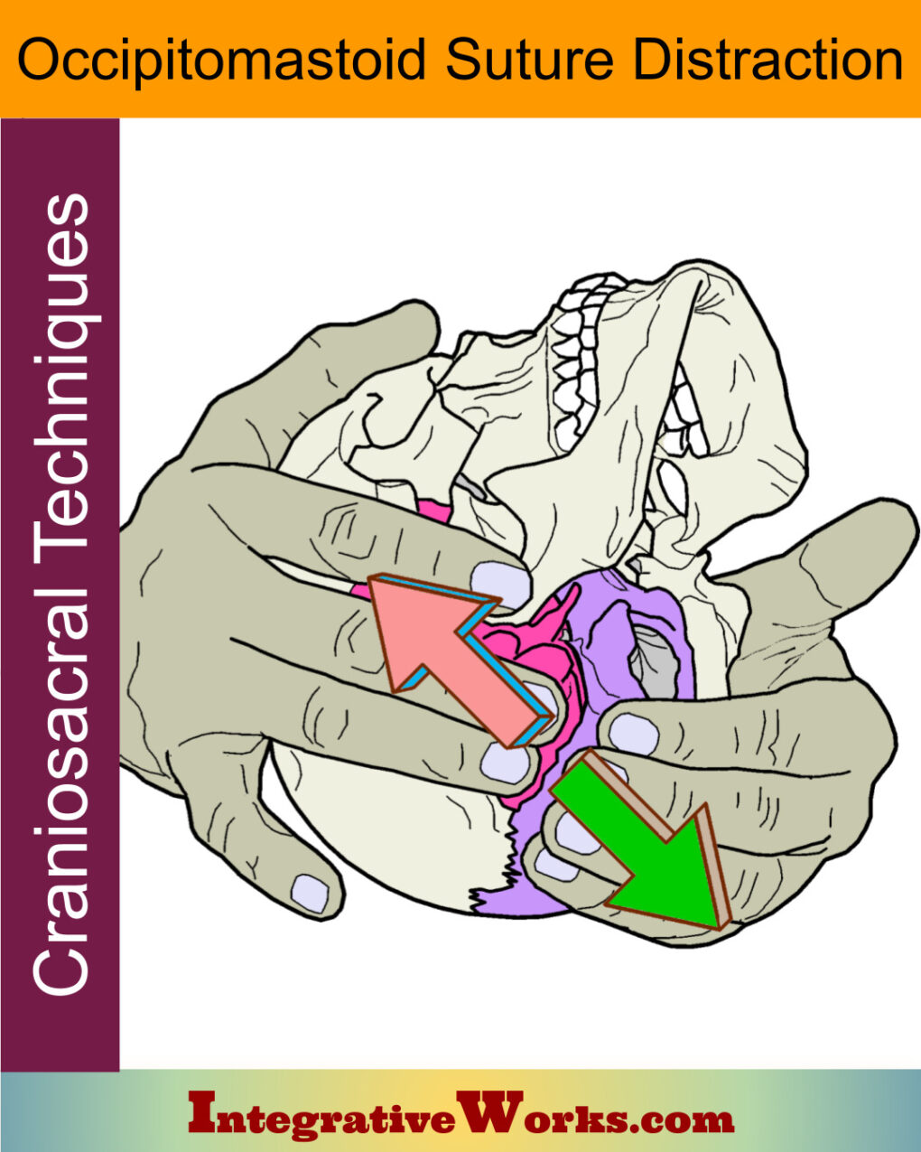
Occipitomastoid Suture Distraction- Craniosacral Techniques
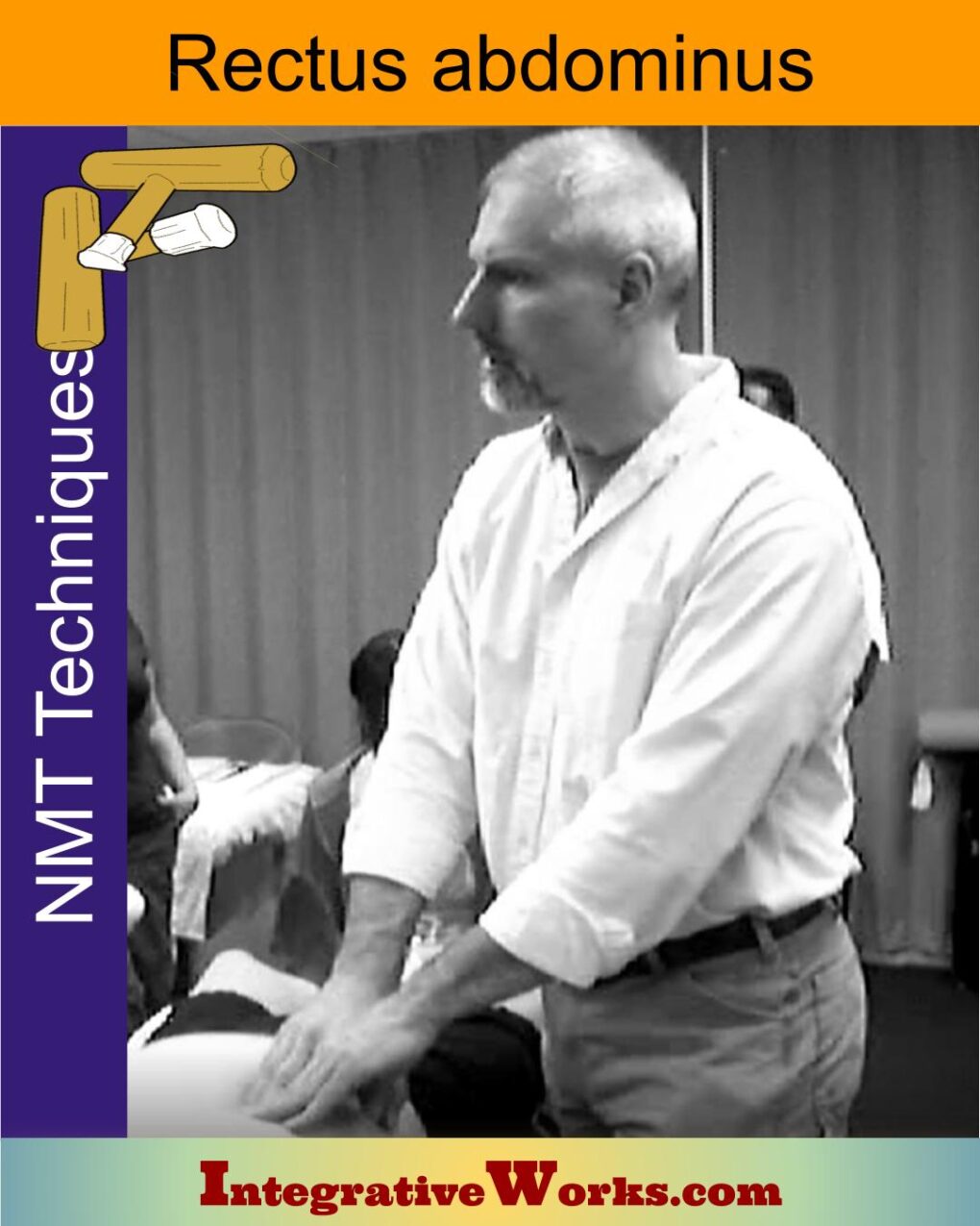
Rectus Abdominus – Neuromuscular Massage Protocol
Rectus Abdominus and Pyramidalis – Massage Therapy Notes
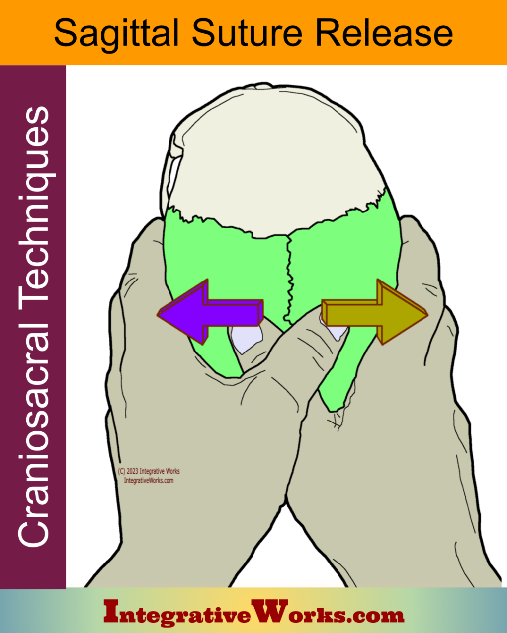
Sagittal Suture Release – Craniosacral Techniques
Self-care – Band of Pain Across Back
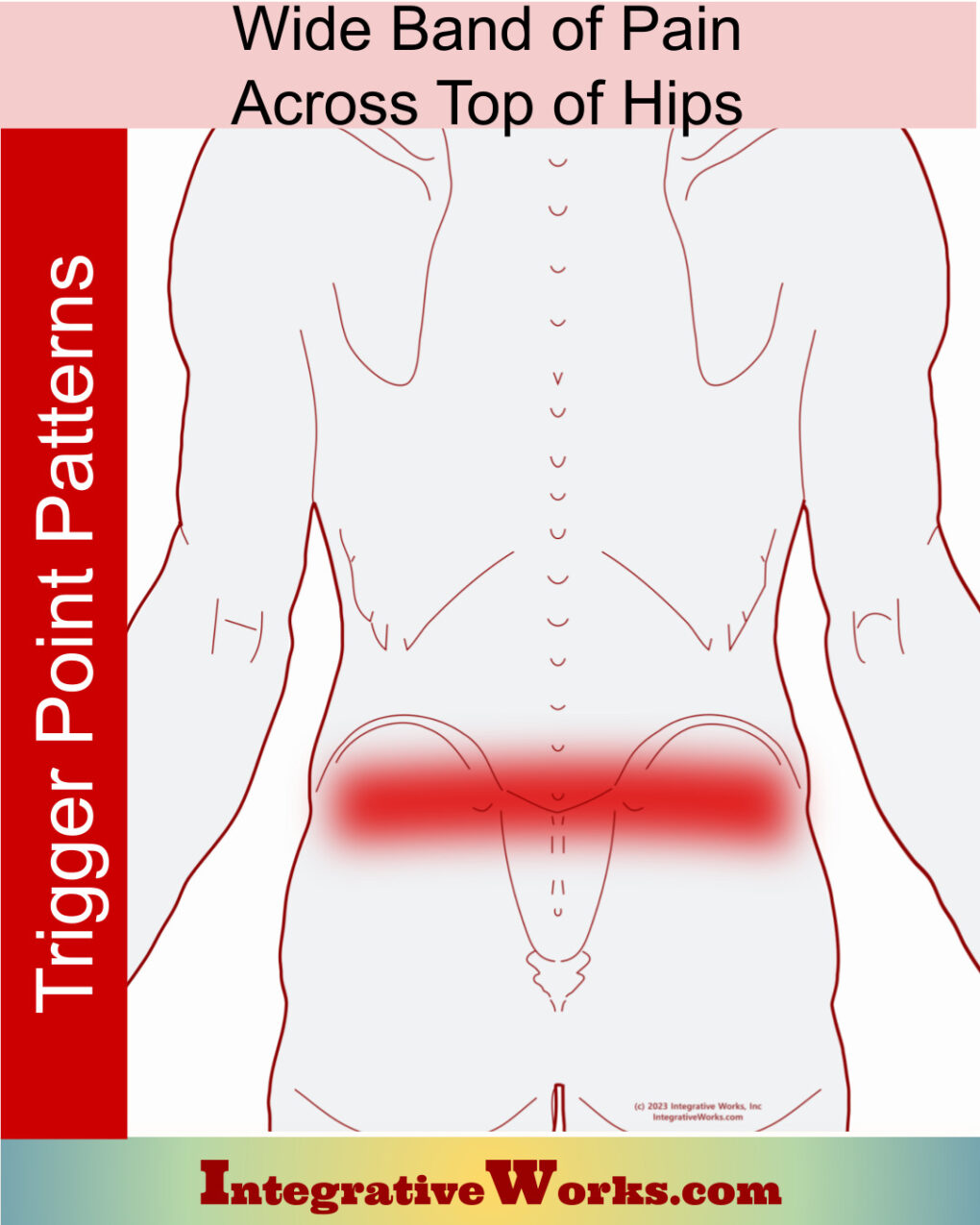
Wide Band of Pain Across the Top of the Hips
Yoga, “that good stretch feeling,” and trigger points
Support Integrative Works to
stay independent
and produce great content.
You can subscribe to our community on Patreon. You will get links to free content and access to exclusive content not seen on this site. In addition, we will be posting anatomy illustrations, treatment notes, and sections from our manuals not found on this site. Thank you so much for being so supportive.
Cranio Cradle Cup
This mug has classic, colorful illustrations of the craniosacral system and vault hold #3. It makes a great gift and conversation piece.
Tony Preston has a practice in Atlanta, Georgia, where he sees clients. He has written materials and instructed classes since the mid-90s. This includes anatomy, trigger points, cranial, and neuromuscular.
Question? Comment? Typo?
integrativeworks@gmail.com
Follow us on Instagram

*This site is undergoing significant changes. We are reformatting and expanding the posts to make them easier to read. The result will also be more accessible and include more patterns with better self-care. Meanwhile, there may be formatting, content presentation, and readability inconsistencies. Until we get older posts updated, please excuse our mess.

