Overview
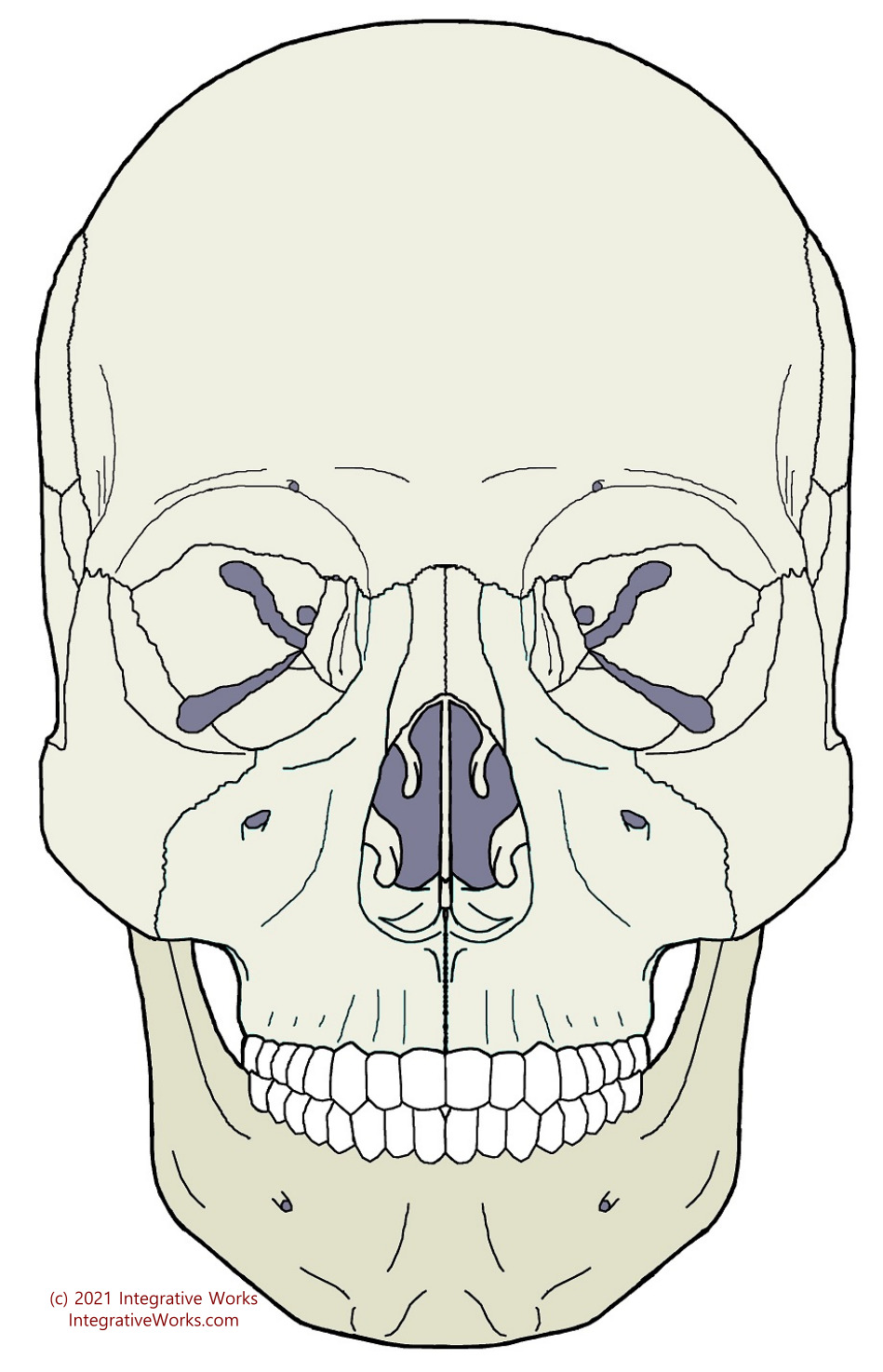
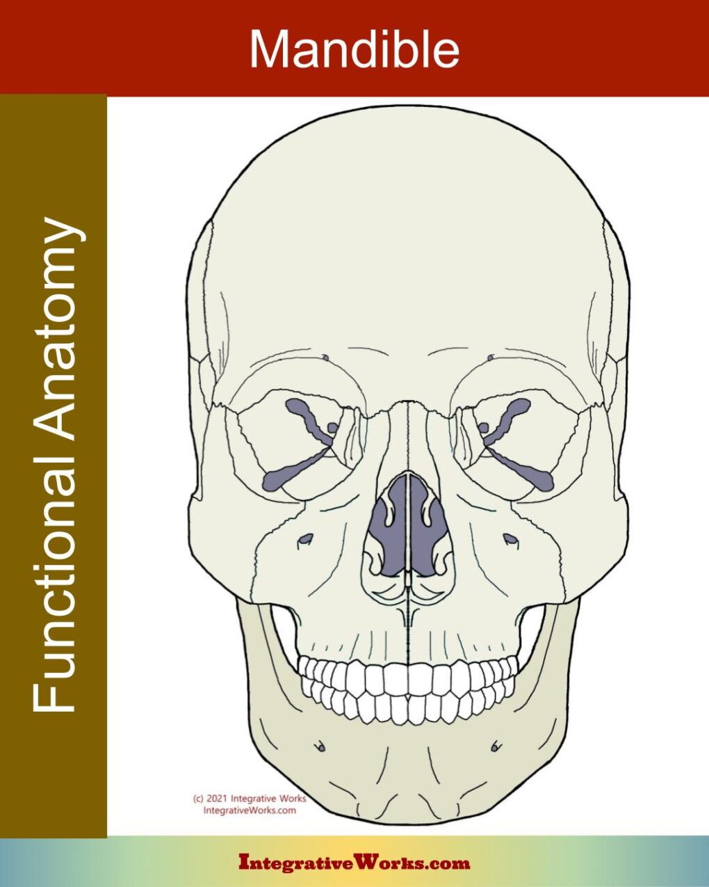
The mandible is symmetrical and forms the jaw. It has two halves joined at the prominence of the chin.
Ossification starts from a central point on each half about the sixth week of pregnancy. These two halves are separate at birth and join during the first year.
Each half has a body, ramus, condyle, and coronoid process.
Features
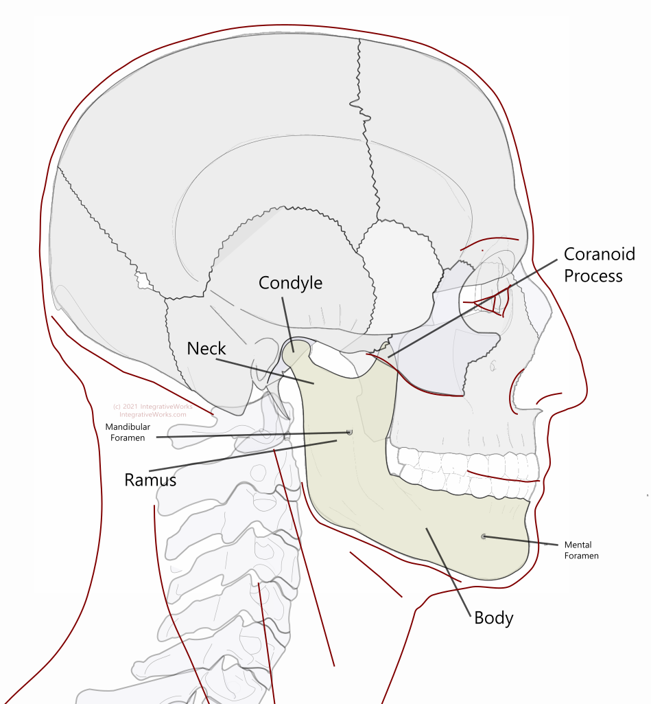
The body is the lengthy section extending posteriorly and laterally from the chin to the angle of the mandible. The body is a hollow section of bone that houses the lower teeth along the alveolar margin of the upper border. The lower edge is a thicker section that gives the bone strength. The oblique line extends along the superior, lateral portion of the body, just below the teeth, and continues up the anterior edge of the ramus. The mental foramen is a port for the mental nerve on the lateral surface of the body. The mylohyoid line along the interior surface of the body extends range-of-motion just behind the last molar, extending anteriorly and downward until it fades into the bone as it nears the inferior mental spine.
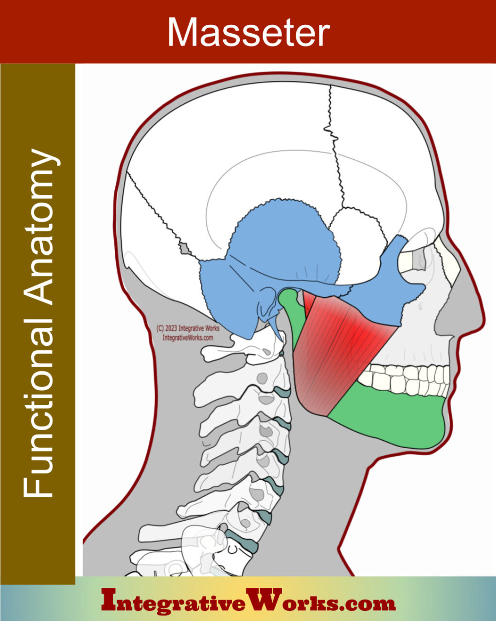
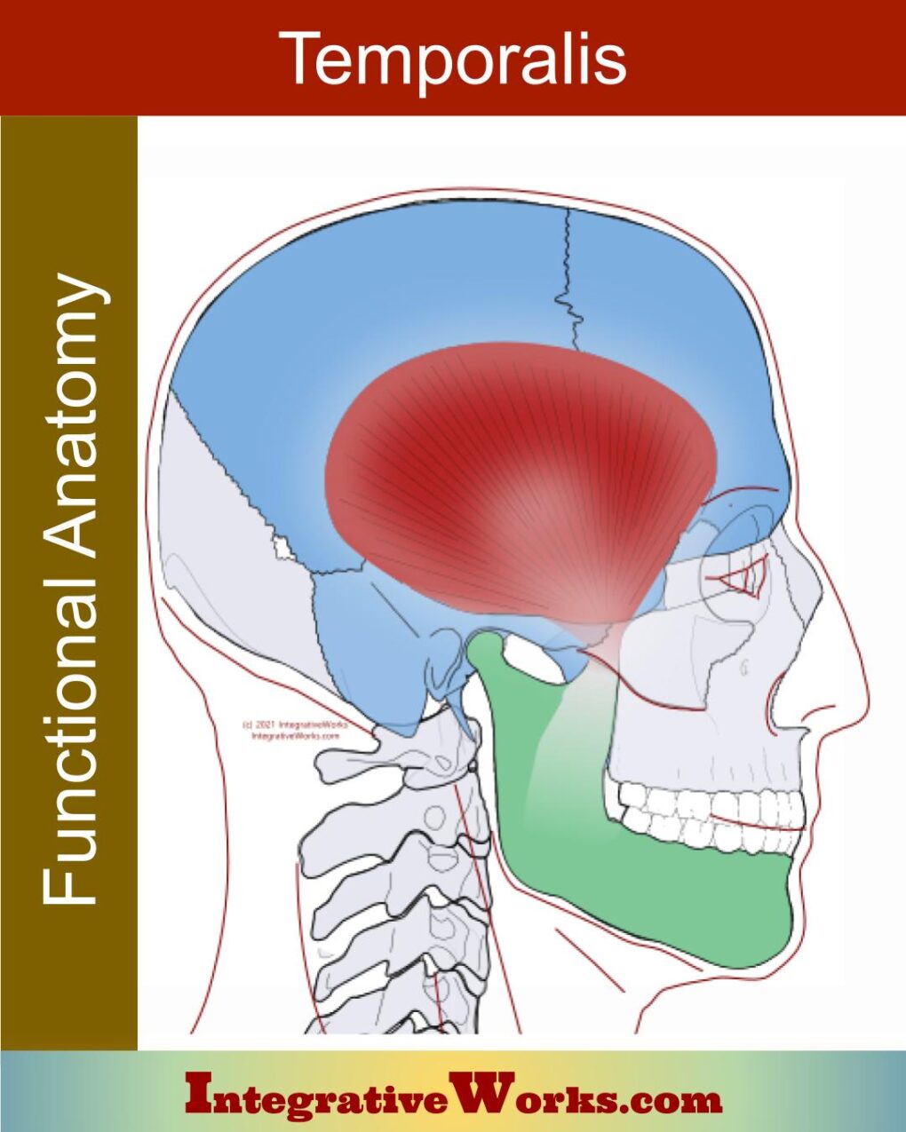
The ramus is the quadrangular portion that extends upward toward the temporomandibular joint. From the angle, the ramus extends superiorly as a flat section bone. The exterior surface is a flat area for the attachment for the masseter. On the interior surface is the mandibular foramen and a sharp, spike-shaped covering called the lingula. The lingula is the attachment for the sphenomandibular ligament. The medial pterygoid attaches on the interior surface near the angle of the mandible where the ramus and body meet.On the anterior aspect of the ramus is the coronoid process. It is a flattened, triangular, upward projection for the attachment of the temporalis muscle. The neck of the mandible also extends upward on the posterior aspect to the ramus to the head, or condyle of the mandible. The head is a convex process that is elongated transversely toward the anterior lip of the foramen magnum.
Craniosacral Elements
Articulations
Functional Anatomy
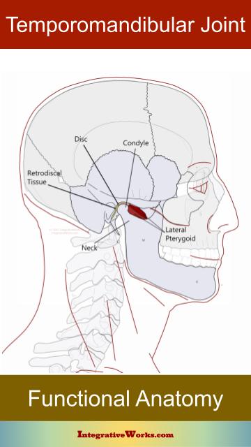
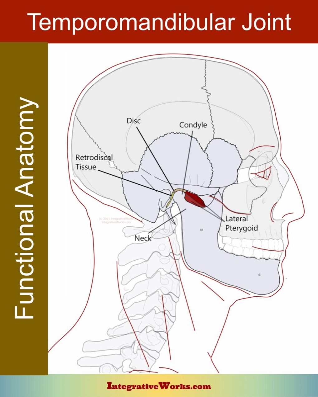
The temporomandibular joint is a complex synovial joint – arguably the most complex joint in the body. Read more about the anatomy in this post.
Muscular Attachments
Muscle attachments include:
- masseter
- temporalis
- medial pterygoid
- lateral pterygoid (inferior belly)
- buccinator
- mentalis
- platysma
- depressor labii inferior
- depressor anguli oris
- genioglossus
- geniohyoid
- mylohyoid
- digastric
Dural Attachments
The mandible is not directly connected to the dural membranes. The temporal bones connect to the dural membranes. In turn, they drive the motion of the mandible.
Motion
As the cranium flexes:
- The mandibular head moves posteriorly and medially as the temporal bones rotate externally.
- The angles move laterally and the mental protuberance moves posteriorly so that the lower and upper teeth move together.
Related Posts
Mandible – Functional Anatomy
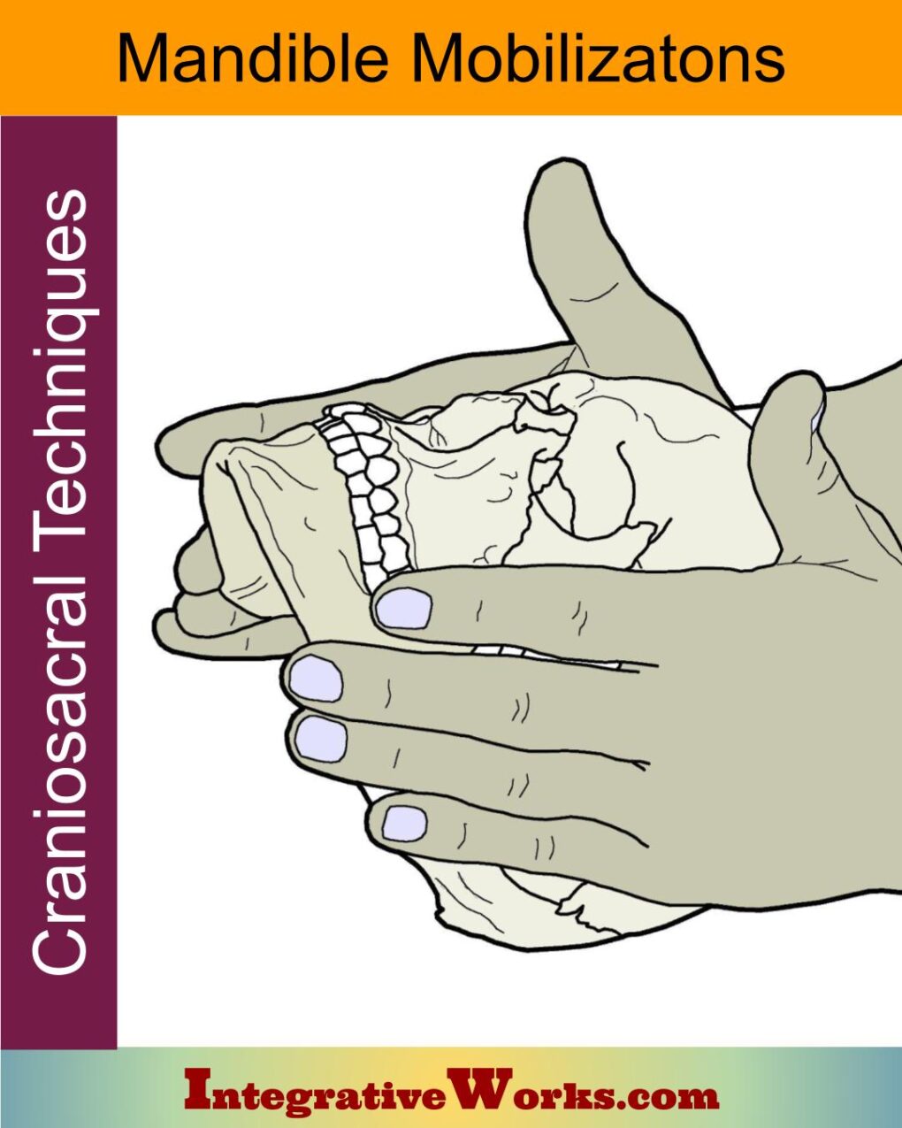
Mandible Mobilizations – Craniosacral Techniques
Masseter – Functional Anatomy
Temporalis – Functional Anatomy
Temporomandibular Joint – Functional Anatomy
Support Integrative Works to
stay independent
and produce great content.
You can subscribe to our community on Patreon. You will get links to free content and access to exclusive content not seen on this site. In addition, we will be posting anatomy illustrations, treatment notes, and sections from our manuals not found on this site. Thank you so much for being so supportive.
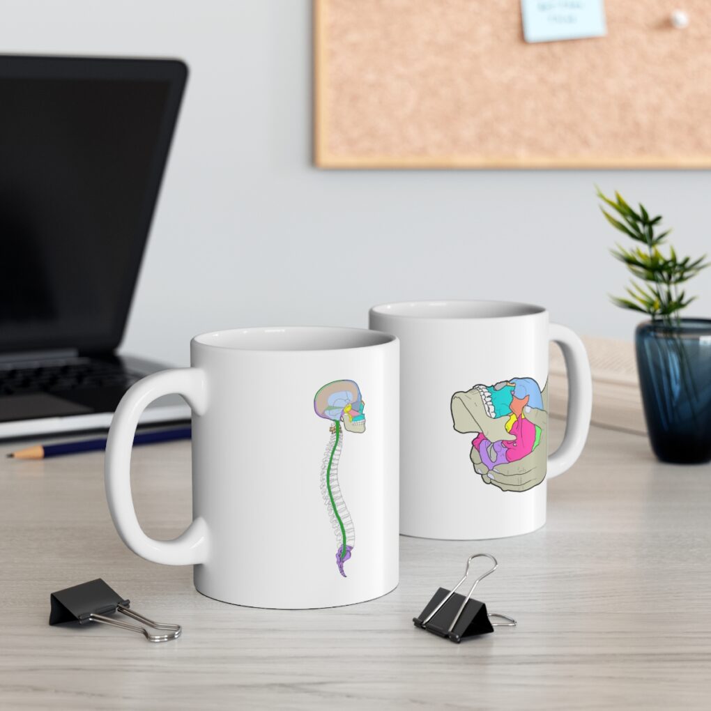
Cranio Cradle Cup
This mug has classic, colorful illustrations of the craniosacral system and vault hold #3. It makes a great gift and conversation piece.
Tony Preston has a practice in Atlanta, Georgia, where he sees clients. He has written materials and instructed classes since the mid-90s. This includes anatomy, trigger points, cranial, and neuromuscular.
Question? Comment? Typo?
integrativeworks@gmail.com
Interested in a session with Tony?
Call 404-226-1363
Follow us on Instagram

*This site is undergoing significant changes. We are reformatting and expanding the posts to make them easier to read. The result will also be more accessible and include more patterns with better self-care. Meanwhile, there may be formatting, content presentation, and readability inconsistencies. Until we get older posts updated, please excuse our mess.

