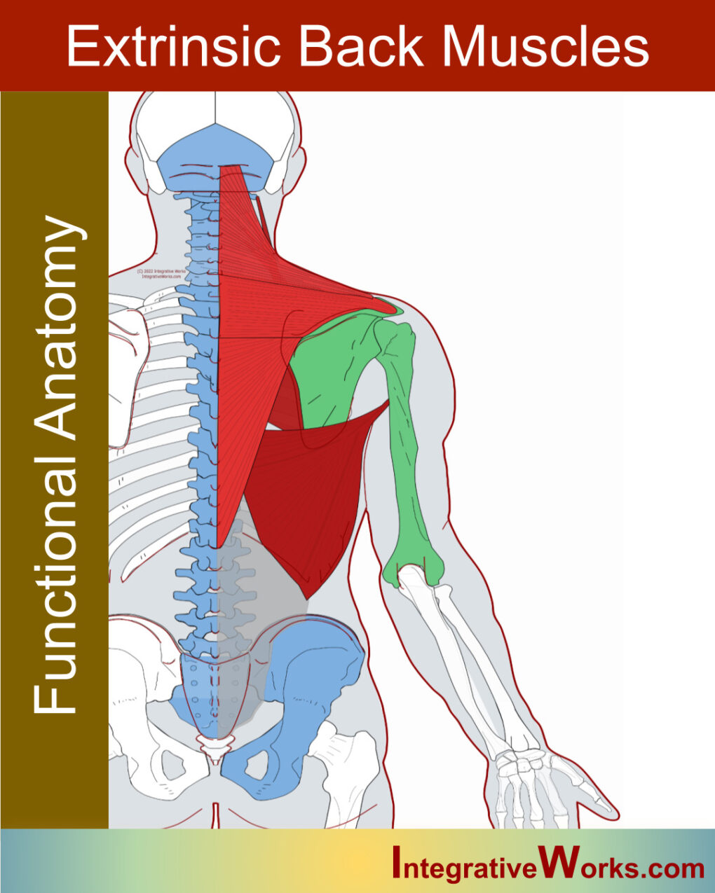
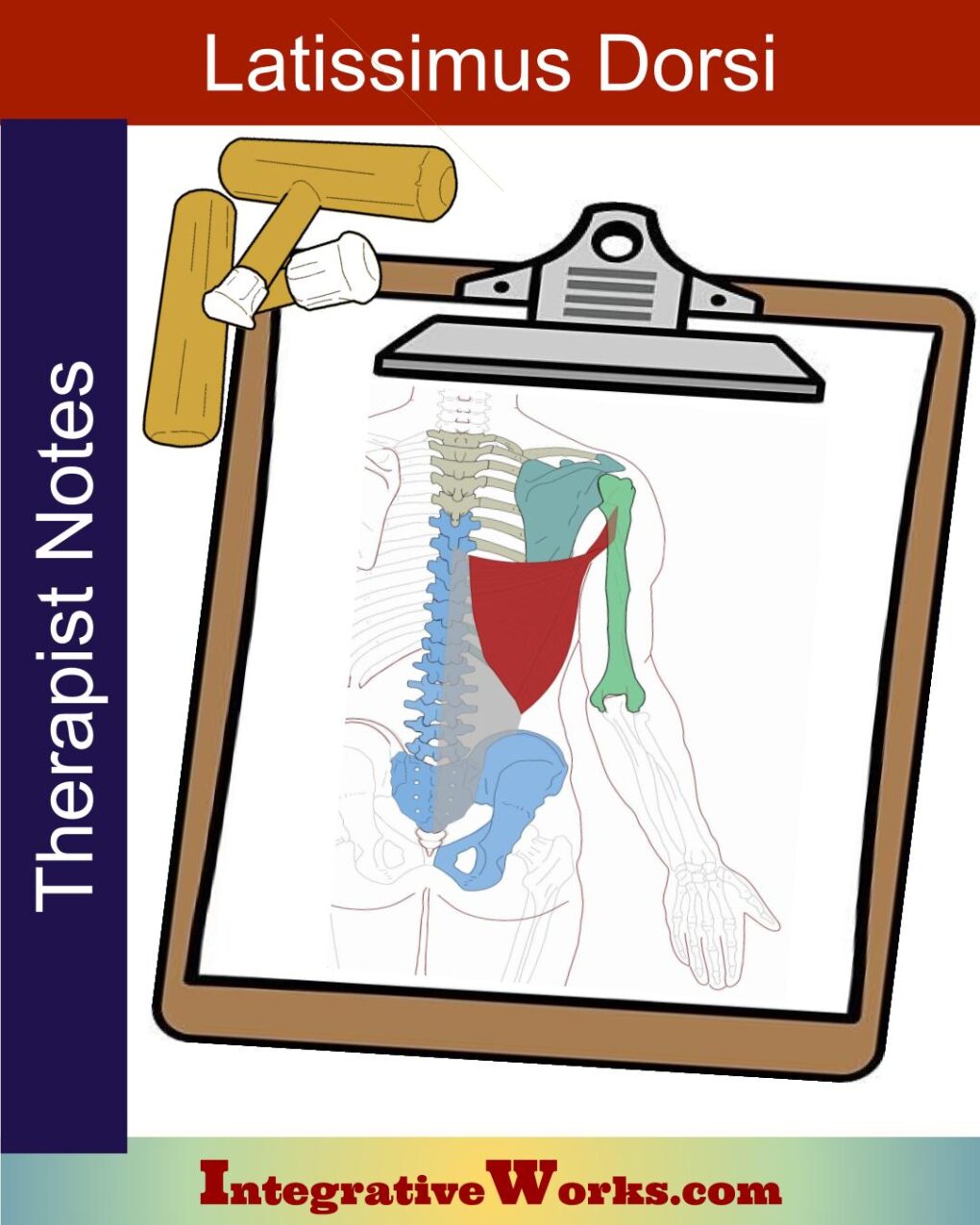
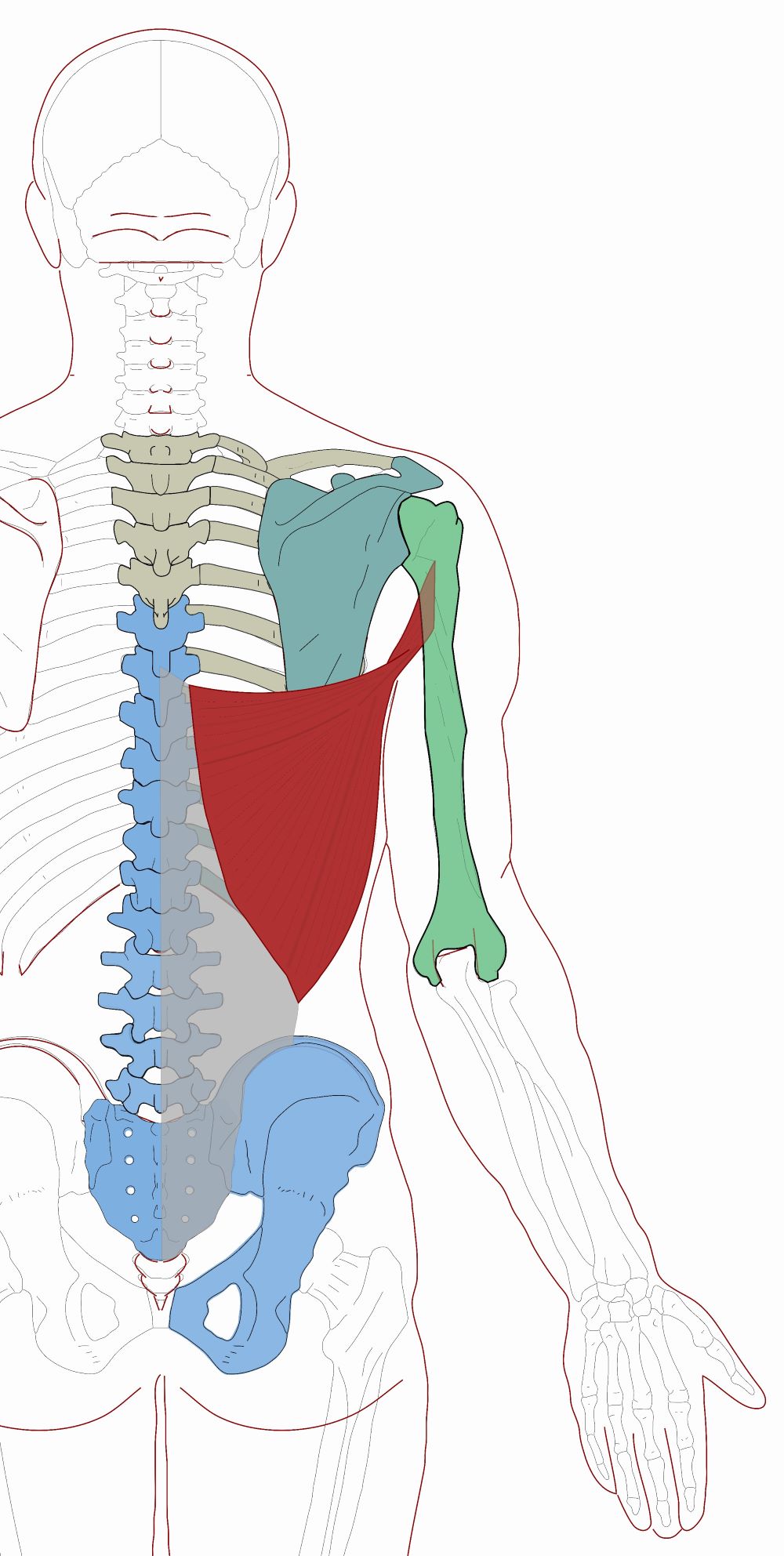
The anatomy of the latissimus dorsi seems simple but ends up attaching to more bones in the back than almost any other muscle. It connects the mid-back, low-back, and pelvis to the humerus through the thoracic vertebrae, clavicle, and scapula.
The latissimus dorsi is a broad, flat muscle covering the mid-back. The name comes from its size and location. It has three compartments and twists nearly 180 degrees around the teres major. The distal end of latissimus dorsi is often in the same compartment as teres major. It is usually innervated in six separate sections by the thoracodorsal nerve. It originates along the axial skeleton and attaches to the humerus, making it an extrinsic back muscle.
Overview of Anatomy
The functional anatomy of the latissimus dorsi is often surprising. For example, it attaches to many more bones than expected at first glance.
Origin
- T6-L5 spinous processes
Sacrum, Ilium
via Thoracolumbar fascia - Ribs 9-12
Insertion
- lateral Lip of bicipital groove
- Interior angle of the scapula (~75% of the time)
Function
- extension of the humerus
- adduction of the humerus
- assists internal rotation
- depression the scapula
- extension of the thoracic spine
Nerve
- the femoral nerve of the lumbar plexus (L2-L4)
Anomalies, Etc.
It has statistically significant variations in its attachments. First, it may cross over the bicipital groove and blend with the attachment of pectoralis major. Also, it may blend with the fibers of the triceps brachii. At times, the lateral fibers may extend down to the crest of the ilium. A little more than 40% of the time, it attaches directly to the scapula’s inferior angle. A little less than 40% of the time, it connects via soft connective tissue. However, about 20% of the time, there is no attachment to the scapula.
Underlying Attachments and Trapped Bones
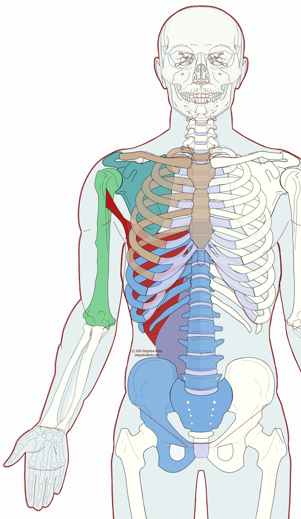
Latissimus dorsi attaches to most of the bones of the axial skeleton below T6, except for a few ribs. It attaches to the spine and pelvis through the thoracolumbar fascia, which originates along the iliac crest, sacrum, lumbar vertebrae, and the last 7 Thoracic vertebrae. The muscle fibers of the latissimus dorsi usually stop short of the iliac crest and lumbar vertebrae.
Fresh Perspective
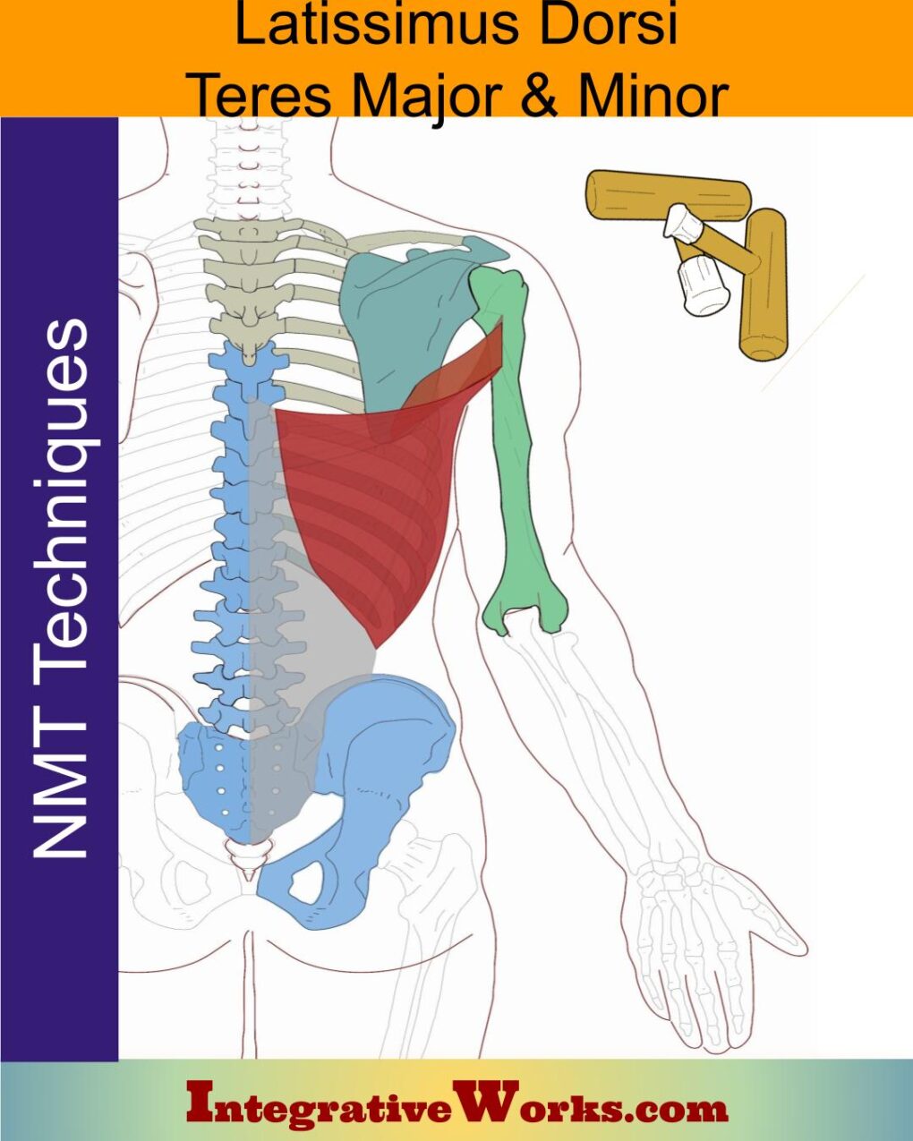
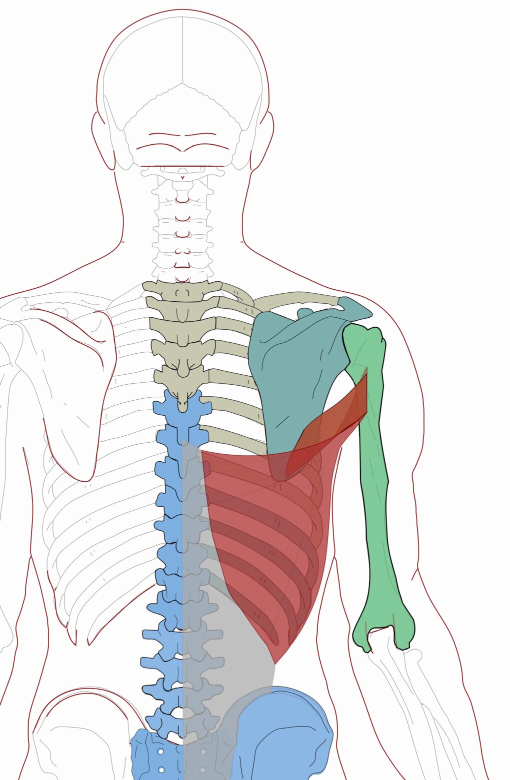
This anterior view shows the complexity of its attachments. You can see how the scapula and lower four ribs get bound between the humerus and spine. Also, there are many bones (in brown)that get trapped between the origin (in blue) and the insertion (in green).
This muscle’s superior border attaches to the scapula’s inferior border directly or by a slip of connective tissue about 75% of the time. It twists around the teres major and often shares the same compartment as it passes along the axilla. It inserts along the medial lip and floor of the bicipital groove in a tendon that usually fuses with the teres major’s fibers.
Functional Considerations
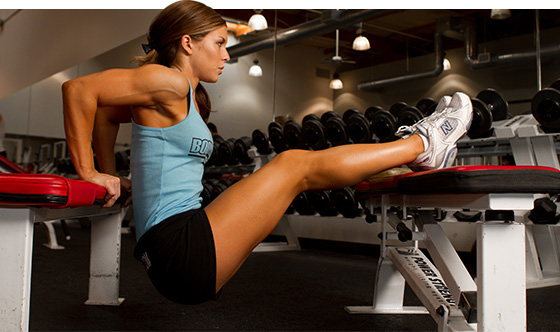
The latissimus dorsi depresses the humerus and shoulder girdle. Because of the mobility of the glenohumeral joint, this can make naming its functions complex. It is mainly dependent on the humerus’ starting position.
When in front of the body, as when chopping wood, it extends the humerus. When the humerus is behind the body, as when dipping on bars, the latissimus dorsi flexes the humerus. When the arm is abducted laterally, as when dribbling a basketball out to the side, it is adducting the arm. It even has some ability to assist in internal rotation.
The lateral compartment is more involved and developed by moving it down from overhead. On the other hand, the transverse compartment is more involved in actions that pull the humerus and shoulder girdle posteriorly, as when rowing.
Swinging
Its broad attachment to the mid and low back make it suited to support the body weight when hanging by the upper extremity. In addition, the biceps slips between the lats and pecs, offering a perfect structure for swinging from a tree branch.
Posts Related to Latissimus Dorsi
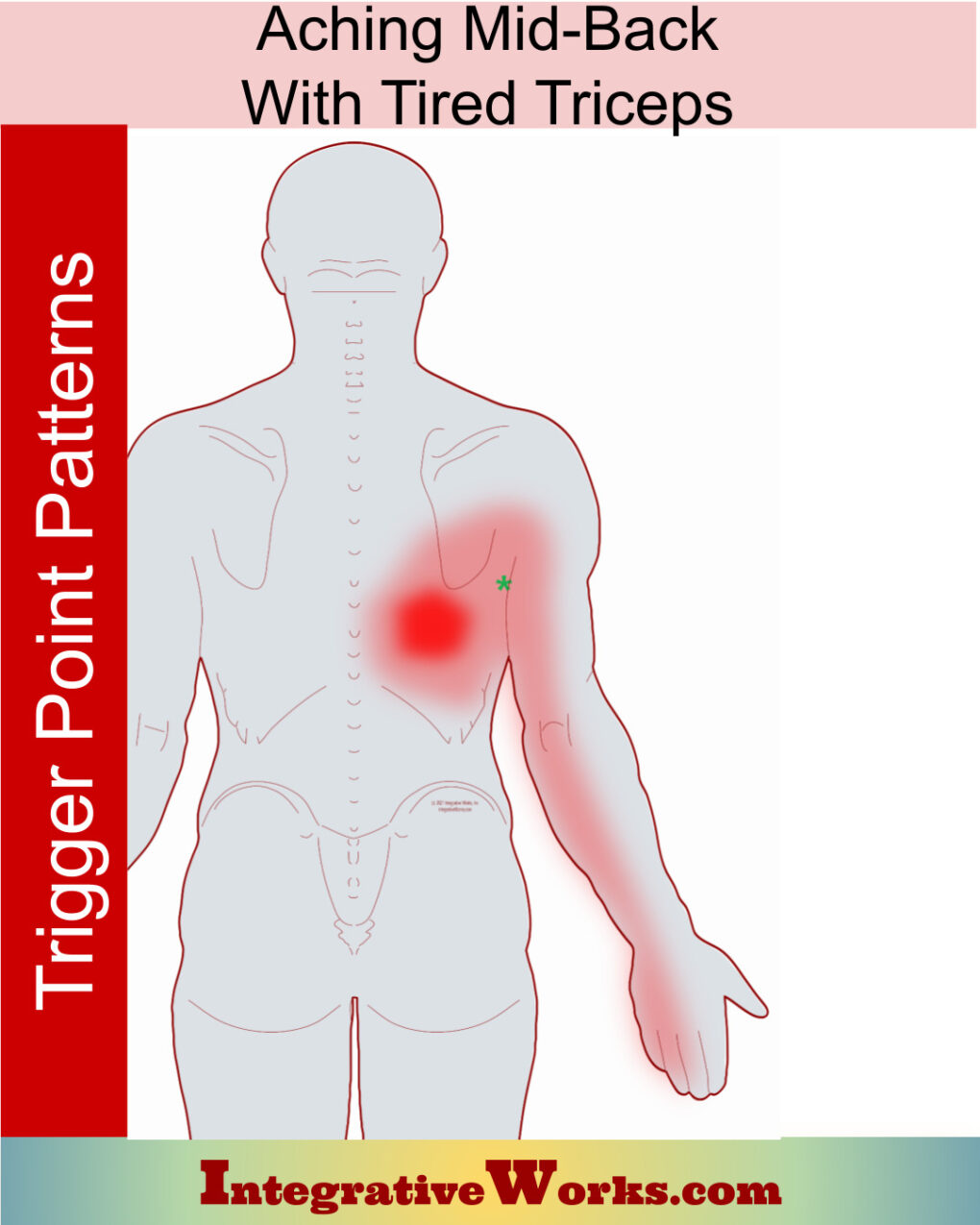
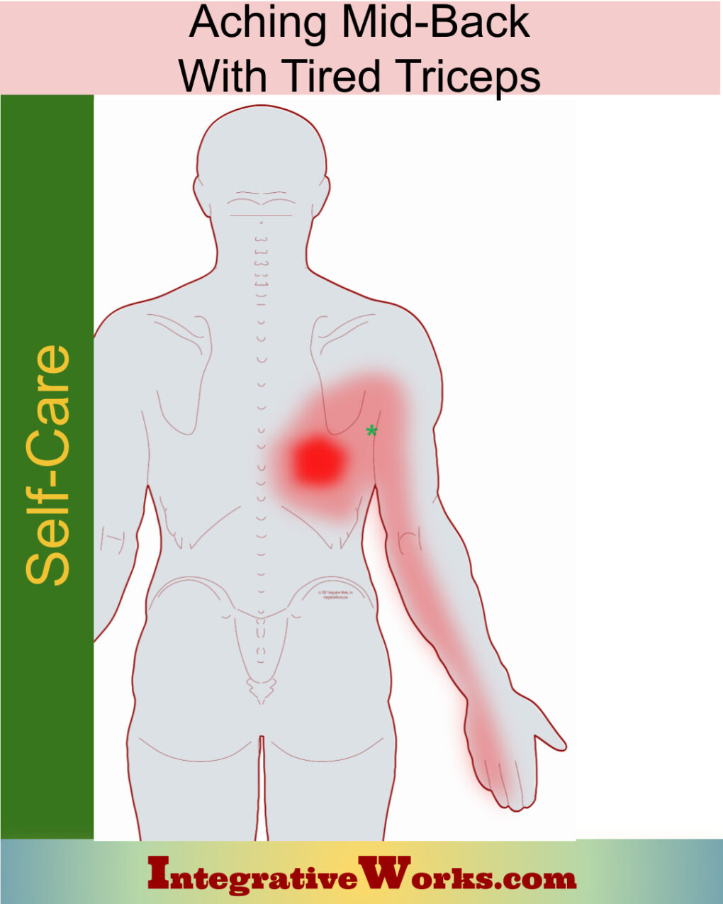
Aching mid-back with tired triceps
Biceps Brachii – Neuromuscular Protocol
Extrinsic Back Muscles – Functional Anatomy
FrontoMax 45 – CranioMuscular Techniques
FrontoMax 45 – Craniosacral – Facial Mobilizations
Latissimus Dorsi – Massage Therapy Notes
Latissimus Dorsi, Teres Major and Teres Minor – Neuromuscular Massage Protocol
Self Care – Aching mid-back with tired triceps
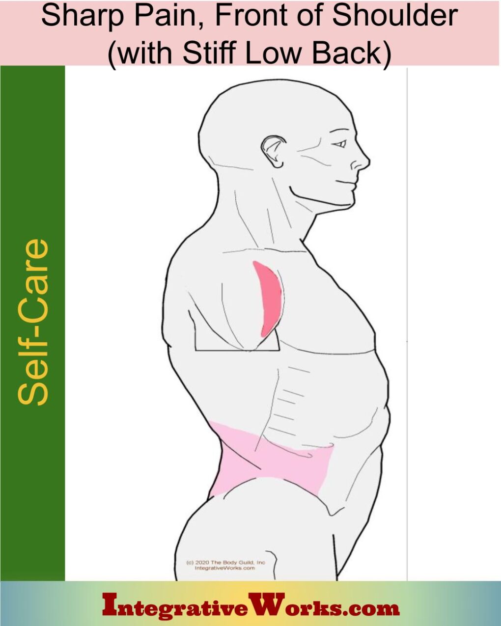
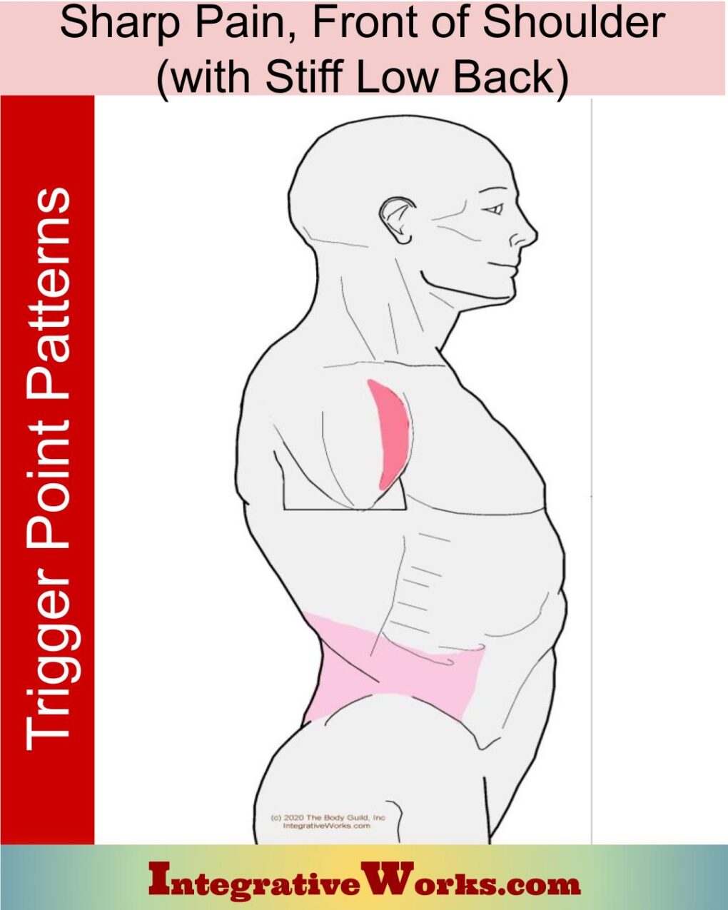
Self Care – Sharp Pain in Front of Shoulder, Tight Low Back
Sharp Pain in Front of Shoulder, with Tight Low Back
Support Integrative Works to
stay independent
and produce great content.
You can subscribe to our community on Patreon. You will get links to free content and access to exclusive content not seen on this site. In addition, we will be posting anatomy illustrations, treatment notes, and sections from our manuals not found on this site. Thank you so much for being so supportive.
Cranio Cradle Cup
This mug has classic, colorful illustrations of the craniosacral system and vault hold #3. It makes a great gift and conversation piece.
Tony Preston has a practice in Atlanta, Georgia, where he sees clients. He has written materials and instructed classes since the mid-90s. This includes anatomy, trigger points, cranial, and neuromuscular.
Question? Comment? Typo?
integrativeworks@gmail.com
Interested in a session with Tony?
Call 404-226-1363
Follow us on Instagram

*This site is undergoing significant changes. We are reformatting and expanding the posts to make them easier to read. The result will also be more accessible and include more patterns with better self-care. Meanwhile, there may be formatting, content presentation, and readability inconsistencies. Until we get older posts updated, please excuse our mess.

