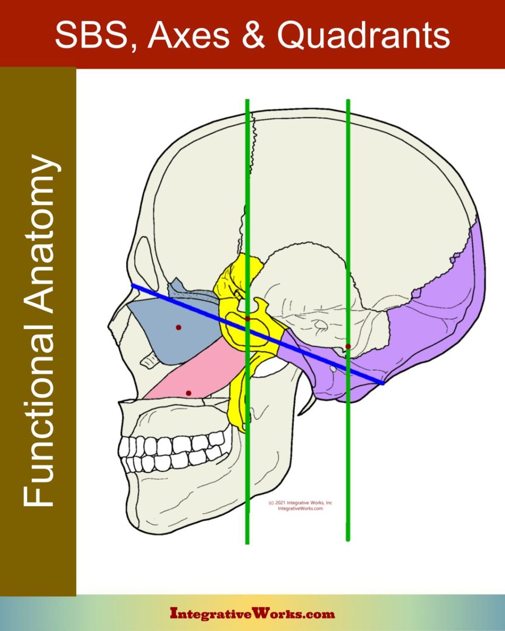
Here you will find a brief overview of the sphenobasilar mechanism and its axes of motion. You should be familiar with an overview of the craniosacral system. You can also find more detail about the nuances of movement of bones as they cycle through flexion and extension in the post on craniosacral motility.
Overview of Structure
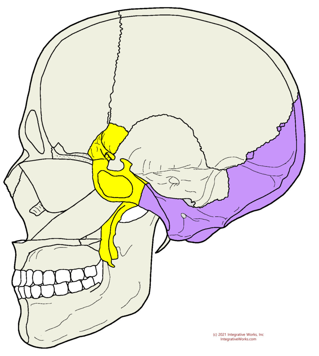
The sphenobasilar mechanism includes the sphenoid bone, the occipital bone, and the sphenobasilar synchondrosis (SBS). The SBS is the joint between the sphenoid and occiput. It starts as a synchondrosis. Eventually, usually between ages 18 and 25, the cartilaginous place is ossified, creating a “sphenocciput.”
In the traditional biomechanical osteopathic model, the SBS is considered the central point of motion in the craniosacral cycle.
The center of this mechanism cradles the pituitary in the cavernous sinus of the venous sinuses. Thus, the pumping action supports the CNS centers for regulation and the cavernous sinus.
Axes of Motion
Overview of Axes
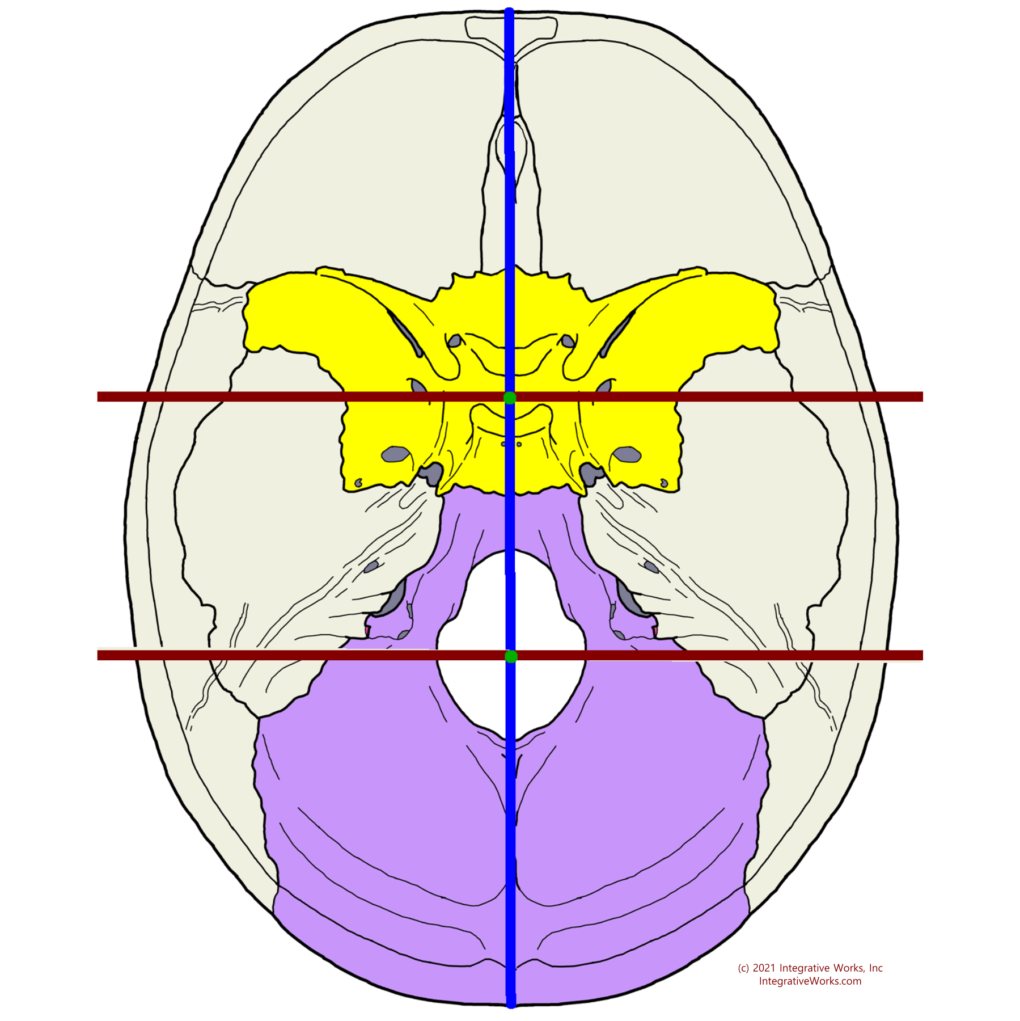
The craniosacral motility is primarily seen as tidal motion that ebb and flow through flexion and extension. You can read more about the details of that in this other post.
There are three pairs of axes:
- Transverse axes
- Anterior to Posterior axes (A/P)
- Verticle axes
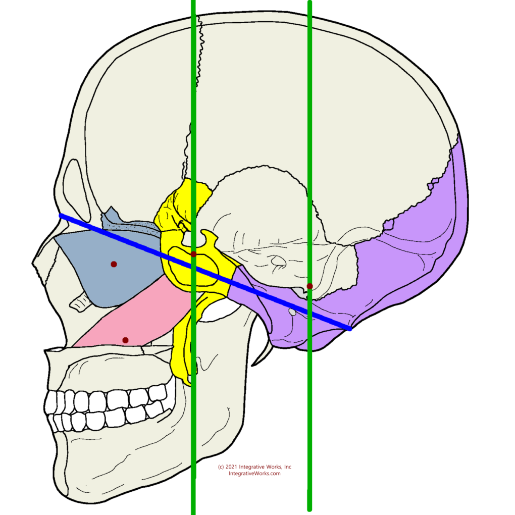
Axes and SBS Patterns
- Transverse axes allow the spenobasilar mechanism flex and extend. When the spenoid and occiput are out of sync, the transverse axis allow superior and vertical strain patterns. As well, the ethmoid and vomer rotate on transverse axes.
- Anterior to Posterior (A/P) axis allow the sphenobasilar mechanism to torsion and rotate. Torsion occurs when the spenoid and occiput rotate in different directions on the A/P axis. Rotation generally occurs when at the far end of sidebending patterns.
- Verticle axes allow the spenobasilar mechanism to lateral strain and sidebend. Lateral strain occurs when the spenoid and occiput rotate in the same dorection on the vertical axes. Sidebending occurs when the sphenoid and occiput rotate in different direction on the vertical axes.
- The SBS has combined patterns such as vertical strain with torstion.
- In addition, the SBS has non-axial motions i.e. shearing.
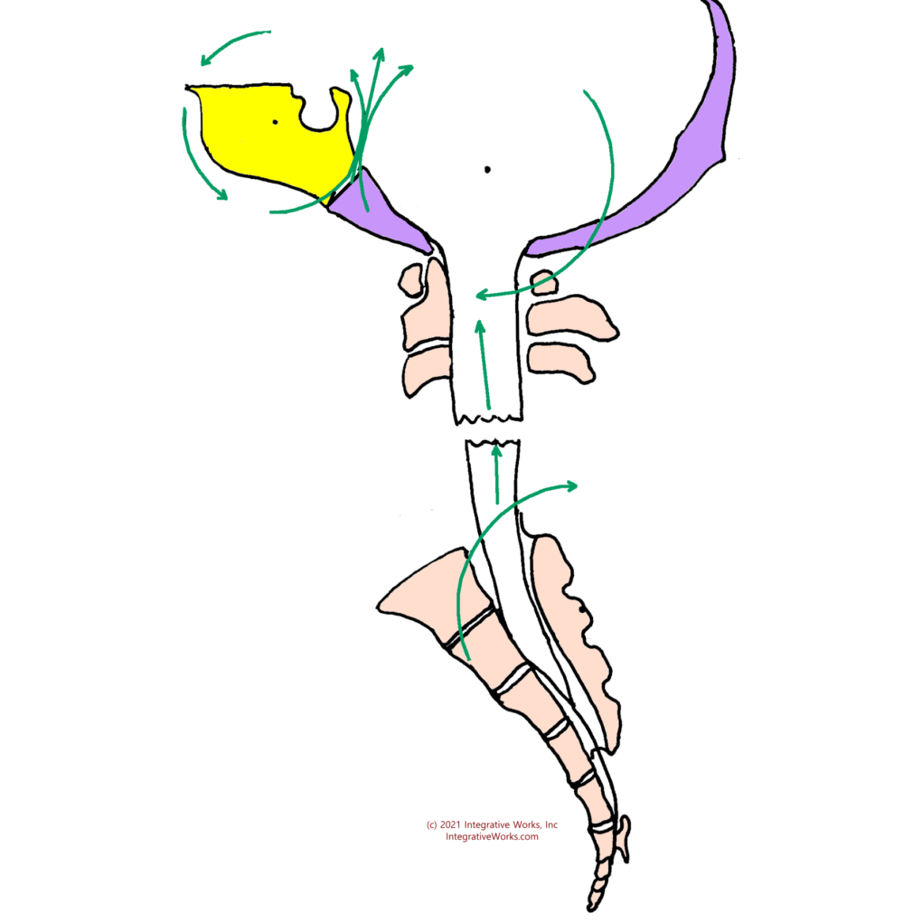
Craniosacral and the Sphenobasilar Mechanism
The word “craniosacral” comes from the direct structural relationship between the sphenobasilar mechanism and the sacrum. The dural tube provides a complex pulley system between the cranium and sacrum.
Although analysis varies a bit from one approach to another, this diagram conveys the basic idea. As the occiput flexes and extends, the sacrum nutates (nods) and counter-nutates. As restrictions create variances in the cranial motility, the sacral motility is altered. In addition, these restrictions and alterations have consequences throughout the pelvis and the entire somatic system.
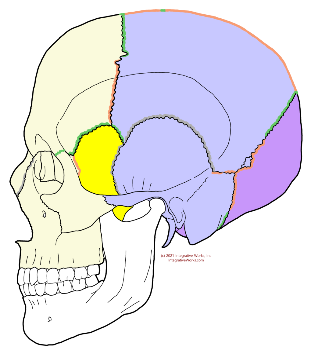
Governing Bones
The sphenoid governs the movements of the frontal bone, ethmoid, and face. On the other hand, the occiput governs the parietal and temporal bones.
This understanding can be critical in assessing and releasing restrictions in the cranial structure.
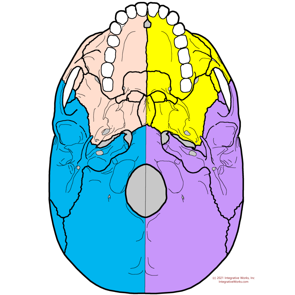
Quadrant Analysis
Quadrant analysis allows the biomechanical craniosacral practitioner to assess and resolve restrictions and altered patterns by smaller sections of the cranial system. It divides the cranium into bones that are governed by the sphenoid or the occiput. Also, it divides those anterior and posterior sections into left and right hemispheres.
This process of quadrant analysis becomes essential in resolving CranioMuscular patterns by quadrant as well. For the craniostructural therapist, quadrant analysis in combination with sutural analysis targets somatic governors. More simply put, this process allows the practitioner to connect cranial restrictions to musculoskeletal patterns.
This exploration of the sphenobasilar mechanism and axes of motion has seemingly endless implications in assessment and treatment. The basic ideas in this document are fundamental to many observations and techniques.
Support Integrative Works to
stay independent
and produce great content.
You can subscribe to our community on Patreon. You will get links to free content and access to exclusive content not seen on this site. In addition, we will be posting anatomy illustrations, treatment notes, and sections from our manuals not found on this site. Thank you so much for being so supportive.
Cranio Cradle Cup
This mug has classic, colorful illustrations of the craniosacral system and vault hold #3. It makes a great gift and conversation piece.
Tony Preston has a practice in Atlanta, Georgia, where he sees clients. He has written materials and instructed classes since the mid-90s. This includes anatomy, trigger points, cranial, and neuromuscular.
Question? Comment? Typo?
integrativeworks@gmail.com
Follow us on Instagram
*This site is undergoing significant changes. We are reformatting and expanding the posts to make them easier to read. The result will also be more accessible and include more patterns with better self-care. Meanwhile, there may be formatting, content presentation, and readability inconsistencies. Until we get older posts updated, please excuse our mess.

