Here, you will find functional anatomy of the temporomandibular joint.
Overview
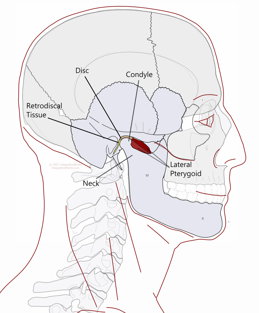
The temporomandibular joint is a complex synovial joint. It has a superior articular surface of the temporal bone, a joint capsule with a disc, and the head of the mandible inferiorly. There are several myofascial attachments to the joint. Fibrocartilage covers the articular surfaces instead of hyaline cartilage
Disc
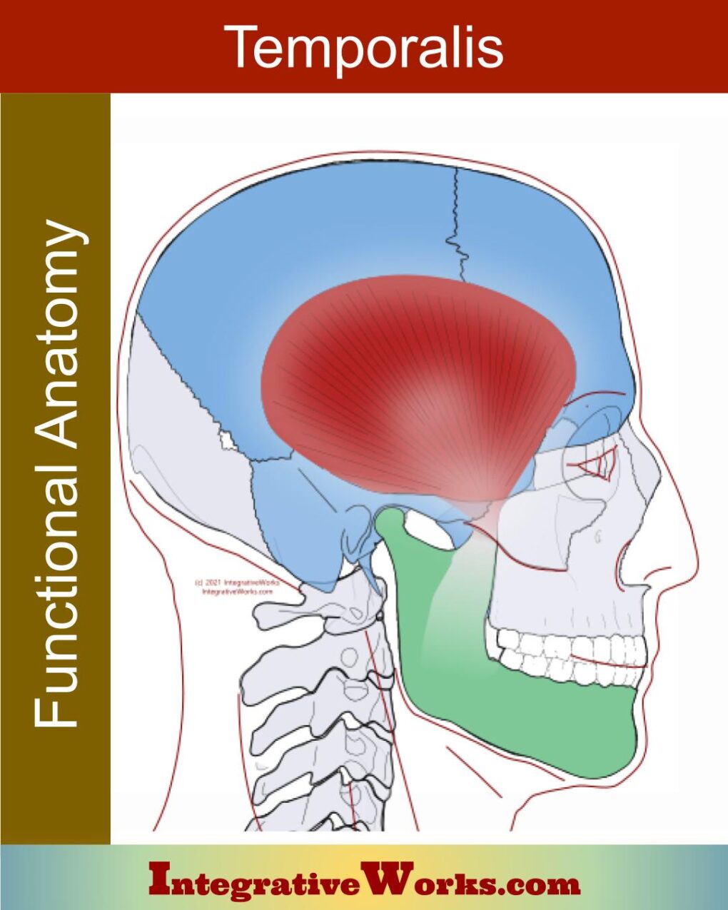
An articular disc, composed of collagen fibers, is positioned between the head of the condyle and the mandibular fossa of the temporal bone. The disc is attached to the medial and lateral portion of the condyle. The superior belly of the lateral pterygoid and middle temporalis fibers attach to the medial and anterior part of the disc.
Retrodiscal Tissue
Just posterior to the disc is the retrodiscal tissue. It is highly vascular and innervated. Posteriorly the capsule is attached to the retrodiscal tissue. This elastic tissue pulls the condyle posteriorly along with the head of the mandible as the jaw closes.
Lateral Pterygoid
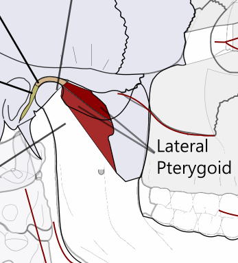
Anteriorly, the capsule is attached to the superior head of the lateral pterygoid. The inferior head of the lateral pterygoid attaches to the head of the mandible to pull it forward with the capsule.
Ligaments
The capsule is thickened laterally for the attachment of the temporomandibular ligament. The sphenomandibular ligament extends along the medial aspect of the joint from the spine of the sphenoid to the lingula of the mandible. The stylomandibular ligament attaches to the styloid process of the temporal bone. It extends downward behind the joint until it attaches to the posterior border of the angle of the mandible.
Function
The TMJ is the only diarthrodial joint in the body. This means it must synchronize both sides, which puts unique demands on the muscles.
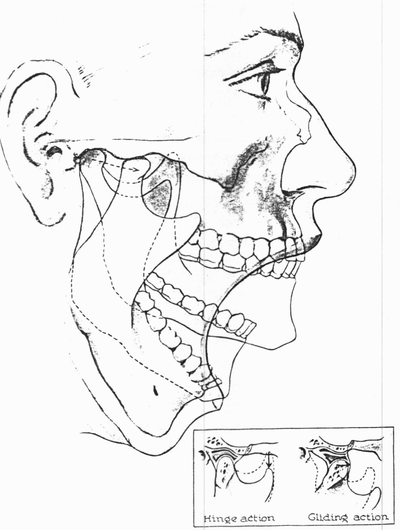
In short, the first ten percent of opening involves rotation of the condyle. Beyond that, the condyle translates (slides) on the articular eminence of the zygomatic process. As well, the head of the mandible rotates on the disc during rotary motions of the jaw.
The condyle carries the disc with it when it translates as the jaws open or close. The condyle glides on the articular eminence, which is heavy articular bone.
Related Posts
Support Integrative Works to
stay independent
and produce great content.
You can subscribe to our community on Patreon. You will get links to free content and access to exclusive content not seen on this site. In addition, we will be posting anatomy illustrations, treatment notes, and sections from our manuals not found on this site. Thank you so much for being so supportive.
Cranio Cradle Cup
This mug has classic, colorful illustrations of the craniosacral system and vault hold #3. It makes a great gift and conversation piece.
Tony Preston has a practice in Atlanta, Georgia, where he sees clients. He has written materials and instructed classes since the mid-90s. This includes anatomy, trigger points, cranial, and neuromuscular.
Question? Comment? Typo?
integrativeworks@gmail.com
Interested in a session with Tony?
Call 404-226-1363
Follow us on Instagram

*This site is undergoing significant changes. We are reformatting and expanding the posts to make them easier to read. The result will also be more accessible and include more patterns with better self-care. Meanwhile, there may be formatting, content presentation, and readability inconsistencies. Until we get older posts updated, please excuse our mess.

Pingback: Mandible - Functional Anatomy - Integrative Works