This post is temporarily available to the public.
Support us & see more at IntegrativeWorks Patreon.
Many of my clients have asked me for a simple explanation of the Craniosacral System. Here is a basic overview of how it is seen in the bio-mechanical model:

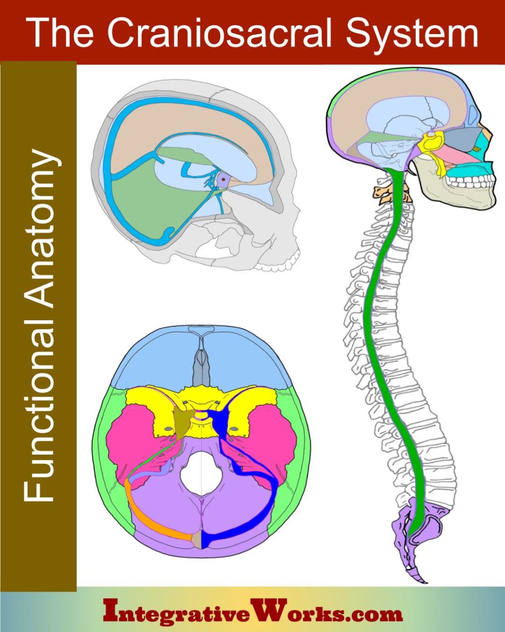
The word “craniosacral” comes from the membrane system
that surrounds the central nervous system
and connects the cranium to the sacrum.
The craniosacral system consists of:
- The cranium
- The sacrum and coccyx
- The meninges
- Cerebrospinal fluid
- The structures involved in the production and re-uptake of CSF
The Cranium
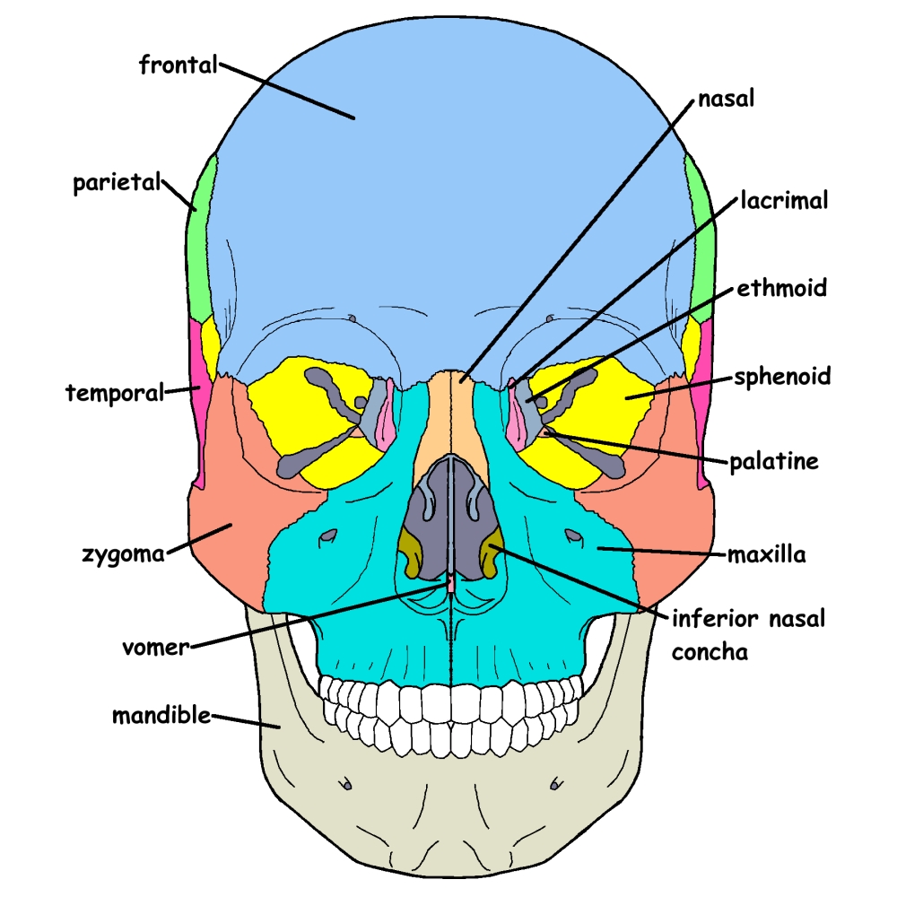
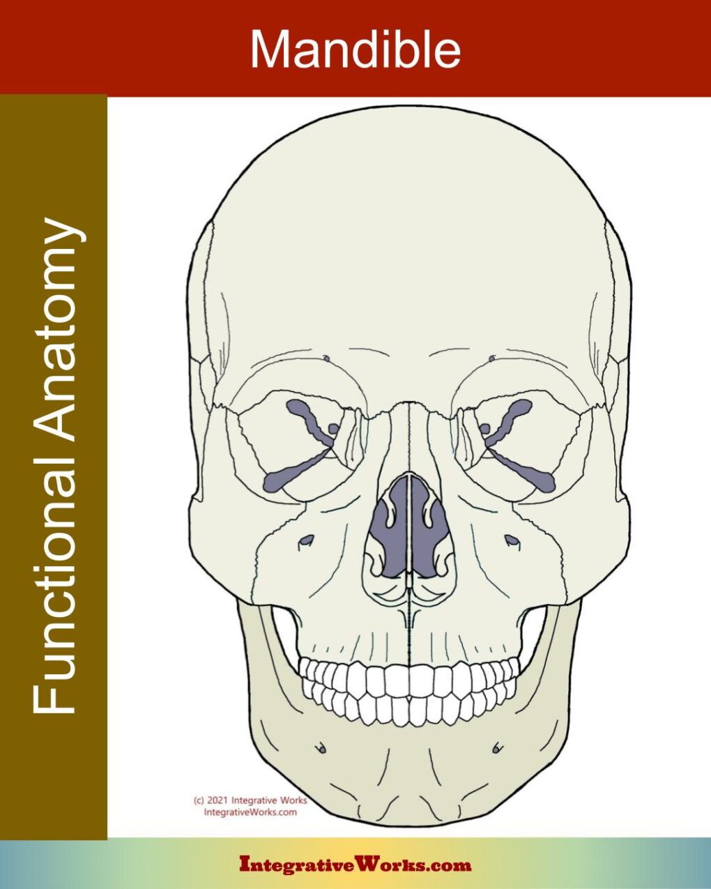
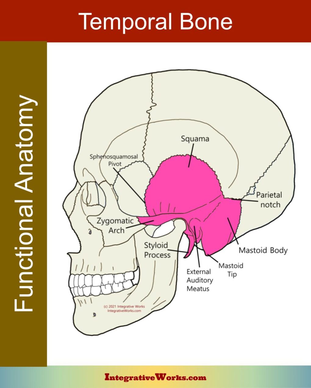
The cranium consists of 22 bones and about 115 or 120 joints, depending on how they form. This bony structure makes a hard shell that protects the brain and allows for some flexibility. In fact, the bones are thinner and more flexible than most people realize. Bones around the temple and in the sinuses are thin enough to be translucent. Think of these bones as being more flexible, like your ribs. Cranial bones are held together by sutures and an internal membrane system.
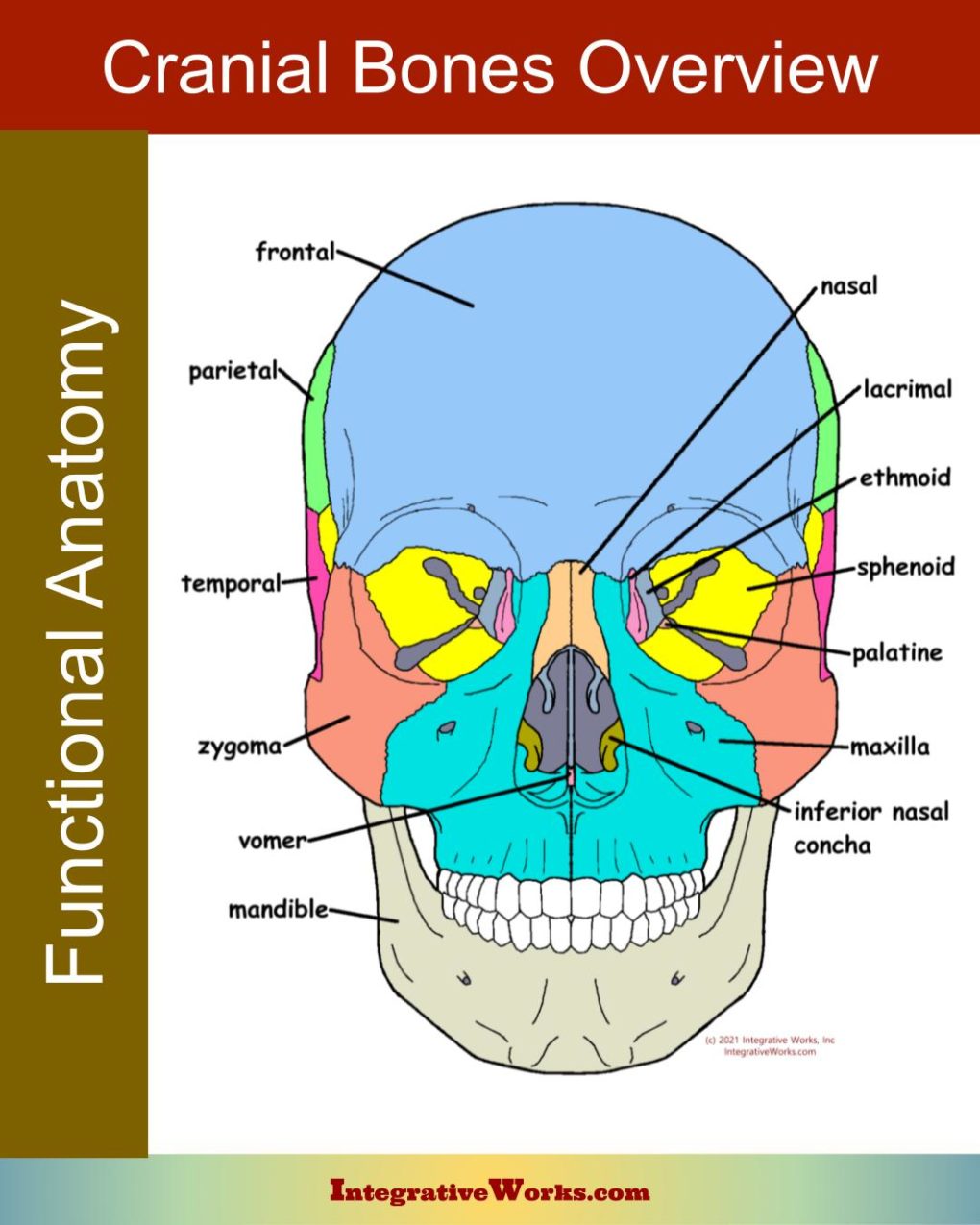
If you’d like an overview of cranial bone anatomy, look at this other post.
Reciprocal Tension Membranes
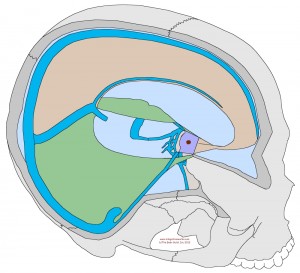
The bones around the brain form the “vault.” Reciprocal tension membranes form partitions dividing the vault into upper, lower, left, and right sections. The reciprocal tension membranes support the brain and provide a balance of tension for the structure. The falx cerebri and falx cerebelli divide the cranium into left and right halves. The tentorium cerebelli forms a tent over the cerebellum, separating it from the cerebrum.
Interestingly, these membranes are heavily populated with proprioceptors, giving feedback about tension and position.
Craniosacral Membranes

Meninges
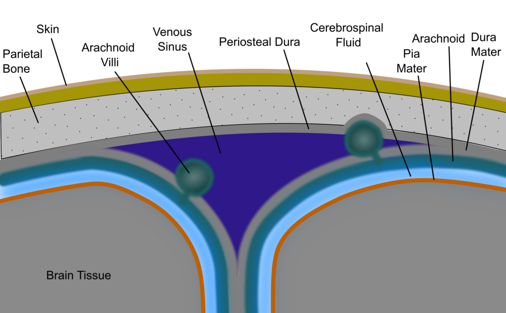
The meninges of the craniosacral system are a triple-layered membrane system that surrounds, supports, and protects the central nervous system.
- The dura mater (“tough mother”) forms the outside layer against the bone.
- The arachnoid (“spider”) is a porous, cob-web-like membrane that adheres to the dura mater.
- The pia mater (“soft mother”) is a thin layer that lines the central nervous system’s surface.
There is room for fluid to flow in the “sub-arachnoid space.” That’s the space between the arachnoid and the pia mater. The membranes provide protective padding and containment of the cerebrospinal fluid in which the central nervous system floats.
Dural Tube
The membranes form a dense ring around the big hole where the spine exits the cranium (foramen magnum). Then, they form a tube that surrounds the spinal cord. This structure is called the dural tube, core link, or thecal sac. That tube attaches firmly to the hole in the cranium and skips the first vertebrae. Next, the membrane attaches to the second and third vertebrae in the neck, bypasses the rest of the spine, and attaches to the sacrum’s second segment. Eventually, this tube narrows to become a filament that attaches to the coccyx.
Sacrum and Coccyx

Sacrum
The sacrum is a large, hollow triangular-shaped bony structure in the center of the posterior pelvis. Five fused vertebrae form its kyphotic curve. It has strong fibrous connections in all directions to provide stability as the torso’s weight is transmitted through this structure while sitting or standing.
Coccyx
The coccyx is formed from 3-5 fused vertebrae with rudimentary transverse processes
Craniosacral System Inlet (Inhalation)
Ventricles
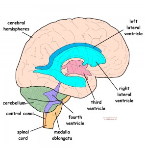
Ventricles are balloon-like structures at the center of the brain. They support the neural tissue of the brain so that it floats above the floor of the cranium. They also create Cerebrospinal fluid (CSF). Hair-like vessels called the Choroid Plexus, filter CSF from the blood into the ventricles. Then, CSF flows out of the ventricles into the subarachnoid space. Once there, CSF offers structural support, protection, nutrition, and waste removal.
The central nervous system, which is a firm gelatinous structure, floats in the cerebrospinal fluid. This cushions and protects the sections of the CNS like stewed tomatoes in a jar of water.
Craniosacral System Outlet (exhalation)
Arachnoid Villi
The arachnoid villi (or granulations) are projections into the venous sinus. They serve as valves to allow cerebrospinal fluid to return to the circulatory system.
These structures tend to be mostly along the sagittal sinus. They create visible indentations in the parietal bone.
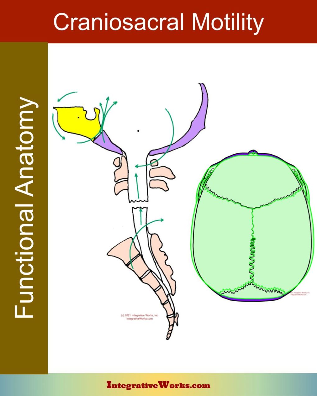
Craniosacral Motility
In the osteopathic model, the valves in the arachnoid villi correspond with craniosacral motility. On one hand, the craniosacral system builds pressure and flexes when the valves are closed. Conversely, when the valves open, the pressure subsides and the craniosacral system flexes.
Additionally, cerebrospinal fluid leaks through the fascial wrapping around spinal nerves.
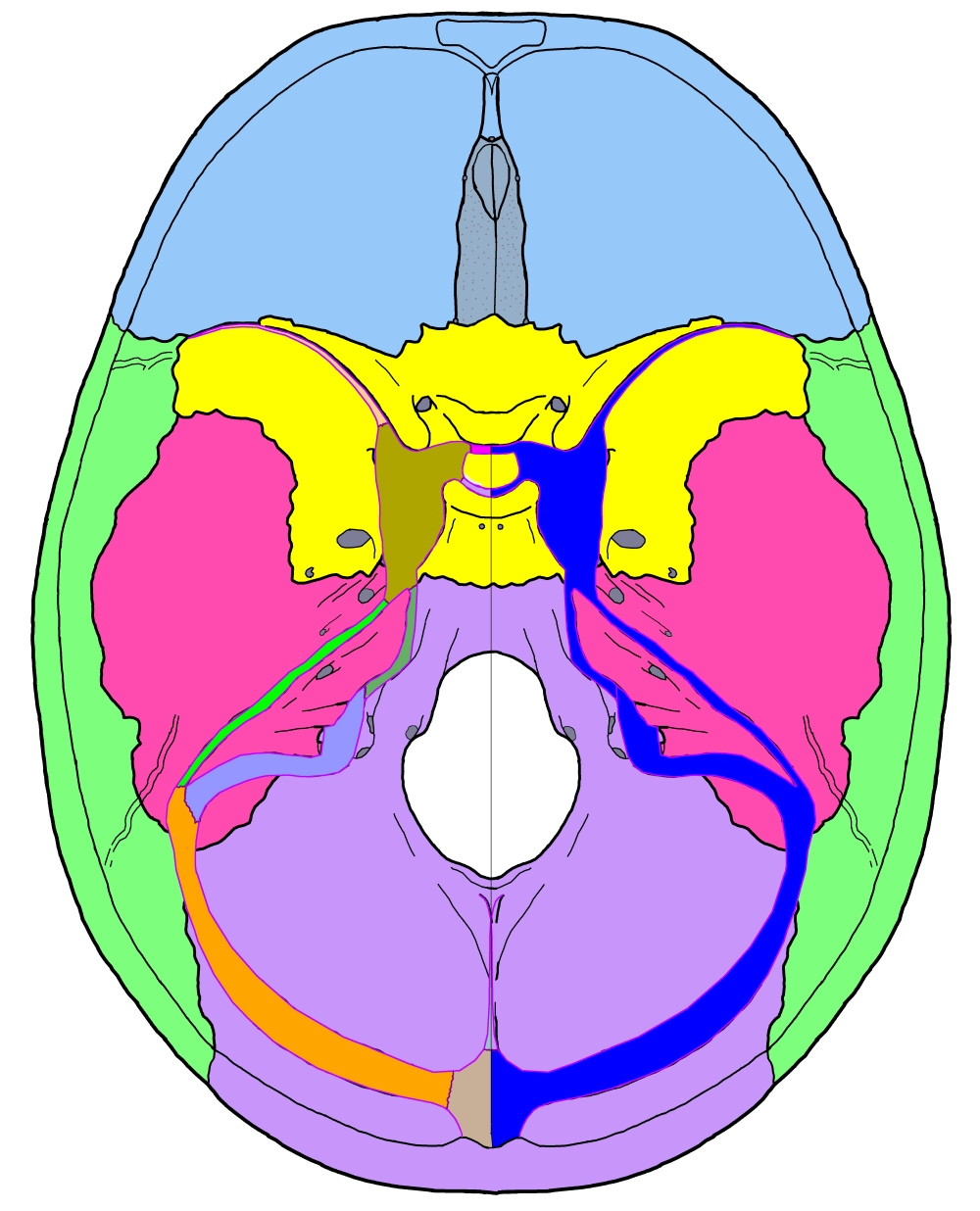
Venous Sinuses
The reciprocal tension membranes join each other and the wall of the cranium. At those connections, they create little channels for blood flow called venous sinuses. On the right side of this illustration of the cranial base, these venous sinuses are shown in blue. The channels form free-flowing exits for blood and cerebrospinal fluid. They are key to the regulation of intracranial pressure.
The pituitary gland sits in a saddle in the center of this structure. It feeds the hormones to the venous sinuses, which carry them to the jugular vein and out to the body.
The detail of its architecture and function is truly amazing. This system provides nourishment, protection, pressure regulation, sensory feedback, and more.
Feel the Craniosacral System
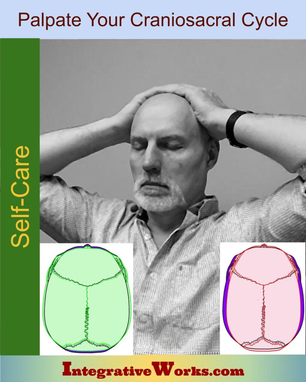
I trained many people to palpate their own craniosacral cycle. You can learn how in this other post.
Volumes have been written about this system and its structures. I will be adding more about this to my blog on a regular basis. If you’d like to see every post related to craniosacral techniques, anatomy, concepts, etc., look at this collection. For now, here are the other posts that I have written about craniosacral anatomy.
Posts on Craniosacral Anatomy
An Overview of Cranial Bone Anatomy
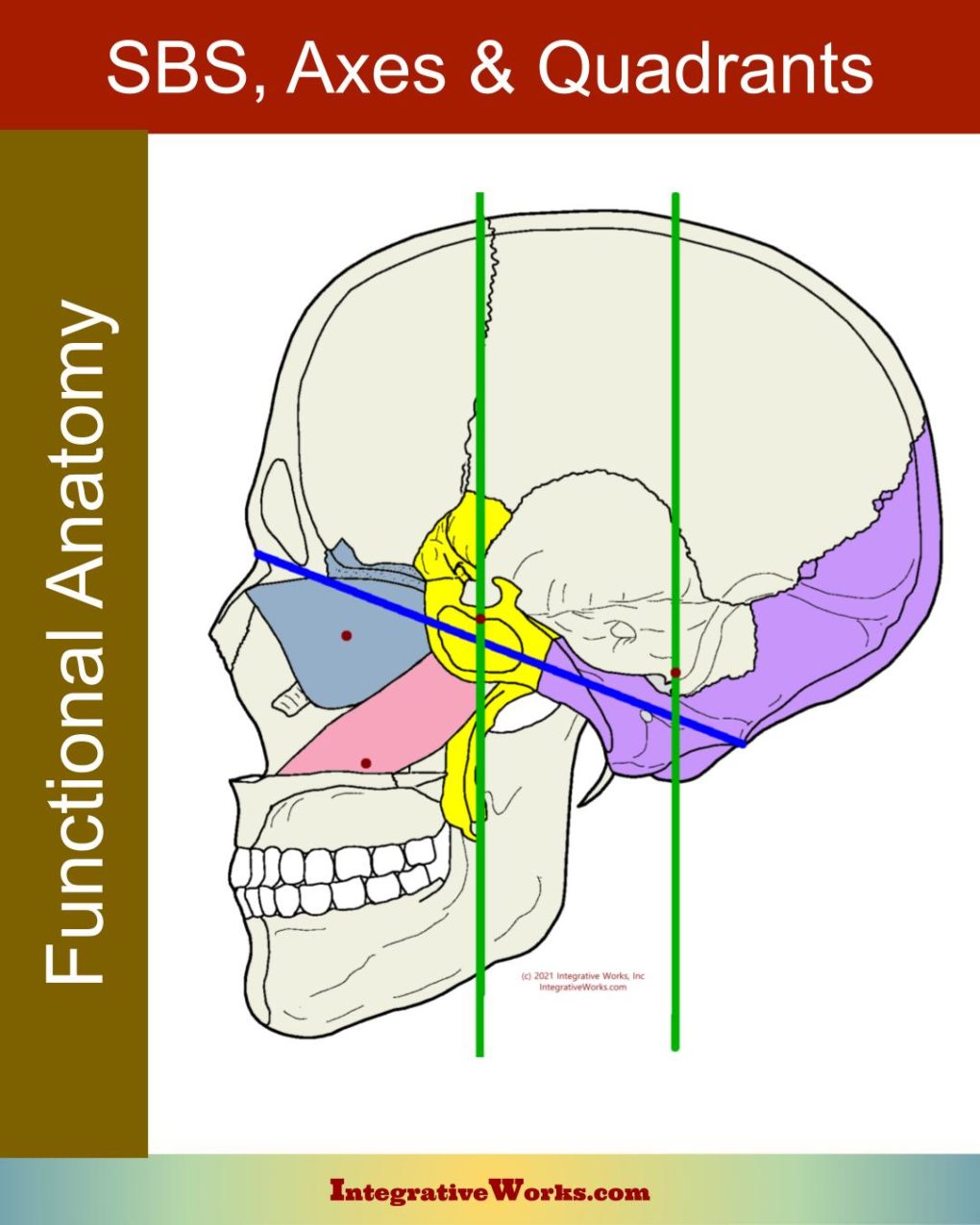
Craniosacral – The Sphenobasilar (SBS) Mechanism, Axes & Quadrants
Craniosacral Motility
Craniosacral System Overview
Mandible – Functional Anatomy
Temporal Bone – Functional Anatomy
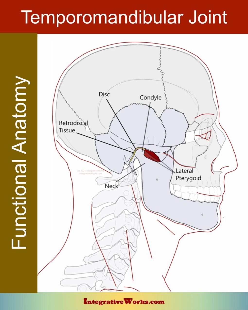
Temporomandibular Joint – Functional Anatomy
Support Integrative Works to
stay independent
and produce great content.
You can subscribe to our community on Patreon. You will get links to free content and access to exclusive content not seen on this site. In addition, we will be posting anatomy illustrations, treatment notes, and sections from our manuals not found on this site. Thank you so much for being so supportive.
Cranio Cradle Cup
This mug has classic, colorful illustrations of the craniosacral system and vault hold #3. It makes a great gift and conversation piece.
Tony Preston has a practice in Atlanta, Georgia, where he sees clients. He has written materials and instructed classes since the mid-90s. This includes anatomy, trigger points, cranial, and neuromuscular.
Question? Comment? Typo?
integrativeworks@gmail.com
Interested in a session with Tony?
Call 404-226-1363
Follow us on Instagram

*This site is undergoing significant changes. We are reformatting and expanding the posts to make them easier to read. The result will also be more accessible and include more patterns with better self-care. Meanwhile, there may be formatting, content presentation, and readability inconsistencies. Until we get older posts updated, please excuse our mess.

Comments are closed.