Overview
Here, you will find a description of the anatomy, physiology, and progression of the sacroiliac joint.
Perspectives on the pelvis and sacroiliac joint have developed over decades, even centuries. There is a long explanation of that development with many supporting links in this meta-study. Extensive research has defined pain mechanisms and the evolutionary transition from four-legged to two-legged locomotion. When exploring the differences based on age and sex, it becomes apparent that studies will vary based on the population’s demographics.
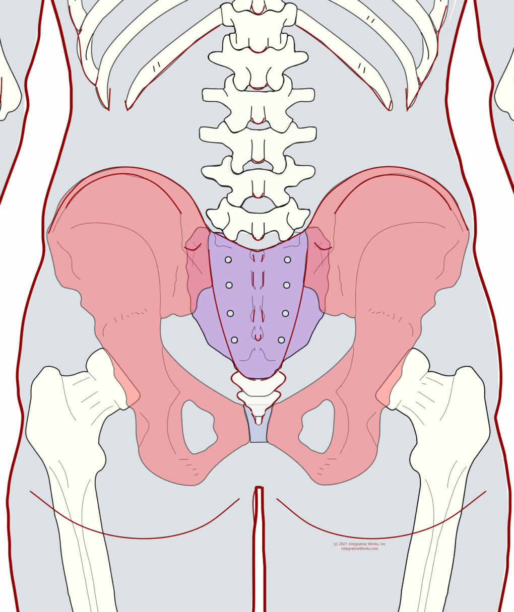
Pelvic Structure
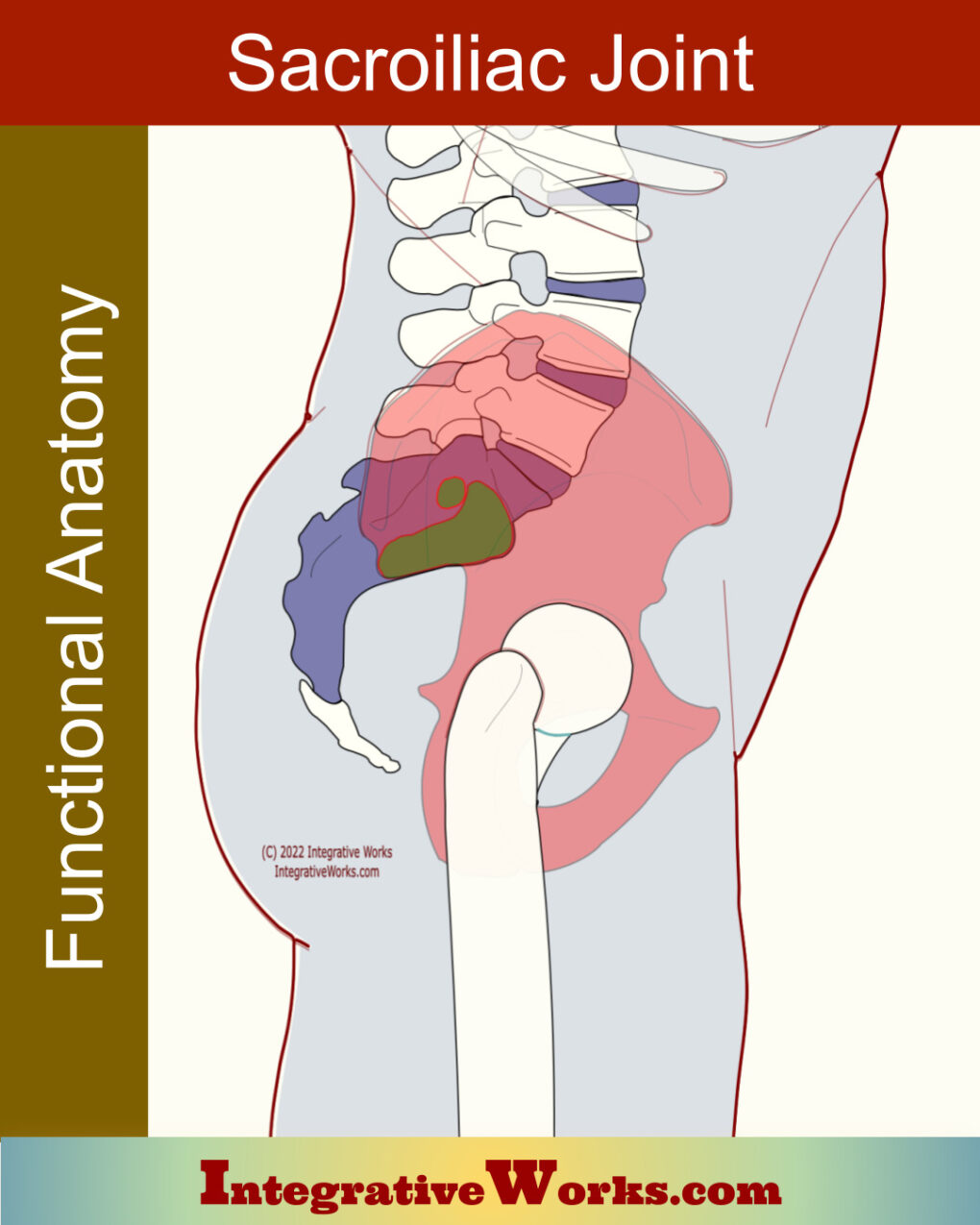
The bony pelvis has two os coxae, shown here in pink. In between, the sacrum is shown here in violet.
Sacral vertebrae and the bones of the os coxae are separate in utero.
- After birth, sacral vertebrae start binding but do not finish until the late 20s.
- The ilium, ischium, and pubis also gradually fuse until they become unified as os coxae, typically between 18 and 25.
As the child begins to locomote, the body of the sacrum changes shape. This new activity torsions the sacroiliac joints and causes the sacrum to expand laterally. Further, this motion also changes the shape of the joint.
Differences by Sex
The male pelvis is larger at twenty-two months, but this difference moderates during childhood. In males, the articulation for L5 is larger in relation to the sacral base. Also, the sacroiliac joint tends to have more surface area in males.
In contrast, females have a wider sacrum and a shorter pelvis. In addition, the base of the female sacrum seats more shallowly between the crests of the ilia. Comparatively, the female sacrum tilts back more and broader. These differences create a female pelvis that is rounder and less cone-shaped.
Sacroiliac Joint Anatomy
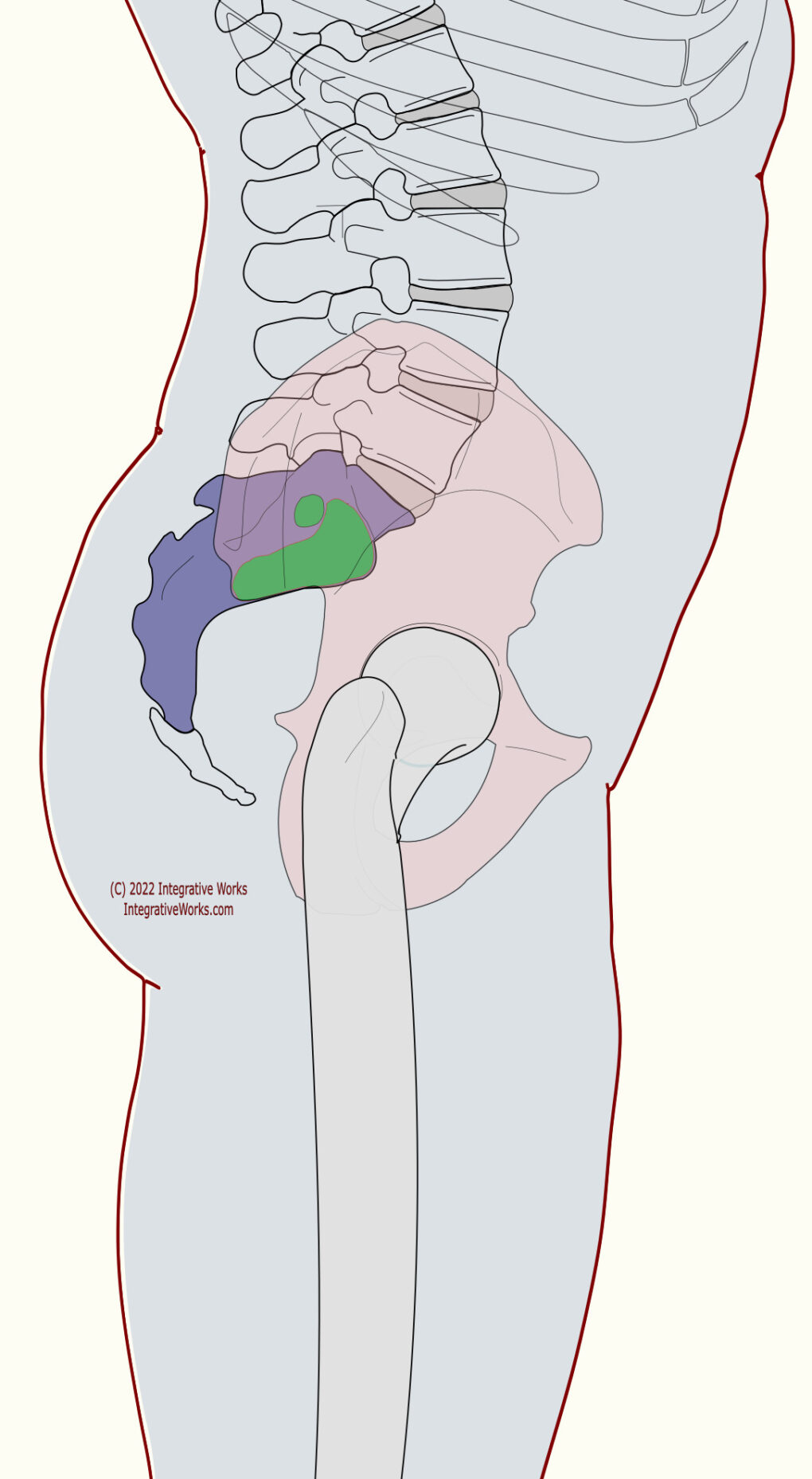
ilium in pink and
joint capsule in green.
Basic Structure
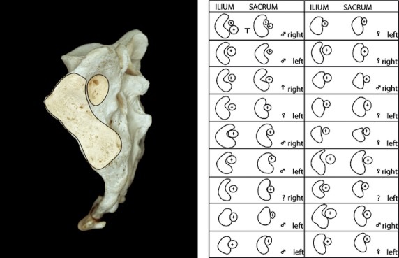
The SIJ has typically formed along the first three sacral segments. The main articulation of the joint is auricular (ear-shaped). It lies along the lateral surface of the sacrum, obliquely stretching from cranial-lateral to caudal-medial. Roughly, its silhouette is C-shaped or L-shaped.
Segments
It can be divided into three parts along the first three sacral segments. The S1 portion (cranial end) tends to be the largest. Conversely, the S3 portion (caudal end) tends to be the smallest.
Naming Conventions
Naming the aspects of the joint can be confusing. In the standing position, as shown here, the cranial section is more ventral, and the caudal portion is more dorsal. In the L-shape, has a shorter cranial leg near the ventral end. So, it has a longer caudal leg that extends toward the dorsal end.
The cranial end of the joint is fibrous. However, most of the joint is synovial. This fibrous section on the cranial end of the SIJ seems to be part of the caudal end of the iliolumbar ligament. On the other end, the caudal end of the SIJ blends with the sacrospinous ligament along the ventral surface.
Accessory Joints
Here’s another area where sacroiliac joint anatomy studies vary. Accessory joints are common but highly variable. Consequently, studies have varied in how they have been defined. This study found accessory joints in all 958 skeletons, on at least one side of East African skeletons. Other studies find accessory joints in as little as six percent of subjects.
More detail was available in this study of a much smaller group. It proposes that an accessory joint that is commonly located above the second segment is the “axial” joint on which the sacrum nutates. Conversely, other studies claim that the surrounding ligament prevents nutation.
These joints have a convex protuberance on the ilium that fits into a concavity on the sacrum. The interosseous portion of the sacroiliac ligaments encapsulates these accessory joints.
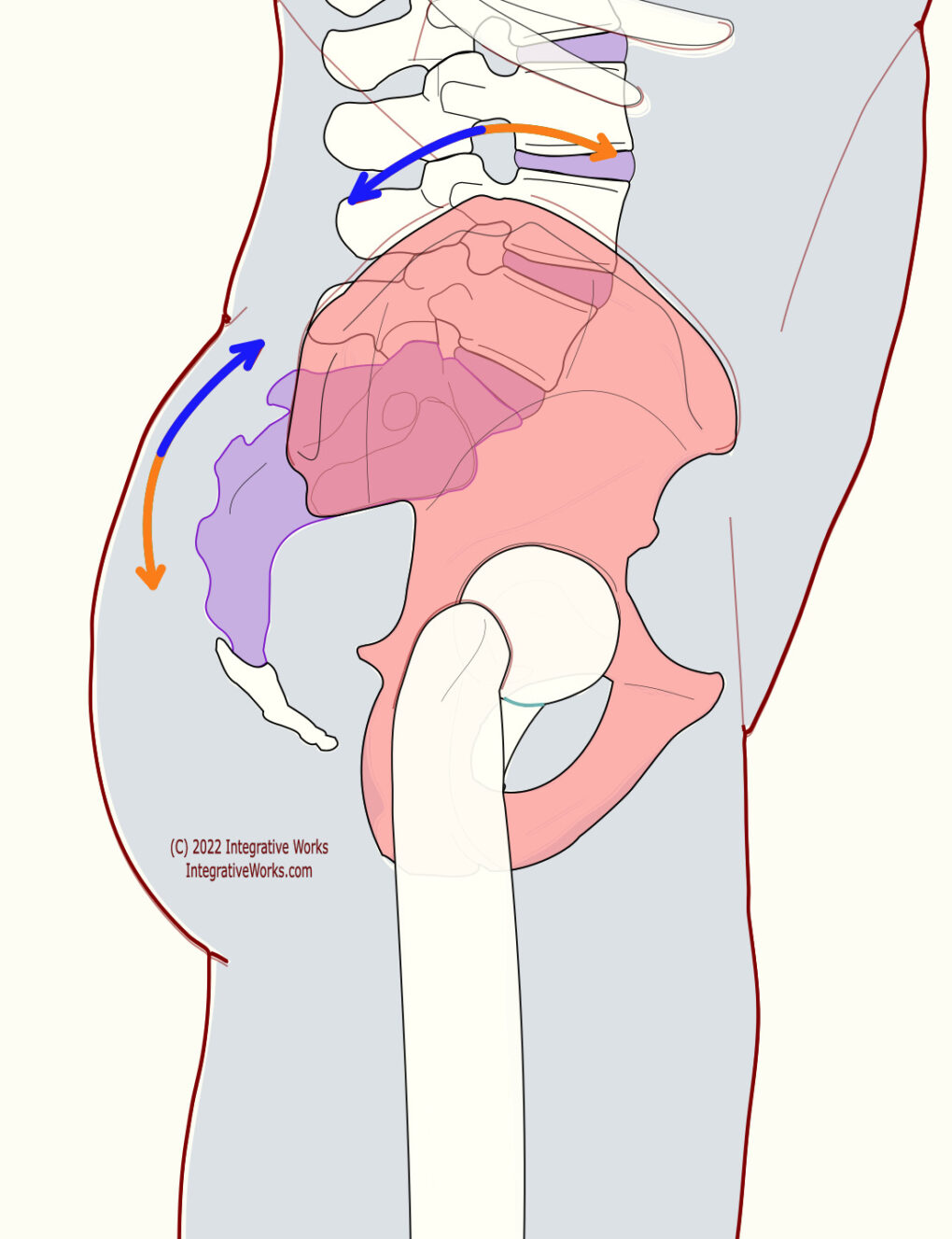
Nutation in blue,
Counternutation in orange
Motion
Sacroiliac joint physiology is somewhat less variable than sacroiliac joint anatomy. However, it is a bit confusing because of the many areas of flexion and extension in the craniosacral mechanism and body. This can be confusing because craniosacral flexion is pelvic extension. So, it has its own terminology.
Therefore, “nutation” and “counternutation” are the terms commonly used to describe SIJ motion, especially among craniosacral therapists.
- Nutation is the forward nodding of the sacrum. Nutation occurs when the sacrum pivots on the ilium so that the coccyx moves posteriorly.
- Conversely, backward nodding is “counternutation.” Historically, medical literature describes motion as very limited, except in the case of pregnancy.
Notably, some practitioners refer to sacral nutation as the forward nod of the sacrum, even when the os coxae rotate with it.
Anomalies and Variations by Age and Sex
Sacroiliac Joint Changes by Decade
- The sacroiliac joint(SIJ) is a recognizable joint from the second month in utero.
- The joint is completed by the eighth month in utero.
- The SIJ resembles the sacroiliac joint of quadrupeds at birth.
- However, the structure shifts as children begin to use their legs.
- Pelvic mobility decreases from birth to puberty.
- There is no notable loss in mobility up to about 50 years.
- Studies on ankylosis (bony joint fusion) are highly variable in their outcomes. However, they largely show that it is more common in males and people over 50.
- Collagenous ankylosis has been studied separately and extensively. Those studies suggest that persistent, excessive compressional forces could contribute by straining the ligamentous and tendonous structures.
Sacroiliac Joint Differences by Male/Female
- Male SIJs are more mobile than females before puberty.
- From puberty, females become more mobile until about age 25, whereas males do not.
- Relaxin (a hormone, not calming activity) increases mobility in the SIJ during pregnancy.
- Beyond age 25, female pelvises are about 40% more mobile than males.
- Beyond the fifth decade, the coccyx and sacrum are about 25% more likely to fuse in males.
Anomalies, Etc.
The surfaces have large variations in size, shape, and contour. Generally, the iliac surface is convex, and the cartilage is rougher. Conversely, the sacral surface is concave and smoother.
- Some are closer to C-shaped, and some are closer to L-shaped.
- The shape of the joint changes notably from the first to the third decade.
- The L5 vertebra has some fusion (facet joint, spinous…) with the sacrum in about 6% of cases
- The coccyx is fused to the sacrum in 25-30% of cases in their fourth decade.
- However, between 44-52% of people beyond their fifth decade are fused between the sacrum and coccyx.
- The ventral SIJ is less formidable and often allows fluid to leak into the surrounding tissues. These tissues include the lumbosacral plexus and dorsal sacral plexus.
- This study shows that 81% of people with SIJ fusion have increased mobility in the lumbosacral joints.
- This Study shows that there are accessory SIJs in about 50% of the cases.
Sacroiliac Ligament
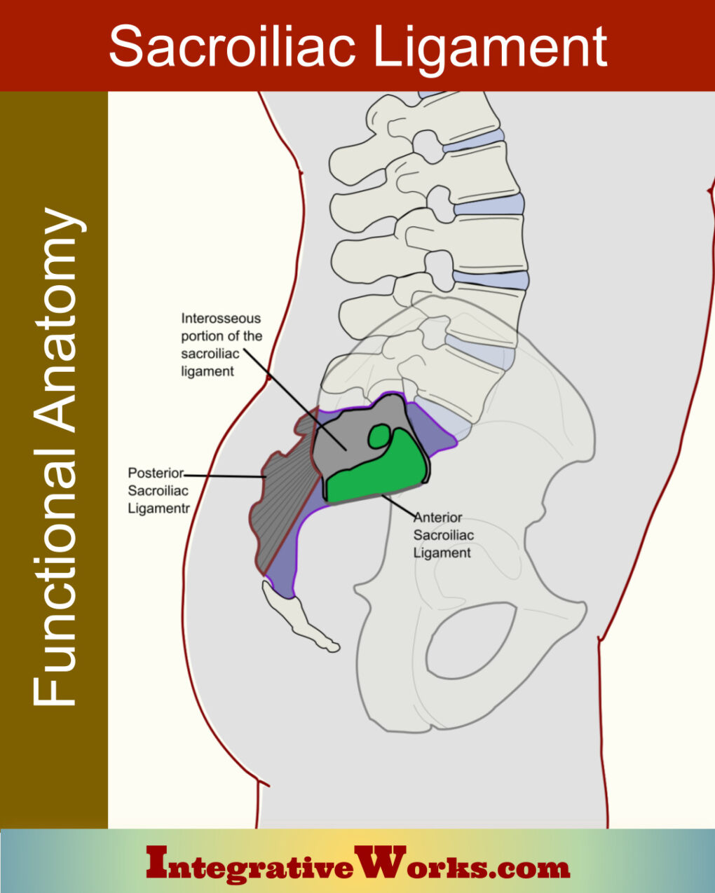
The sacroiliac ligament is an important part of understanding sacroiliac joint anatomy. In fact, it’s not really a ligament. It’s a complicated connective tissue structure with three main sections:
- The interosseous section ties the lateral sacrum to the posterior surface of the ilium.
- The posterior section ties the posterior sacral surface to the posterior iliac spine
- The anterior section veneers the ventral surface of the joint.
Each section has a unique structure. Additionally, they impact the integrity of the sacroiliac joint very differently.
Iliolumbar Ligament
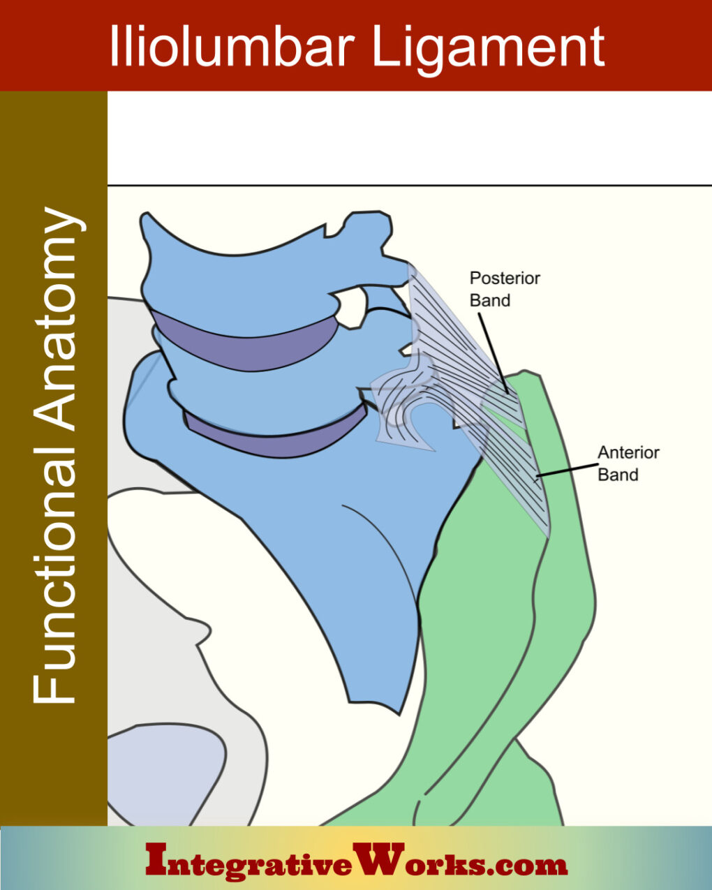
The iliolumbar ligament also has sections that vary by age, sex, and race.
Notably, this ligament is unique to animals that walk on two legs. It stabilizes the lower lumbar vertebrae against the pelvic bones. And, let’s be clear, it is also quite variable.
Support Integrative Works to
stay independent
and produce great content.
You can subscribe to our community on Patreon. You will get links to free content and access to exclusive content not seen on this site. In addition, we will be posting anatomy illustrations, treatment notes, and sections from our manuals not found on this site. Thank you so much for being so supportive.
Cranio Cradle Cup
This mug has classic, colorful illustrations of the craniosacral system and vault hold #3. It makes a great gift and conversation piece.
Tony Preston has a practice in Atlanta, Georgia, where he sees clients. He has written materials and instructed classes since the mid-90s. This includes anatomy, trigger points, cranial, and neuromuscular.
Question? Comment? Typo?
integrativeworks@gmail.com
Interested in a session with Tony?
Call 404-226-1363
Follow us on Instagram

*This site is undergoing significant changes. We are reformatting and expanding the posts to make them easier to read. The result will also be more accessible and include more patterns with better self-care. Meanwhile, there may be formatting, content presentation, and readability inconsistencies. Until we get older posts updated, please excuse our mess.

