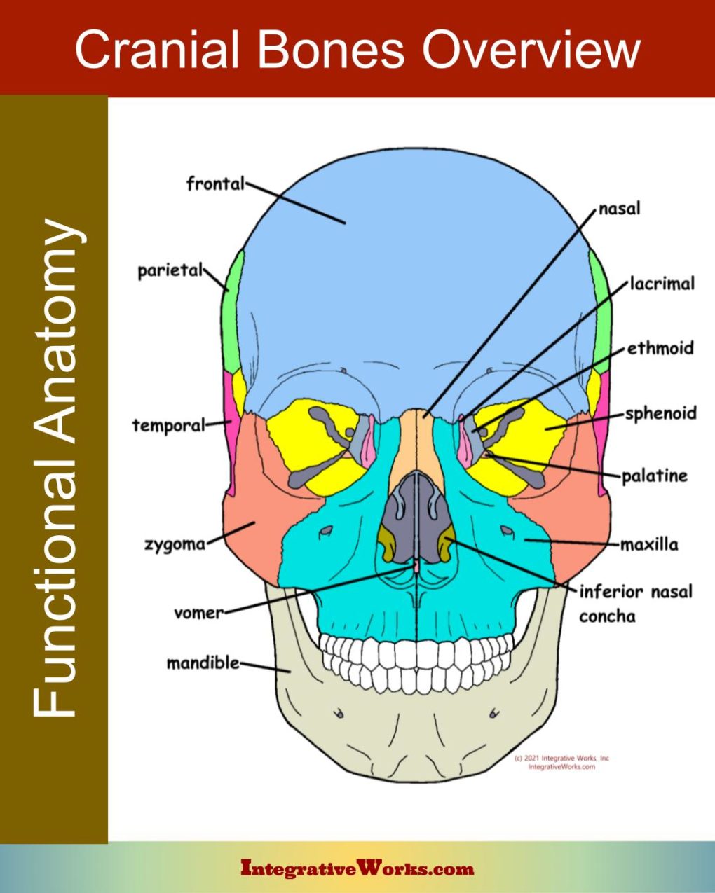
Here, you will find an overview of cranial bone anatomy. If you’re not familiar with the craniosacral system as a whole, look at this other post first.
Vault and Face
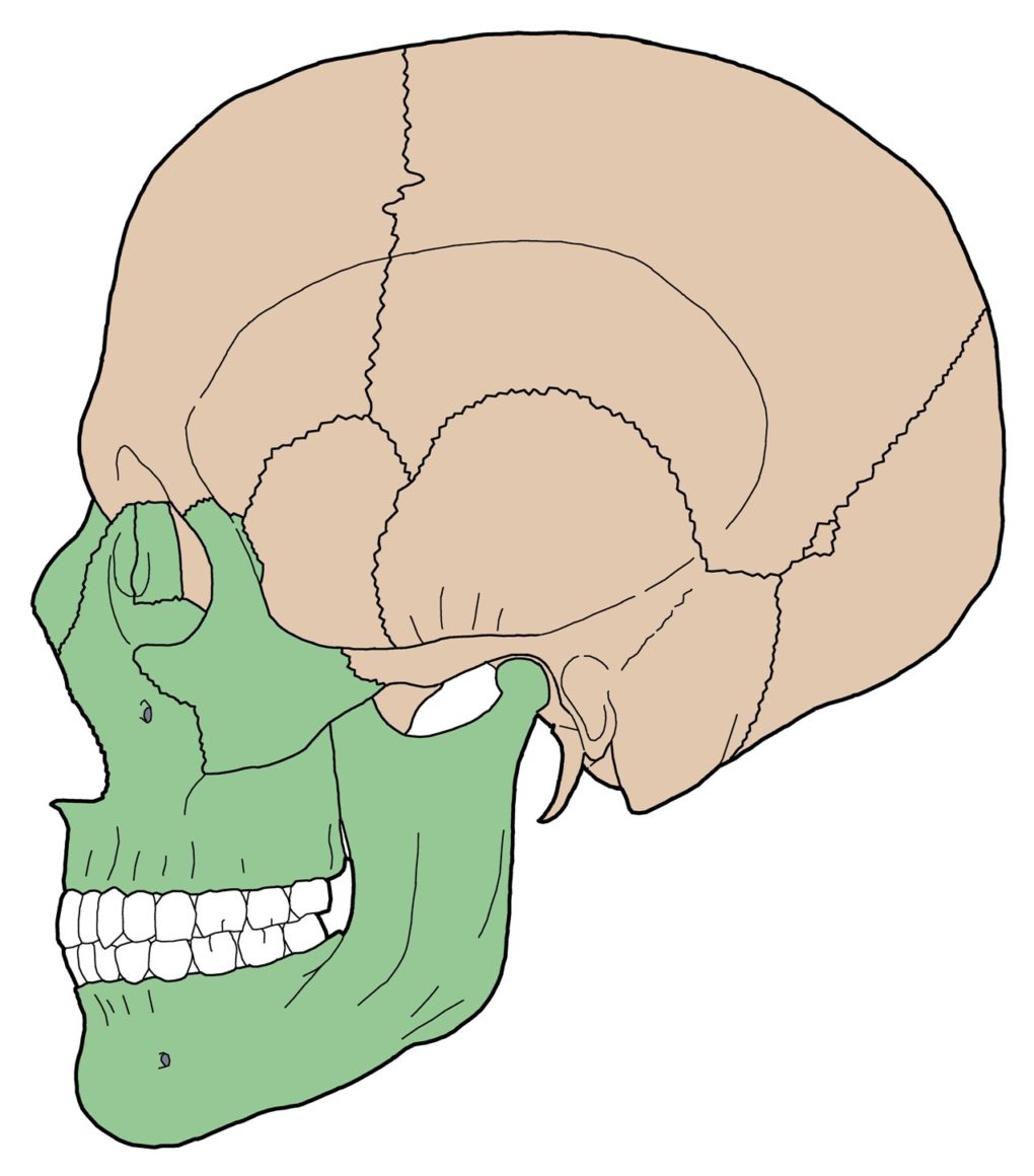
Cranial bone anatomy can be divided into vault bones and facial bones. These sections develop from different embryonic tubes and have different functions.
- Vault bones develop from the neural tube. The vault consists of 8 bones that primarily offer a protective housing for the brain.
- Facial bones develop from the nasopharyngeal tube. The facial ones are mostly involved in sensory and intake functions.
Cranial Base and Upper Vault
Below the external auditory meatus, the cranial base forms in cartilage (endochondral ossification). Accordingly, these sections of bone are more petrous or rock-like. Their rigid structure gives them strength but lacks flexibility. In addition, the cranial base has more articular complexity, which permits more motility.
The upper portions of the vault form in a membrane (intramembranous ossification). making the bones more flexible. In addition, the lateral sutures of the upper vault are pivoted and beveled to allow more movement at the joint as the bone flexes.
Same Bone, Different Formations
A single bone may have different sections that form differently. For example, the petrous portion of the temporal bone forms part of the cranial base. Accordingly, it is one of the hardest bones in the body. However, the temporal squama is formed in a membrane and is more flexible than ribs. The occiput, sphenoid, and other cranial bones have sections that form differently and later fuse.
Mid-line and Paired
The cranial bone anatomy can by divided into mid-line and paired bones. They have different overall motions.
- Mid-line bones tend to flex and extend. They act like cogs in a wheel, driving the motion of flexion and extension along the sagittal plane.
- Paired bones tend to rotate externally and internally. The act like louvers on pivots. They have flexible sections that allow remodeling throughout craniosacral motion.
Some bones, like the occiput, flex while externally rotating and extend while internally rotating.
Pivots and Bevels
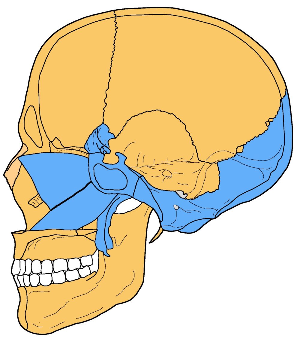
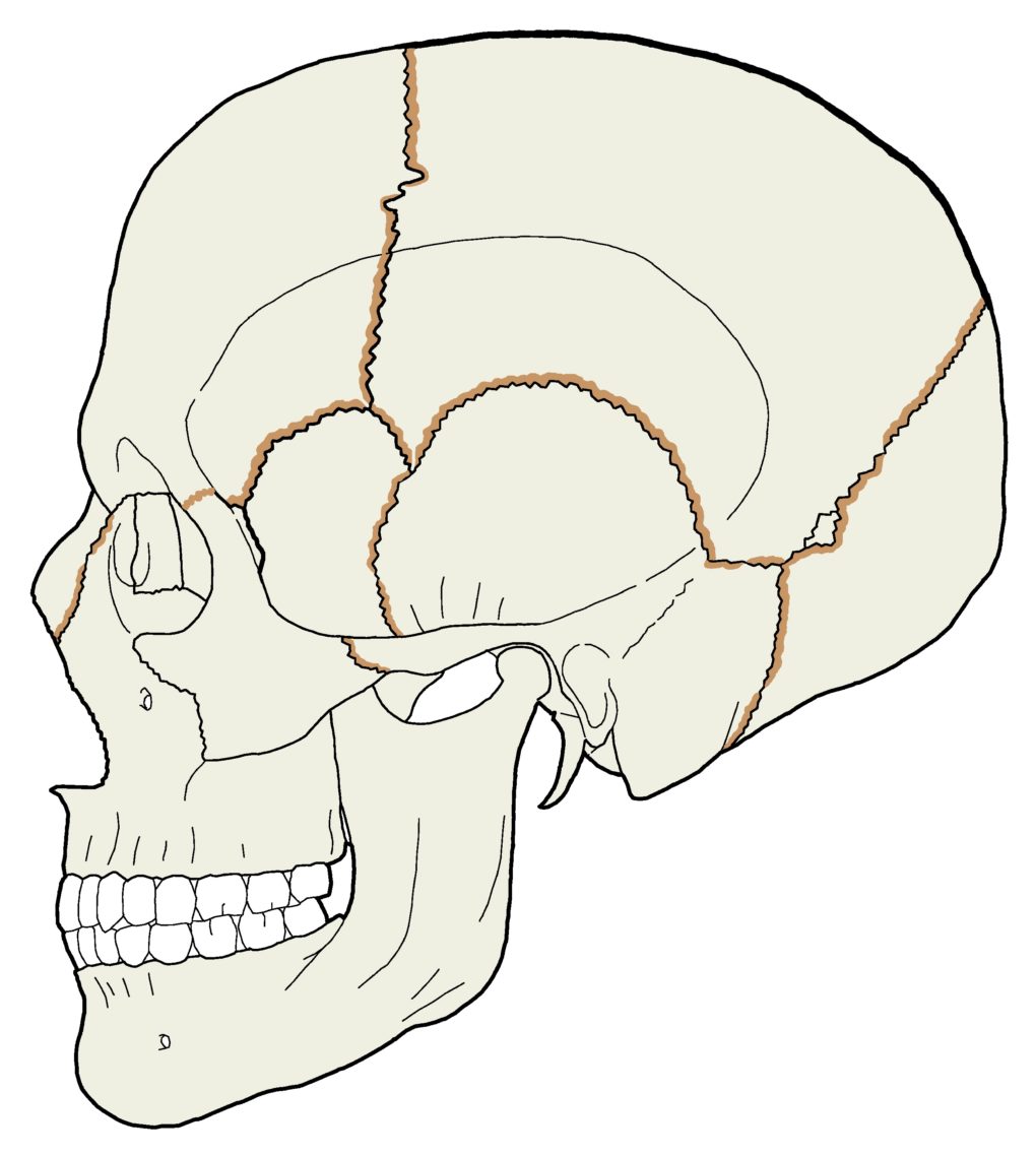
Cranial bevels are an essential part of the articular structure. They allow specific movements and prohibit others. It is also essential to understand which bevels underlie and overlie other bevels when
distracting sutures.
- External bevels (shown in brown) have the superficial edge compromised so that the slanted surface faces outward and underlies the articulating bone.
- Internal bevels have the deep edge compromised so that the slanted surface faces inward and overlaps the articulating bone.
- The point where the bevel changes from internal to external forms a pivot on which the bone rotates.
Quadrants
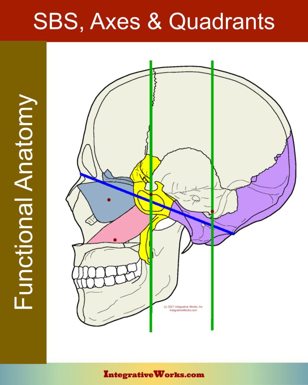
The sphenobasilar mechanism divides the cranium into bones controlled by the sphenoid or occiput. Further, those anterior and posterior sections can be divided into left and right hemispheres. You can read more about those details in this other post.
Visual Overview of Cranial Bone Anatomy
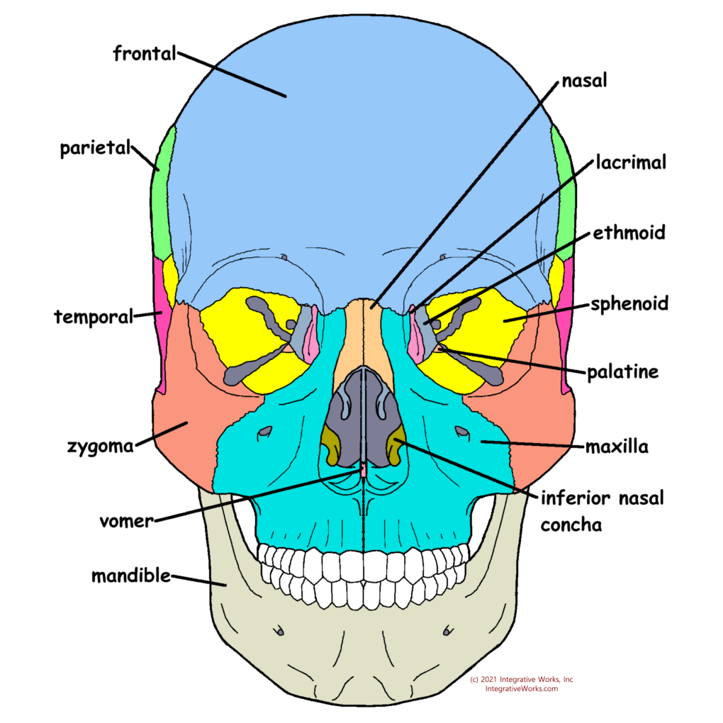
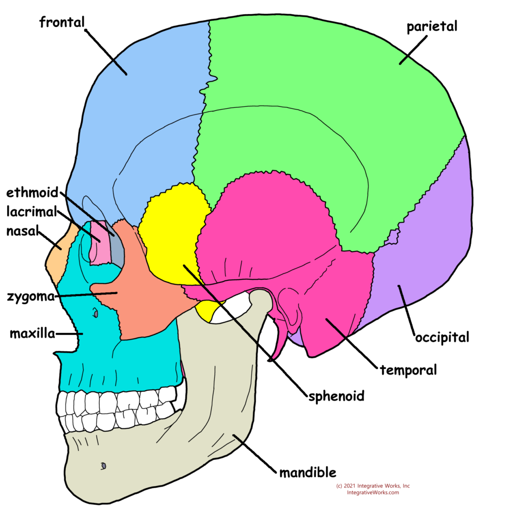
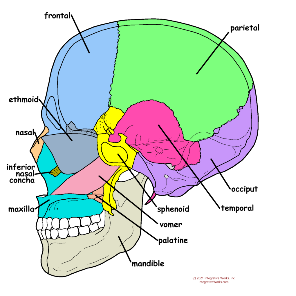
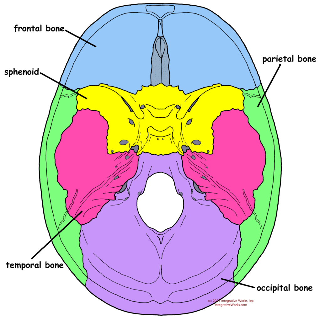
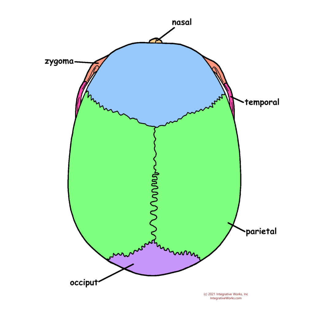
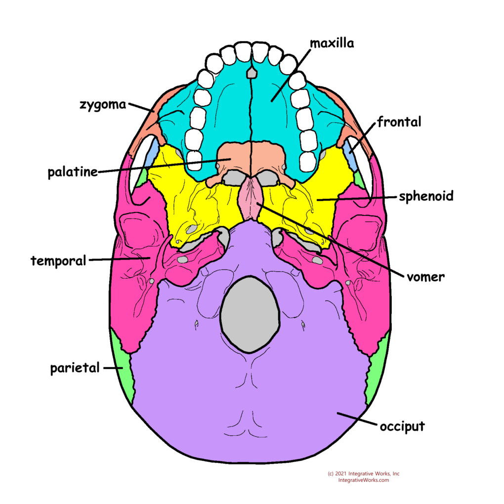
This post is a simple overview of the cranial bones. More detailed posts on each bone, each view, and other structures that bind them will be included in that post with an overview of the craniosacral system.
Support Integrative Works to
stay independent
and produce great content.
You can subscribe to our community on Patreon. You will get links to free content and access to exclusive content not seen on this site. In addition, we will be posting anatomy illustrations, treatment notes, and sections from our manuals not found on this site. Thank you so much for being so supportive.
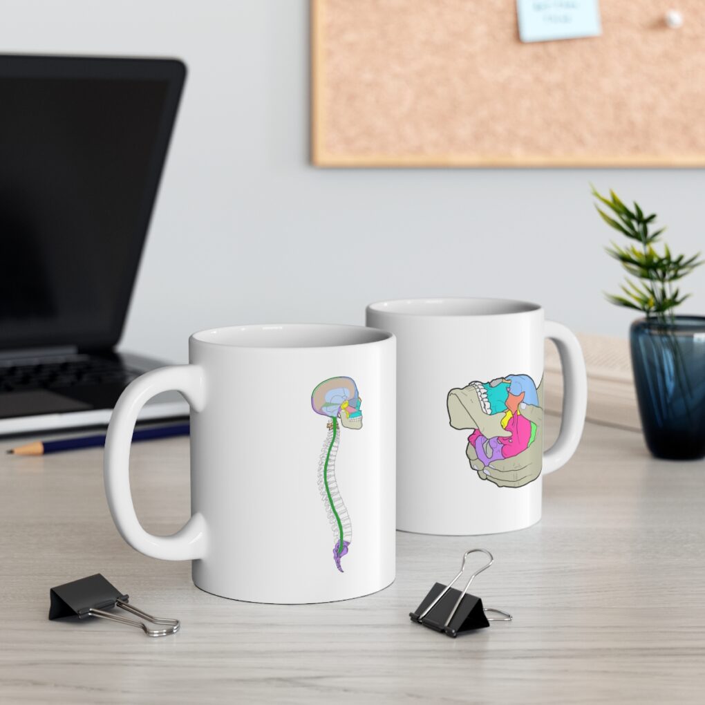
Cranio Cradle Cup
This mug has classic, colorful illustrations of the craniosacral system and vault hold #3. It makes a great gift and conversation piece.
Tony Preston has a practice in Atlanta, Georgia, where he sees clients. He has written materials and instructed classes since the mid-90s. This includes anatomy, trigger points, cranial, and neuromuscular.
Question? Comment? Typo?
integrativeworks@gmail.com
Interested in a session with Tony?
Call 404-226-1363
Follow us on Instagram
*This site is undergoing significant changes. We are reformatting and expanding the posts to make them easier to read. The result will also be more accessible and include more patterns with better self-care. Meanwhile, there may be formatting, content presentation, and readability inconsistencies. Until we get older posts updated, please excuse our mess.

