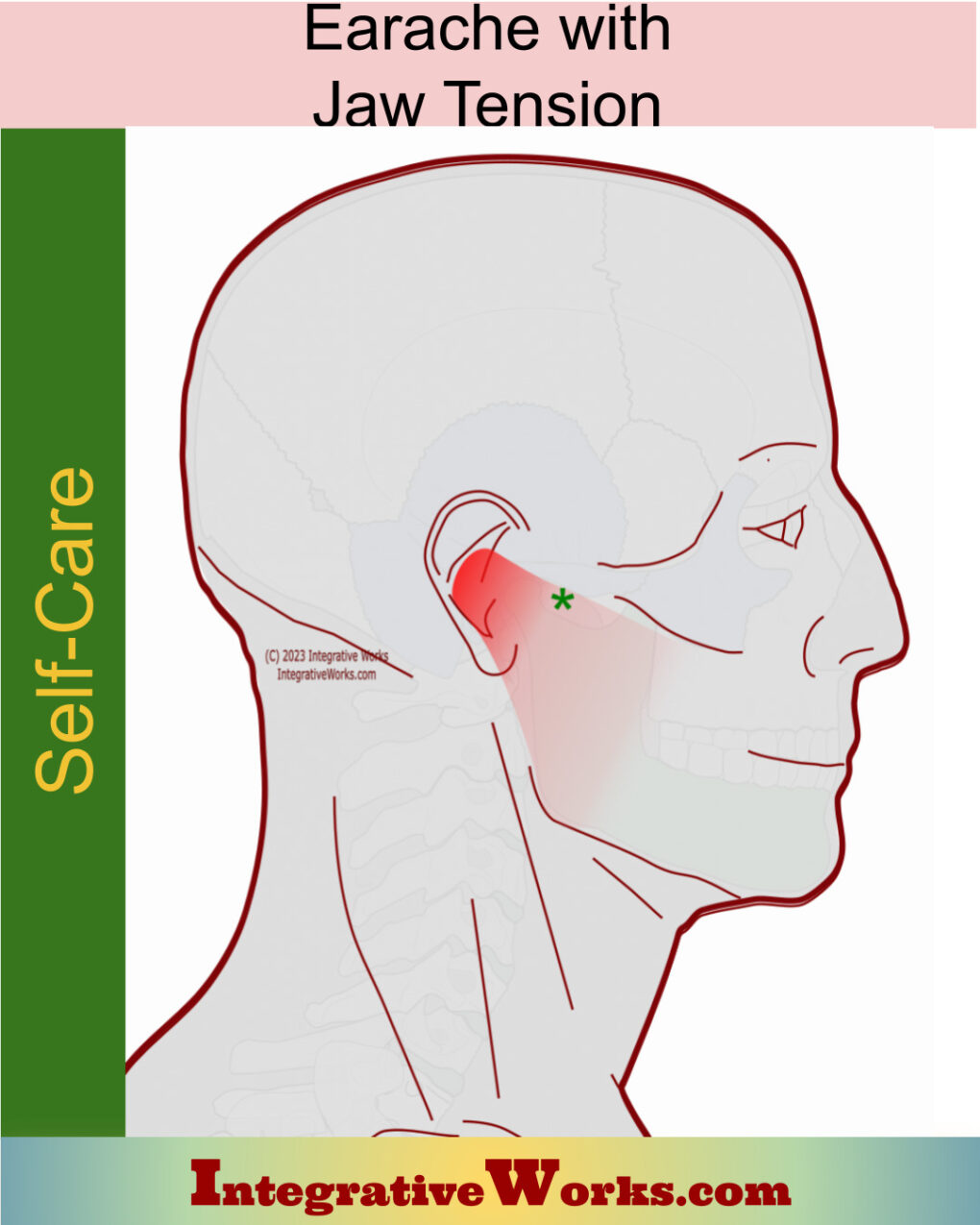
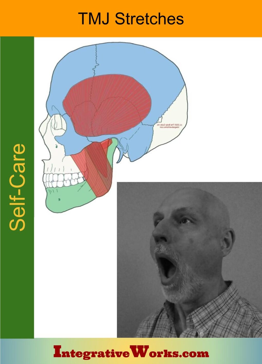
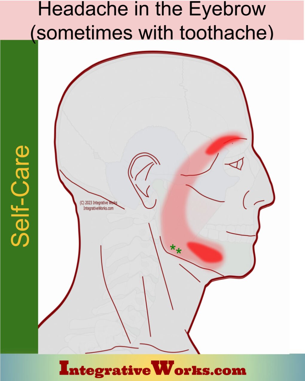
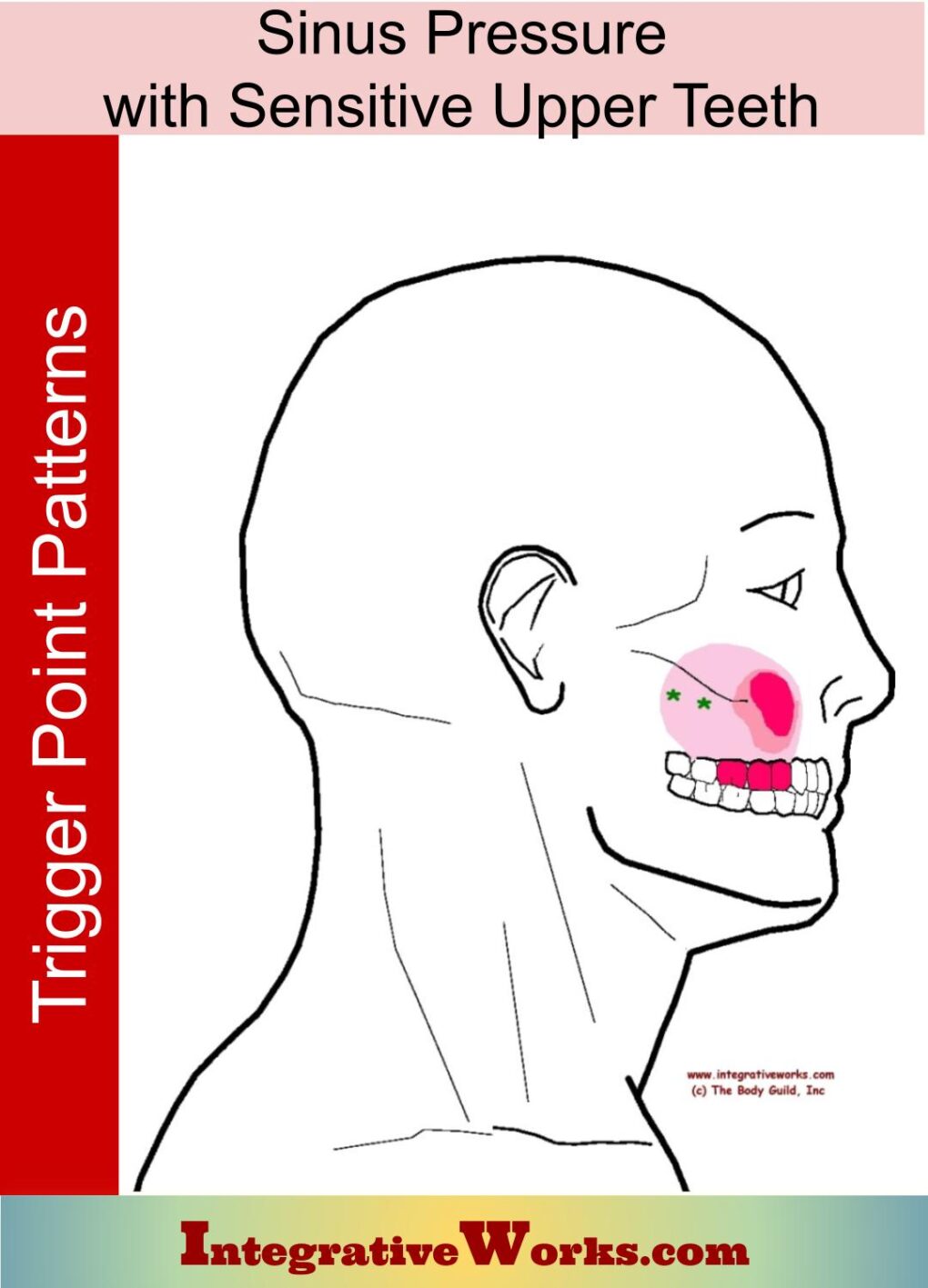
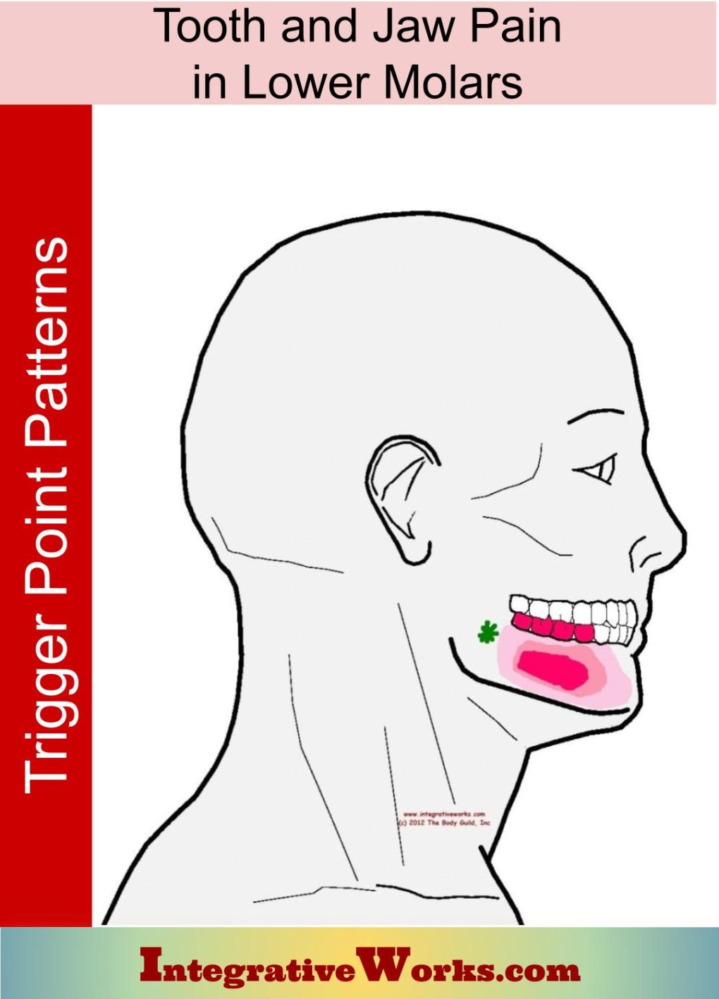
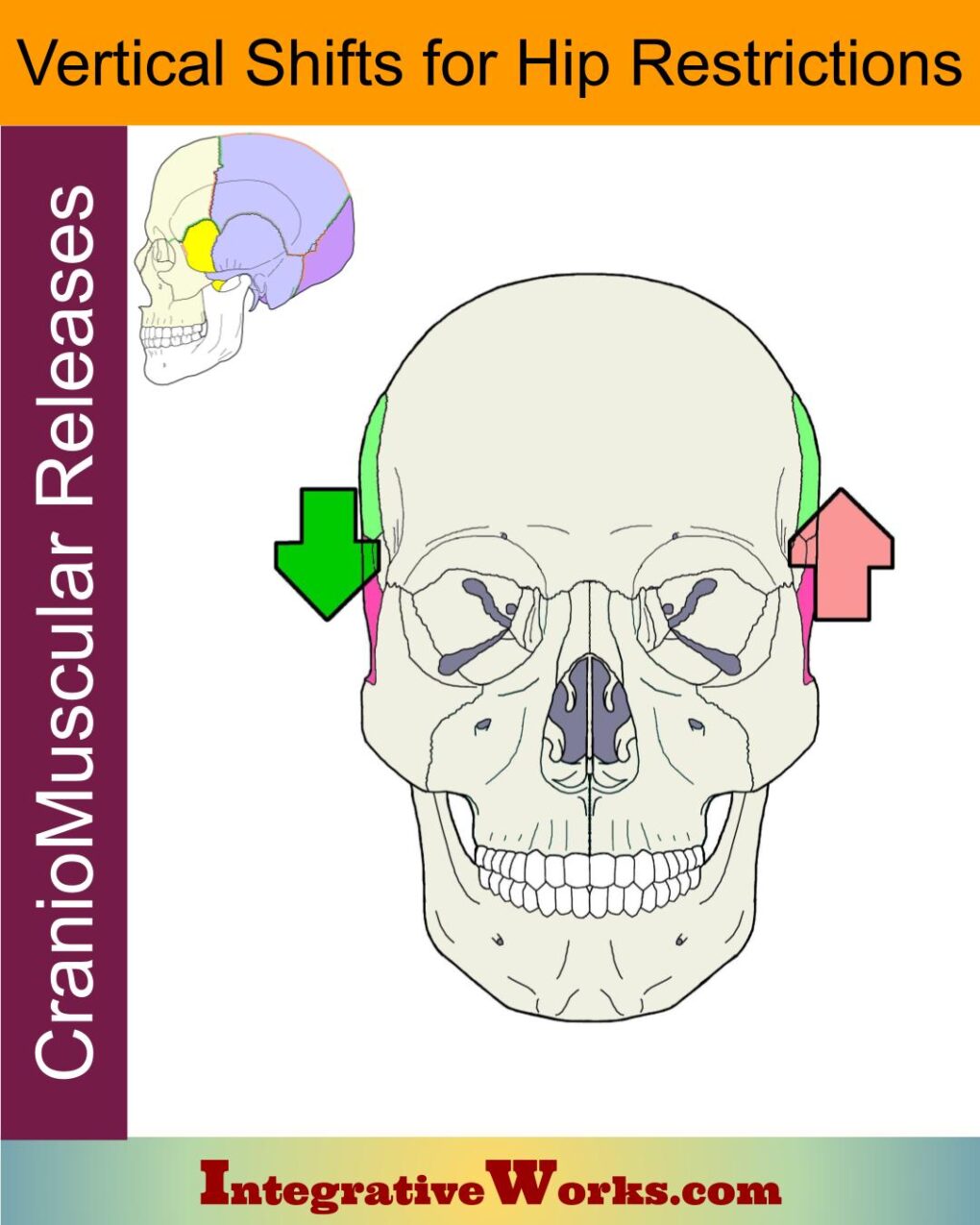
Overview
The masseter anatomy is similar to other three-bellied muscles with a strong fascial covering, like gluteus medius and infraspinatus. Also, like those muscles, it balances and stabilizes a joint that needs both strength and flexibility. Pound for pound, it is considered to be the strongest muscle in the body.
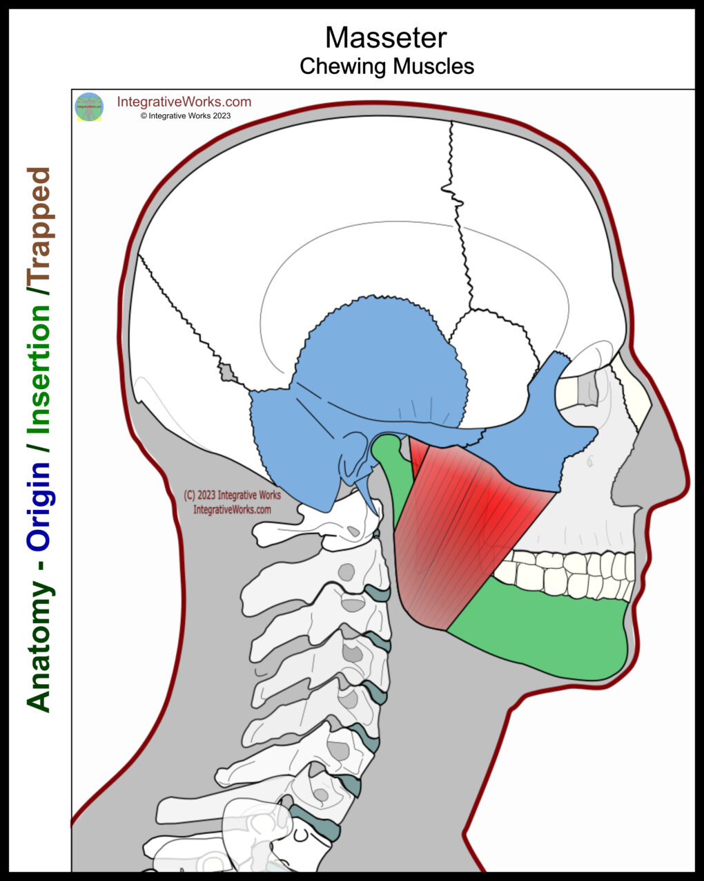
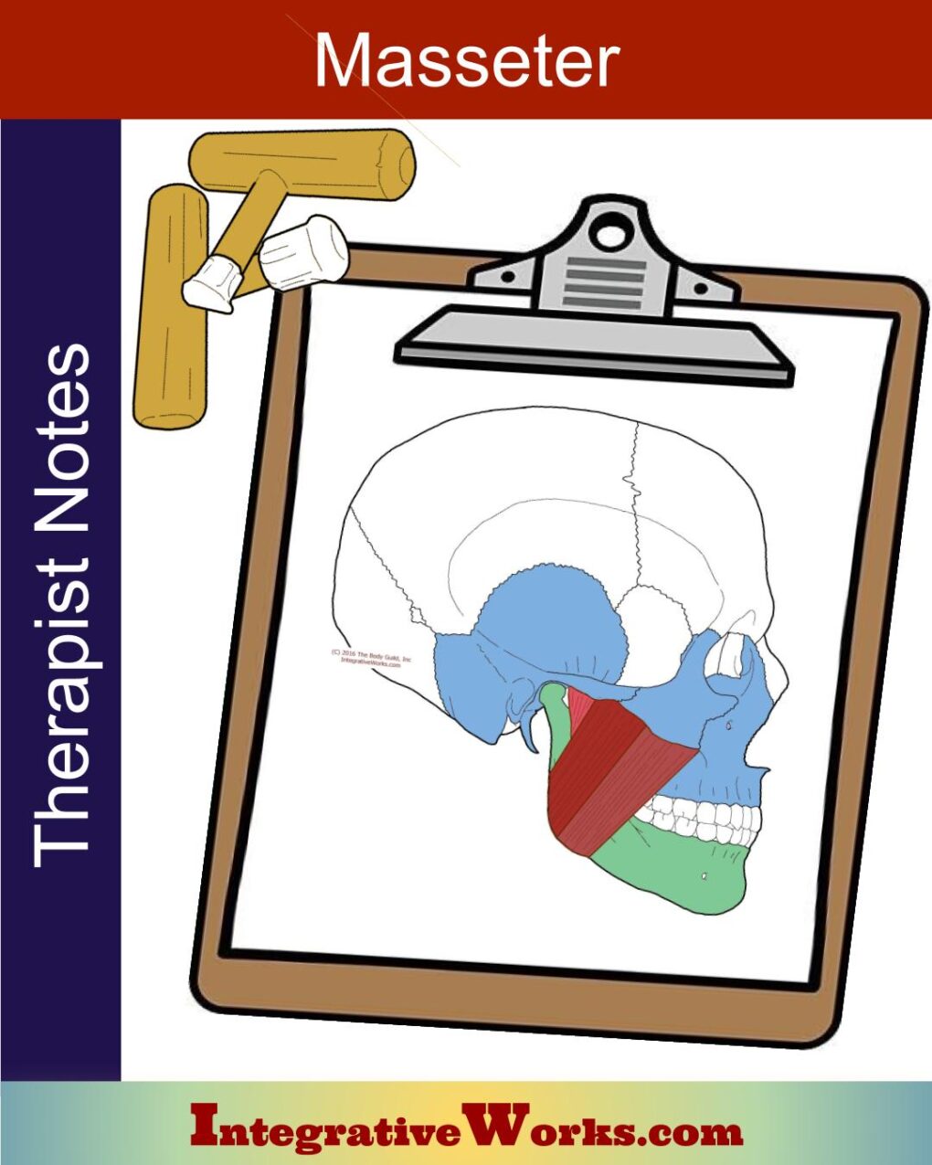
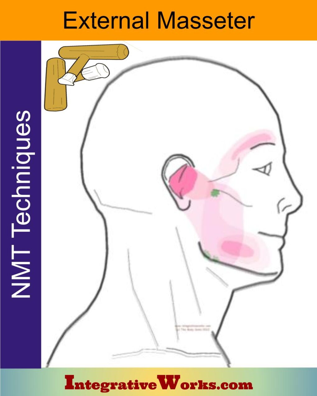
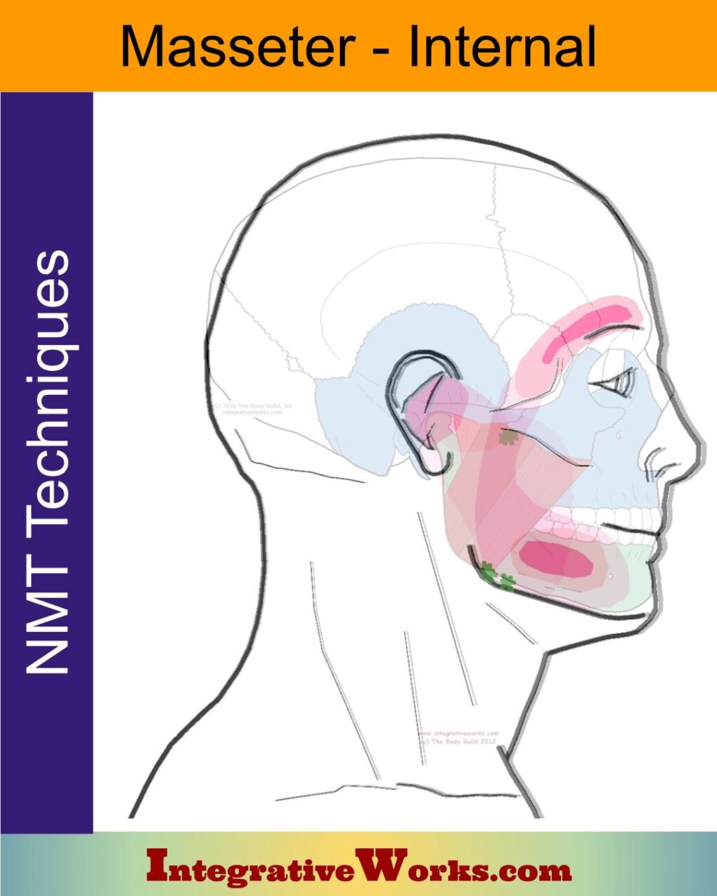
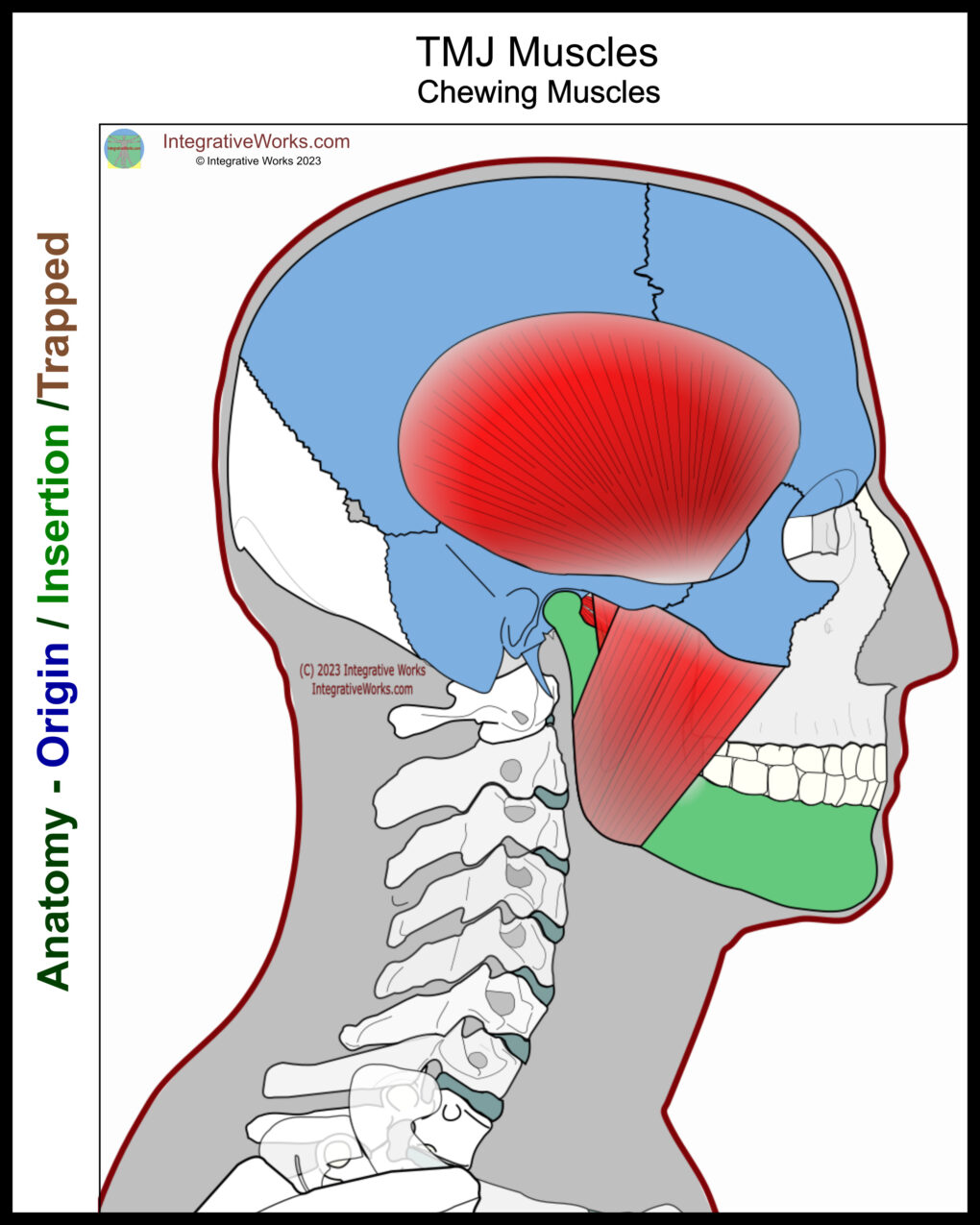
The masseter is a three-bellied muscle on the lateral jaw. It is often regarded as having the strongest muscle fibers in the body.
Superficial bellies:
Origin
- the anterior third of the zygomatic arch of the temporal bone.
- the inferior and medial surface of the zygoma including the maxillary process
Insertion
- posterior belly attaches to the surface of the ramus posterior to the attachment of the deep belly
- anterior belly attaches along the anterior surface of the mandible in front of the attachment of the deep belly.
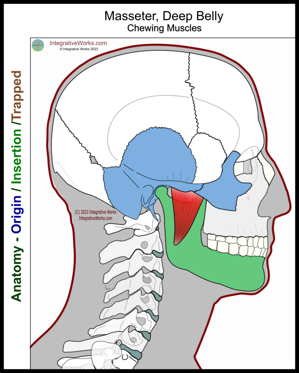
Deep belly:
Origin
- posterior third of the zygomatic arch along the zygomatic process of the temporal bone.
Insertion
- the lateral surface of the coronoid process of the mandible.
Function
- elevate the mandible and close the jaw.
- some protraction of the mandible
Innervation
- trigeminal nerve, mandibular division
The masseter is another one of those three-bellied muscles with a strong fascial covering, like gluteus medius and infraspinatus. Also, like those muscles, it balances and stabilizes a joint that needs both strength and flexibility.
Functional Considerations
The masseter muscle is one of the four muscles responsible for the action of mastication (chewing).
This meta-study shows that the internal tendon architecture divides the muscle into partitions. Further, those divide into compartments representing small motor unit territories within each anatomical partition. The individual compartments have unique biomechanics that can generate a wide range of torques relative to the temporomandibular joint. Regional masseter muscle activation studies show that individual compartments can be uniquely activated depending on the task. However, they more commonly activate in discrete groups with other jaw muscles.
Anomalies, Etc.
Masseter anatomy is fairly consistent. Occasionally, there are anomalous slips that extend to the coronoid process. It is reported that the most common anomaly is masseteric hypertrophy.
Related Posts
Support Integrative Works to
stay independent
and produce great content.
You can subscribe to our community on Patreon. You will get links to free content and access to exclusive content not seen on this site. In addition, we will be posting anatomy illustrations, treatment notes, and sections from our manuals not found on this site. Thank you so much for being so supportive.
Cranio Cradle Cup
This mug has classic, colorful illustrations of the craniosacral system and vault hold #3. It makes a great gift and conversation piece.
Tony Preston has a practice in Atlanta, Georgia, where he sees clients. He has written materials and instructed classes since the mid-90s. This includes anatomy, trigger points, cranial, and neuromuscular.
Question? Comment? Typo?
integrativeworks@gmail.com
Interested in a session with Tony?
Call 404-226-1363
Follow us on Instagram

*This site is undergoing significant changes. We are reformatting and expanding the posts to make them easier to read. The result will also be more accessible and include more patterns with better self-care. Meanwhile, there may be formatting, content presentation, and readability inconsistencies. Until we get older posts updated, please excuse our mess.

