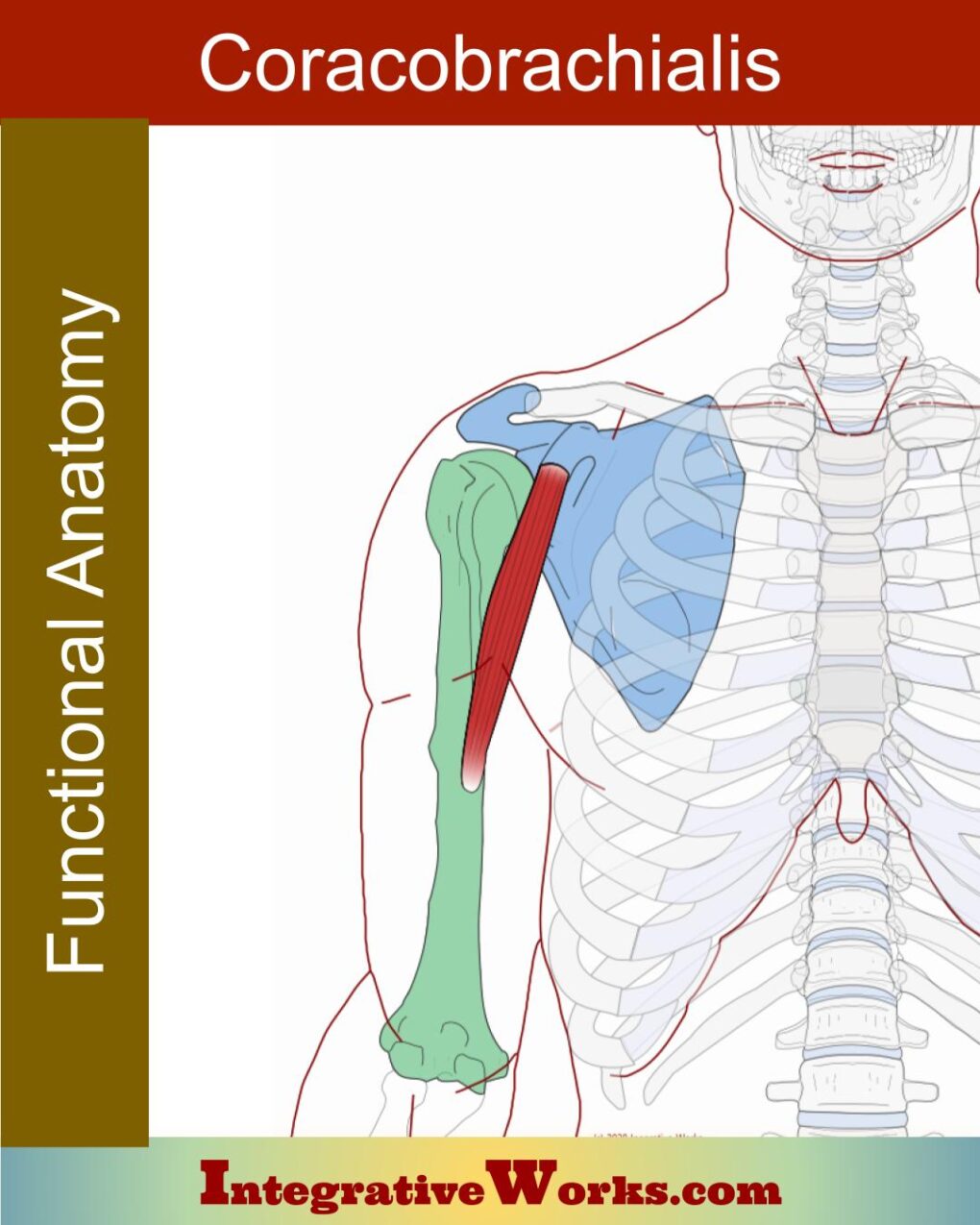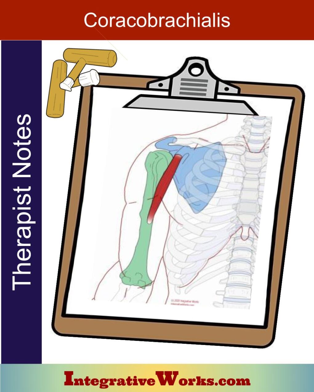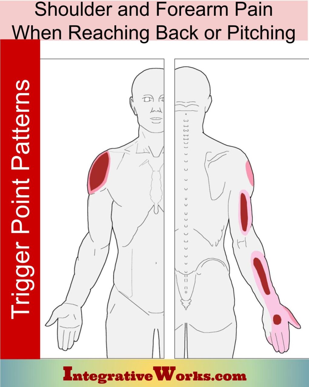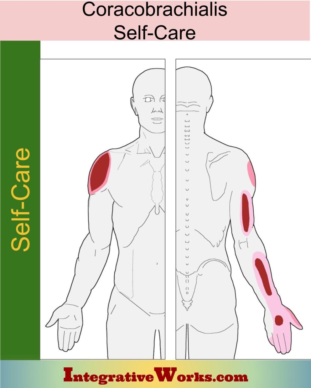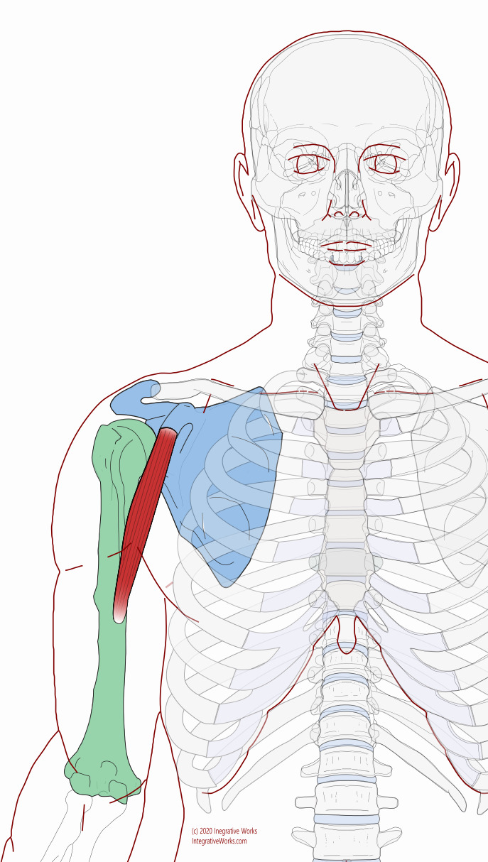
The coracobrachialis is a long muscle on the upper, inside edge of the upper arm.
The Musculoskeletal Anatomy Behind Your Pain
Origin
- tip of the coracoid process of the scapula
Insertion
- medial shaft of the humerus
Function
- flexes the shoulder (glenohumeral joint)
- adducts the shoulder
- some medial rotation of the shoulder
Innervation
- musculocutaneous nerve (C5-7)
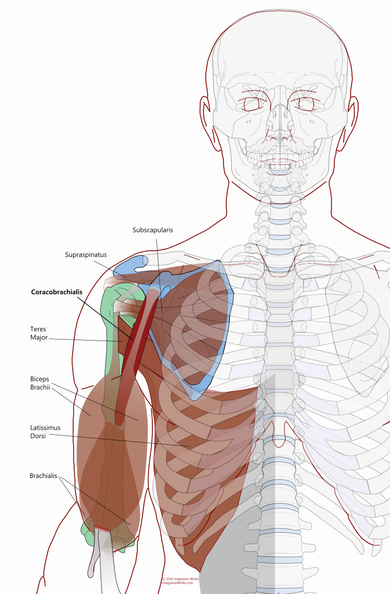
The coracobrachialis is typically a long single belly but does have clinically significant variations.
Attachment Details
The coracobrachialis is one of three muscles that attach to the coracoid process of the scapula. It usually attaches to the apex of the process. When there are two heads, the superficial head tends to attach to the medial aspect with the short head of the biceps. The pectoralis minor usually attaches along the medial border, superior to the coracobrachialis.
The insertion attaches along the medial humerus just above the medial, superior attachments of the brachialis.
Anomalies, Etc.
Anomalies are statistically significant. They are more likely to occur as multiple bellies in the same muscle. One study shows how the muscle often has a deep and superficial belly. See more about this in the discussion about on details of attachments.
Based on innervation and attachments, cadavers are more likely to have extra heads of the biceps and less likely to have extra heads of the coracobrachialis. Also, the musculocutaneous nerve splits and varies when the muscle has more than one belly.
Wikipedia entry for coracobrachialis.

