Overview
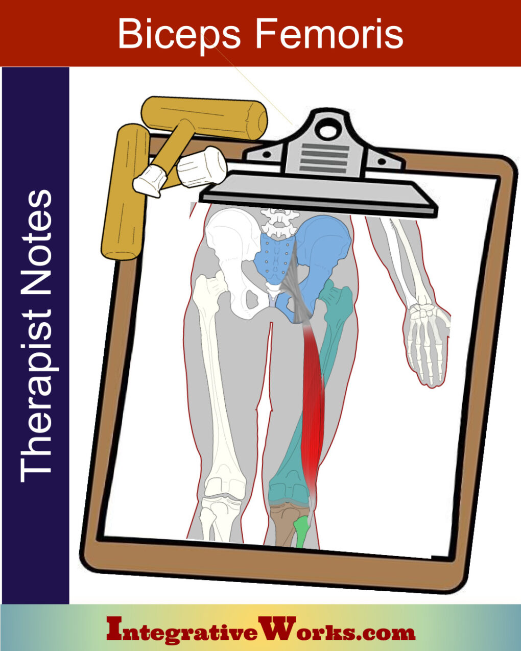
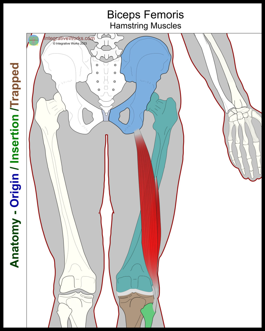
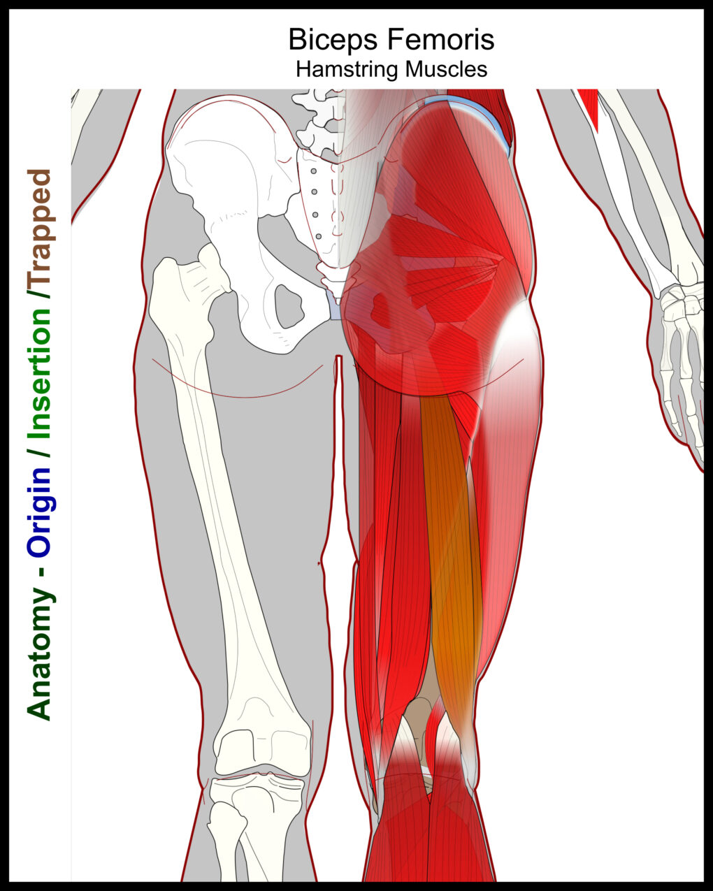
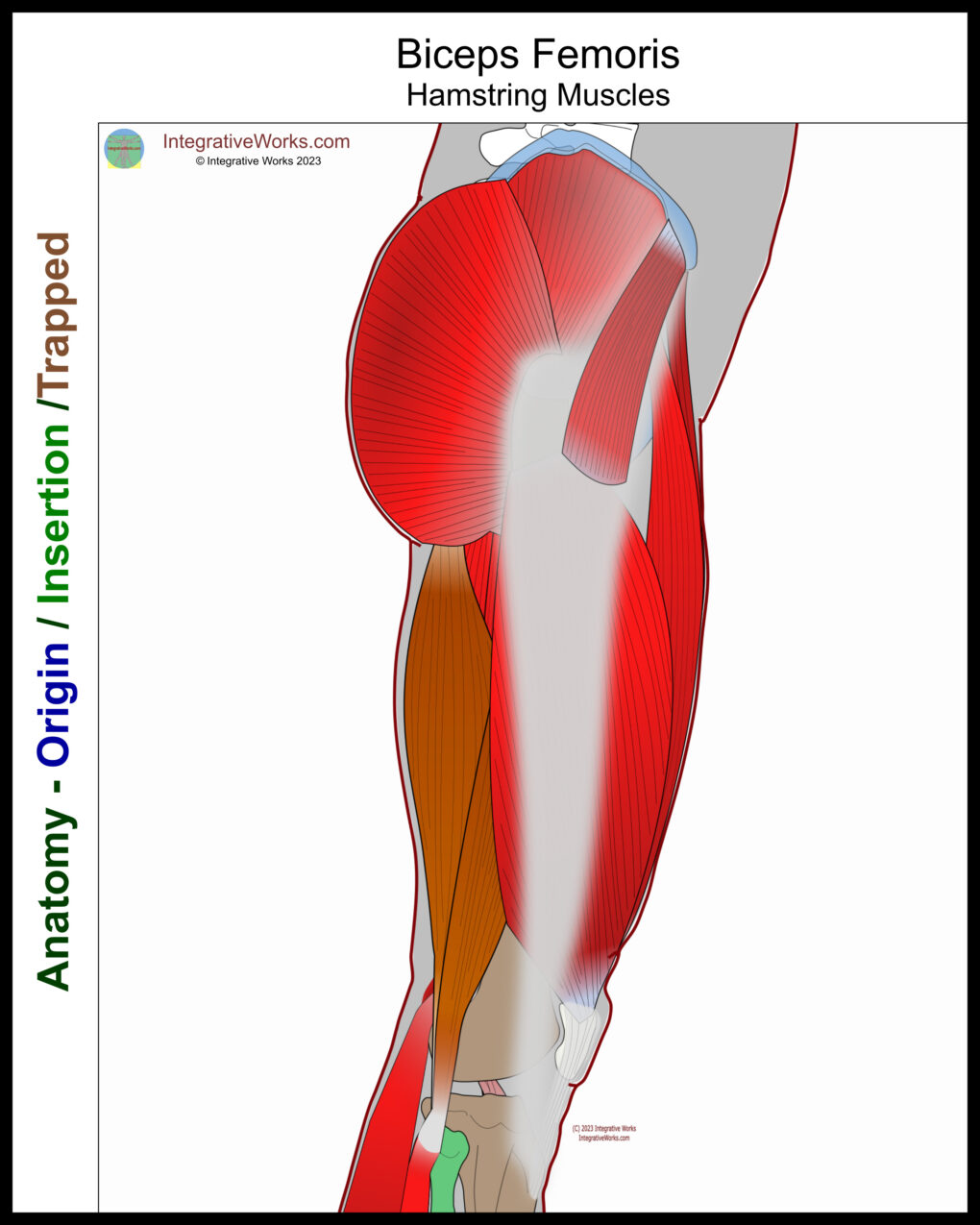
Here, you will find the basic anatomy and Physiology of the biceps femoris muscle. The biceps femoris is a two-headed muscle on the lateral thigh. It belongs to the hamstring group.
Origin
- ischial tuberosity
Insertion
- fibular head
- (tibia, femur)
Function
- flex knee joint
- extend hip
- (external rotation at the knee)
Nerve
- sciatic nerve
Functional Considerations
Involvement in Exercise
There are a multitude of studies about hamstring function, especially related to exercise. This meta-study is particularly extensive.
Knee Balance
This meta-study reviews the anatomy of distal attachments of the biceps femoris. It asserts that the biceps femoris is significant in balancing the knee. My clinical experience indicates the same.
Quadriceps Balance
Many seminars on the clinical treatment of the thigh explore the balance between the quadriceps and hamstrings. Typically, they conclude that when one is overpowering, it creates pain in the antagonist, hip, and low back. Again, my clinical experience supports this.
Low Back Balance
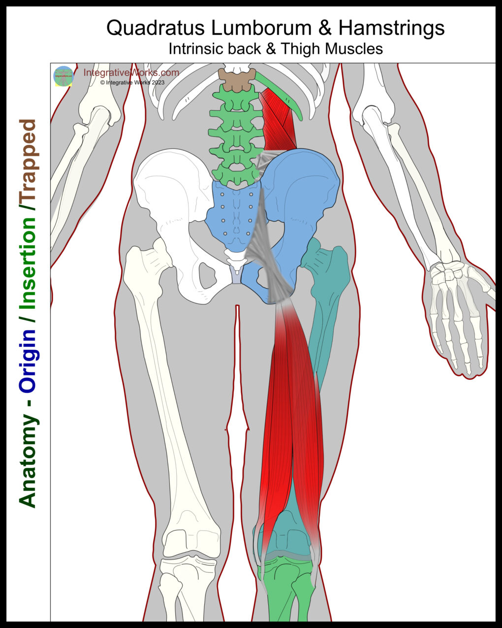
The low back is strongly impacted by hamstring length. Its connection to the base of the pelvis can restrict pelvic rotation, creating stress on the low back. Muscles like the quadratus lumborum, which is known for its pain patterns, are directly torqued through their mutual attachment to the pelvis. Of course, many other muscles, such as multifidi and erector spinae, are impacted.
Anomalies, Etc.
Short head
This study reveals notable variations in the short head of the biceps femoris. More specifically, it varies in size, attachment, and absence. Variations are more common in the Korean Population. This study discusses the impact on the lateral peroneal nerve because of variations in the structures for its passage.
Fibular Attachment
Studies on this vary. However, they focus on the attachments to the femur and tibia. This meta-study finds that attachment to the tibia and femur is the norm. Therefore, standard anatomical descriptions of the attachment of the biceps femoris should be revised.
Related Posts
Support Integrative Works to
stay independent
and produce great content.
You can subscribe to our community on Patreon. You will get links to free content and access to exclusive content not seen on this site. In addition, we will be posting anatomy illustrations, treatment notes, and sections from our manuals not found on this site. Thank you so much for being so supportive.
Cranio Cradle Cup
This mug has classic, colorful illustrations of the craniosacral system and vault hold #3. It makes a great gift and conversation piece.
Tony Preston has a practice in Atlanta, Georgia, where he sees clients. He has written materials and instructed classes since the mid-90s. This includes anatomy, trigger points, cranial, and neuromuscular.
Question? Comment? Typo?
integrativeworks@gmail.com
Interested in a session with Tony?
Call 404-226-1363
Follow us on Instagram

*This site is undergoing significant changes. We are reformatting and expanding the posts to make them easier to read. The result will also be more accessible and include more patterns with better self-care. Meanwhile, there may be formatting, content presentation, and readability inconsistencies. Until we get older posts updated, please excuse our mess.

