
Overview
The anatomy of the teres major is both complicated and straightforward. At first glance, it is a rounded (“teres”) structure with two attachments. However, variations in the muscles and neurovascular bundle that surround it vary its role in the shoulder.
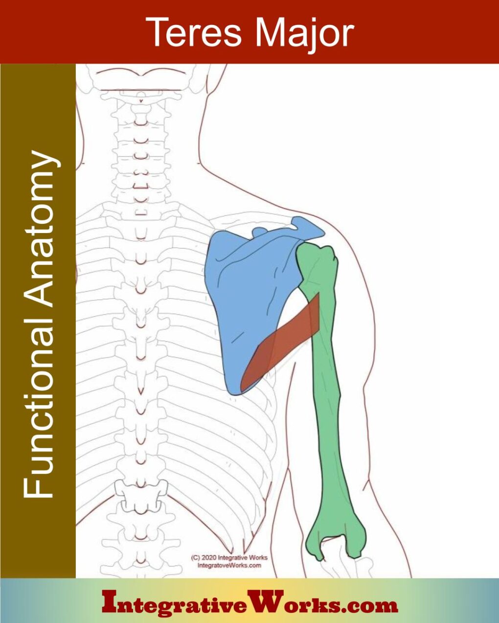
Origin
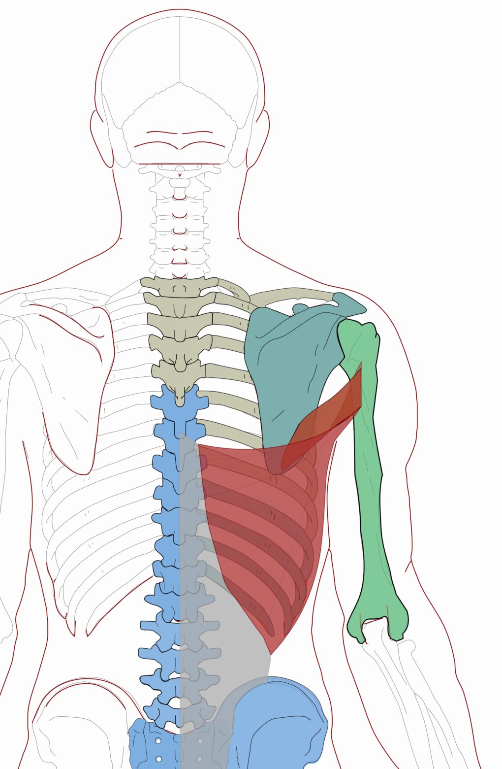
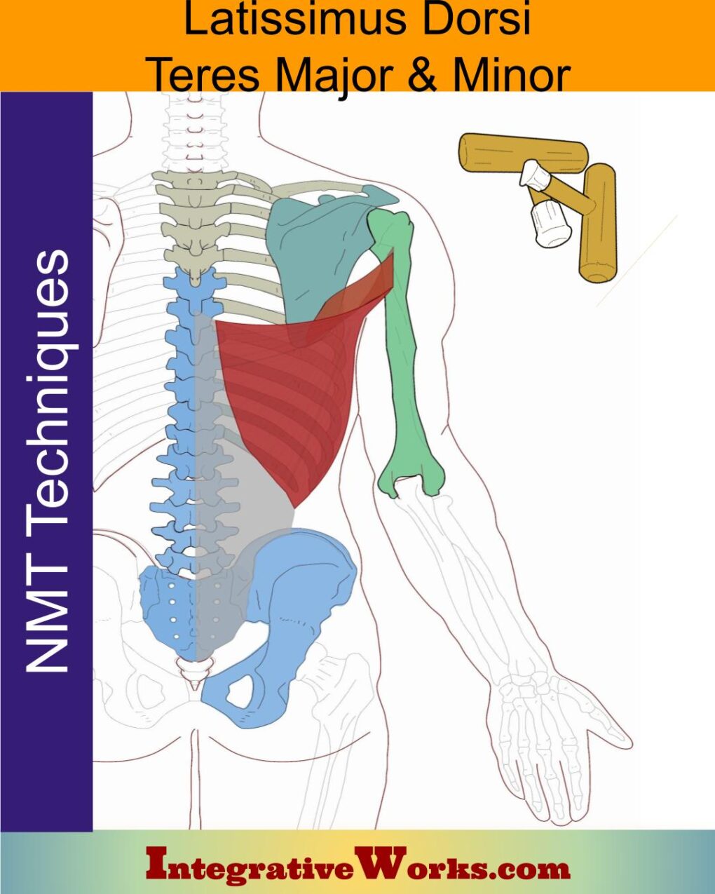
- the posterior, inferior edge of the lateral border of the scapula
Insertion
- the medial lip of the bicipital groove of the humerus
Function
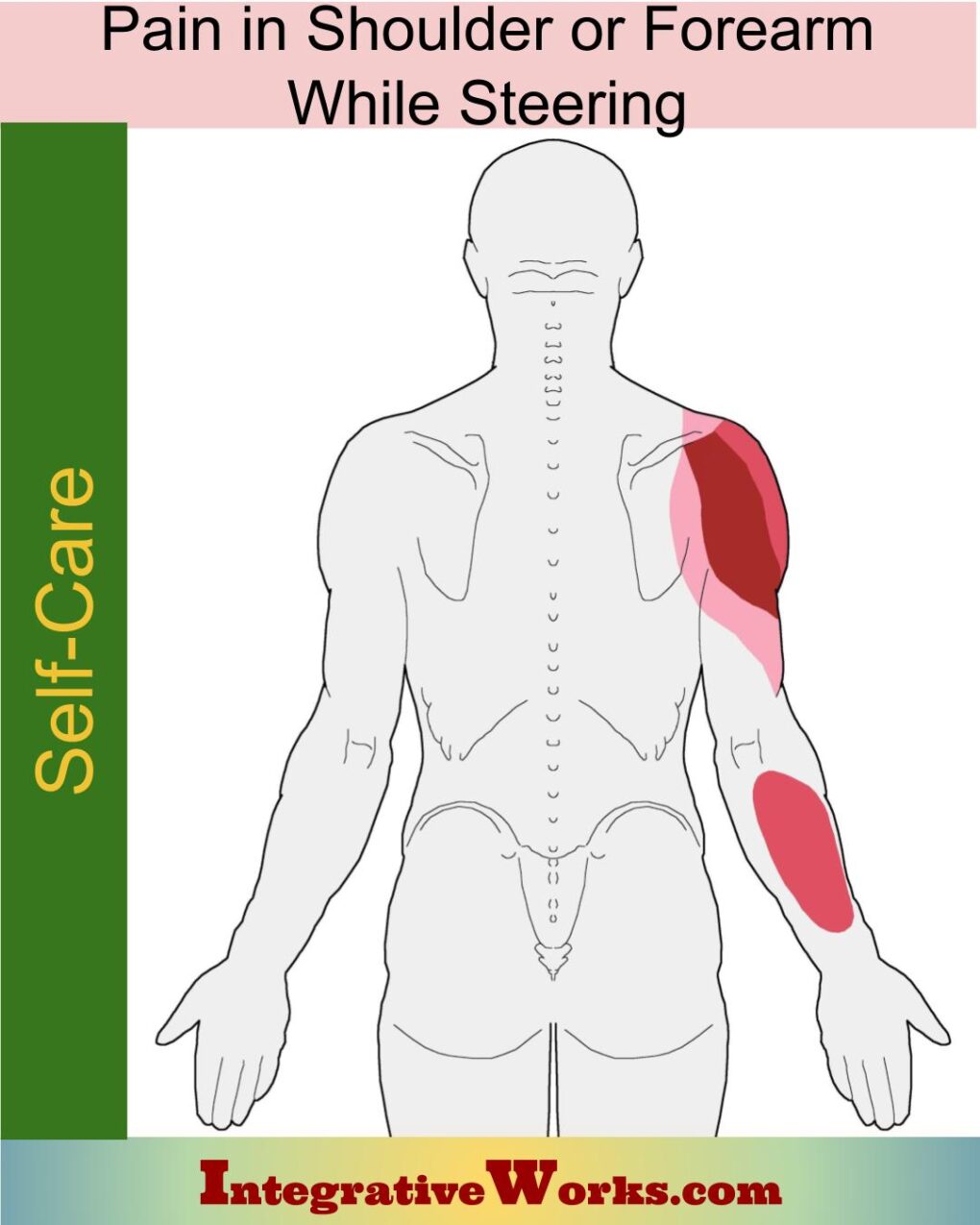
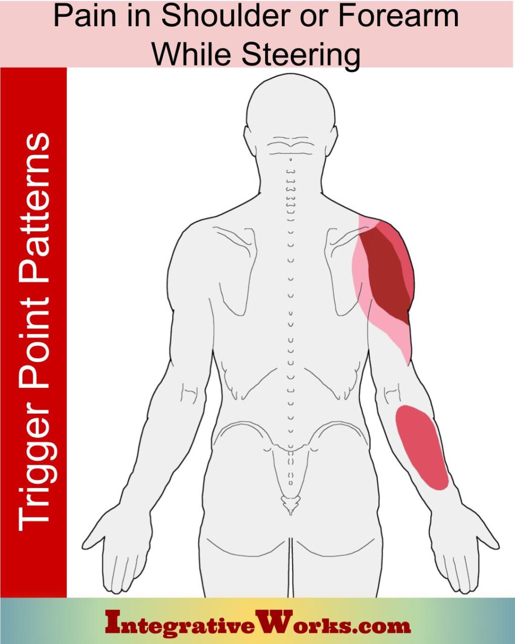
- adducts and medial rotates the humerus when it is lateral to the body,
- extends the humerus when it is in horizontal flexion (in front of the body
Innervation
- lower subscapular nerve – C5-C7
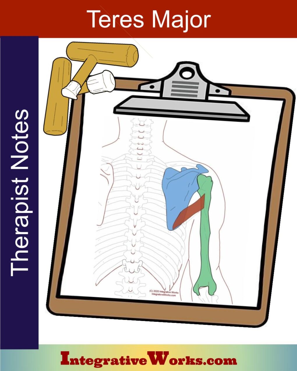
Attachment Details
It is a simple rounded muscle that attaches to the scapula’s lower border and inserts on the bicipital groove.
Its relationship with latissimus dorsi makes it more complicated. Often, but not always, it lives in a sheath with the latissimus dorsi. A bursa usually separates the tendon attachments. Teres major typically attaches by a separate tendon more medially in the bicipital groove.
Functional Considerations
The physiology is also interesting. Teres major works closely with latissimus dorsi in moving the humerus toward the scapula. It may extend the humerus when it is in front of the body. Conversely, it may flex the humerus when it is behind the body. As well, it assists in the medial rotation of the humerus, especially when the humerus is in front of the body.
It is not considered a rotator cuff muscle because it does not attach to the humeral head. However, one study suggests that teres major has a major role in stabilizing the glenohumeral joint with an attachment to the humeral head.
Neurovascular Complications
The neurovascular bundle containing the brachial plexus complicates the anatomy of the teres major muscle. The entire bundle lies close to the humeral attachment, and most of the nerve fibers pass anteriorly. However, others pass over and posteriorly to the teres major. At times, the nerves weave through the muscle fibers.
Anomalies, Etc.
About half of the cadavers in this study had “teres major accessorius.” It is a small slip of muscle that attaches along the superior border of the teres major on the scapula. The insertion tendon is separate from the teres major on the humerus.
The humeral attachment of teres major and latissimus dorsi is quite variable. They are usually separate, but not always. This study finds a connection in about 25% of cases. Additionally, they vary in their overlap. According to this study, there is a small percentage of complete overlap or no overlap.
Posts Related to Teres Major
Wikipedia entry to teres major.
Support Integrative Works to
stay independent
and produce great content.
You can subscribe to our community on Patreon. You will get links to free content and access to exclusive content not seen on this site. In addition, we will be posting anatomy illustrations, treatment notes, and sections from our manuals not found on this site. Thank you so much for being so supportive.
Cranio Cradle Cup
This mug has classic, colorful illustrations of the craniosacral system and vault hold #3. It makes a great gift and conversation piece.
Tony Preston has a practice in Atlanta, Georgia, where he sees clients. He has written materials and instructed classes since the mid-90s. This includes anatomy, trigger points, cranial, and neuromuscular.
Question? Comment? Typo?
integrativeworks@gmail.com
Interested in a session with Tony?
Call 404-226-1363
Follow us on Instagram

*This site is undergoing significant changes. We are reformatting and expanding the posts to make them easier to read. The result will also be more accessible and include more patterns with better self-care. Meanwhile, there may be formatting, content presentation, and readability inconsistencies. Until we get older posts updated, please excuse our mess.