Overview
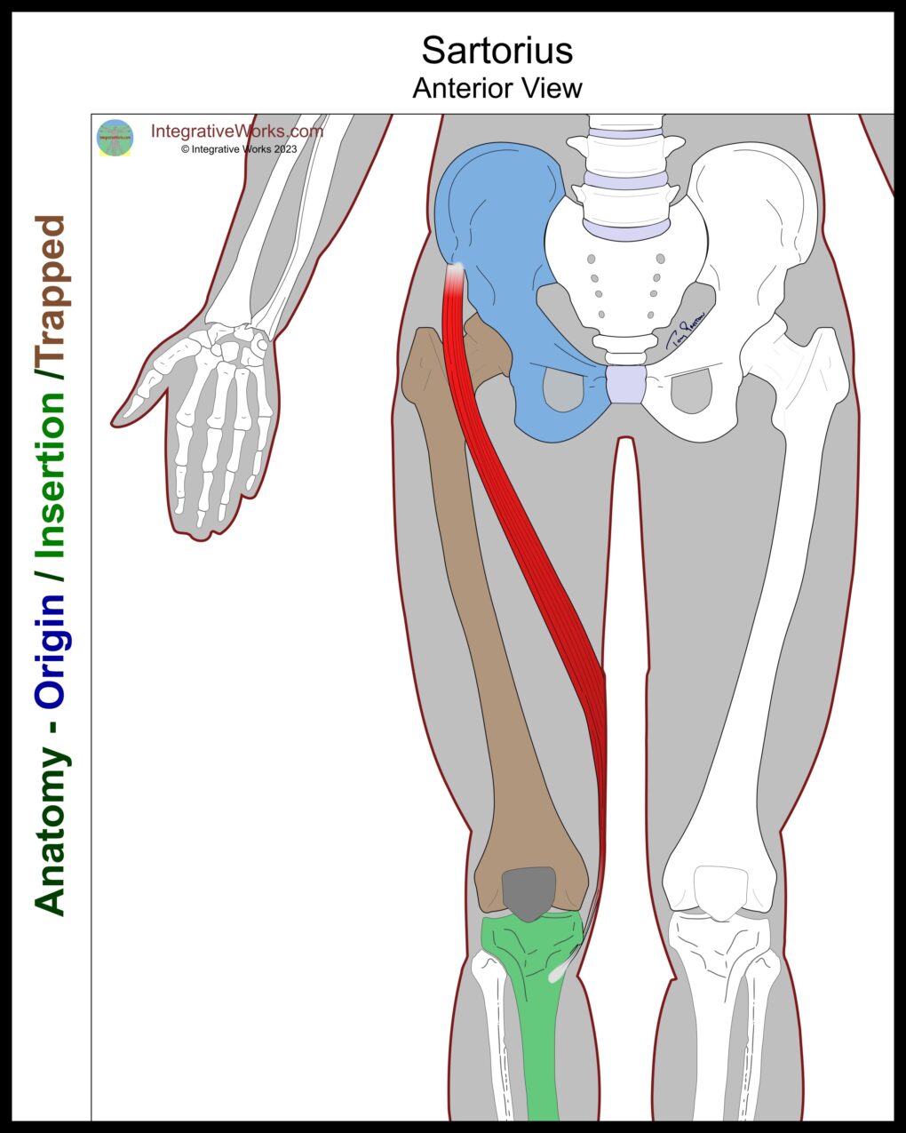
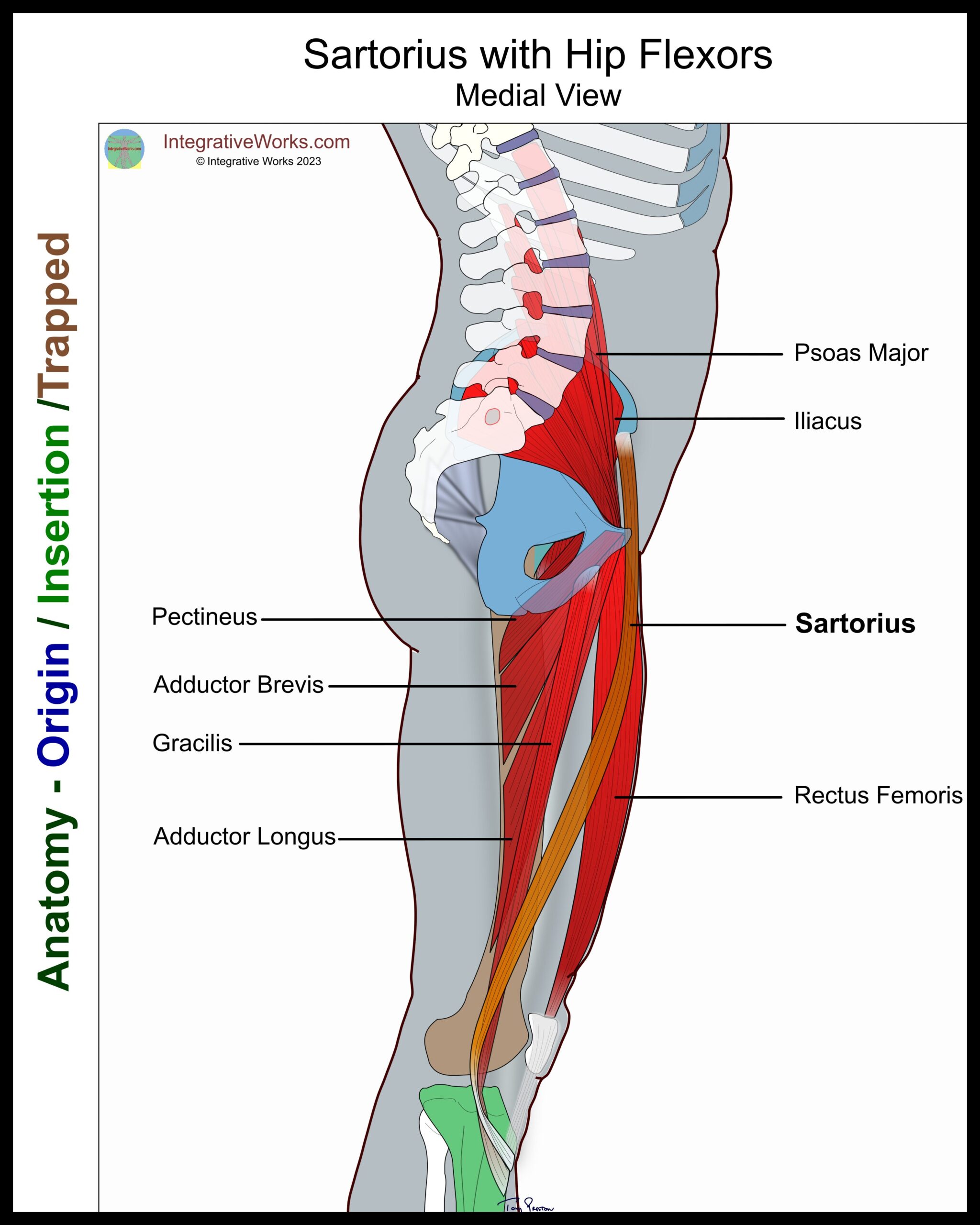
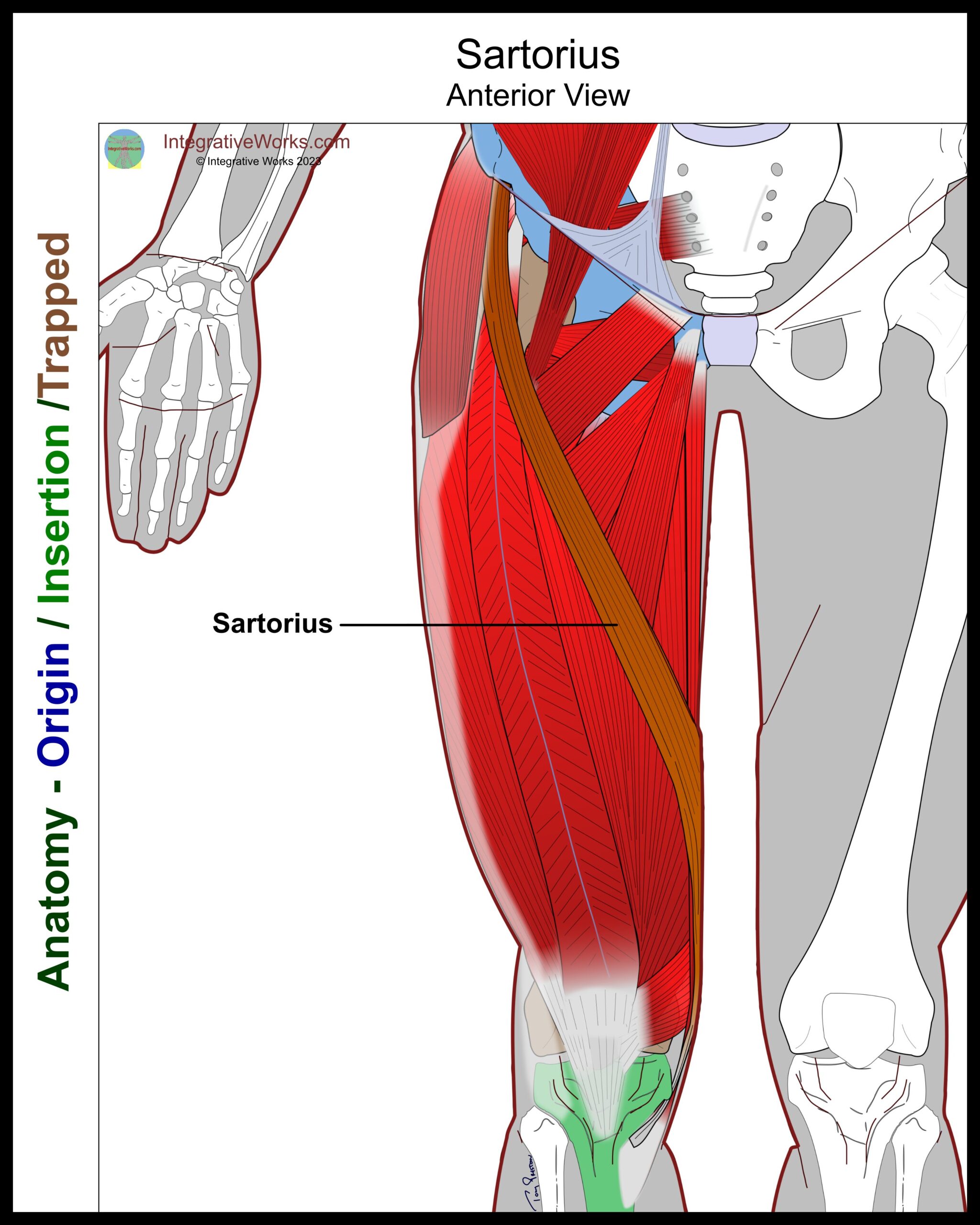
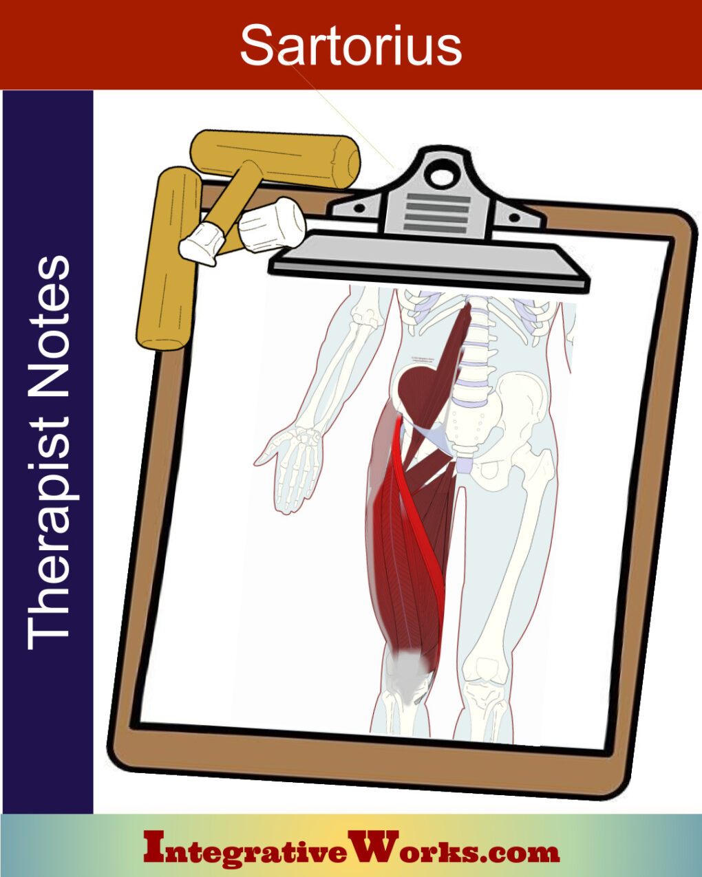
The functional anatomy of the sartorius muscle is complicated and often argued. The muscle is used when adopting a cross-legged position associated with a tailor’s sewing posture. Consequently, its name comes from the Latin sartor, meaning ‘tailor.’
Notably, it is the longest muscle in the body.
Origin
- anterior superior iliac spine
Insertion
- pes anserine on the superior medial tibia.
Function
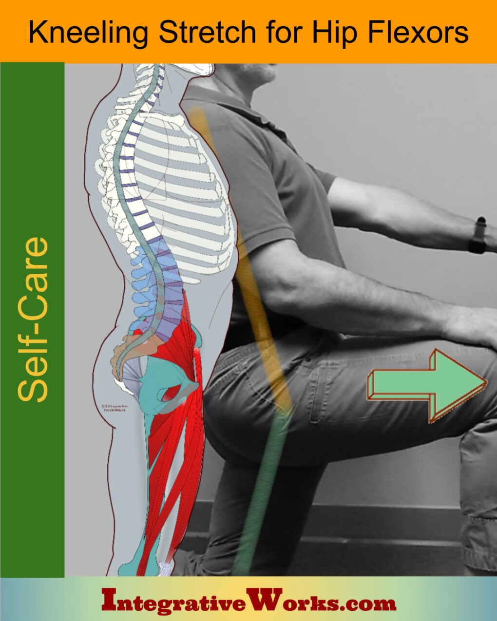
- flexion of the hip
- external rotation of the hip
- internal rotation of the knee
Nerve
- femoral nerve, anterior division – L2-L4
Functional Considerations
In discussions of sartorius’ anatomy and physiology, therapists often consider this muscle a “jack of all trades, master of none.” However, it seems most prevalent in crossing the leg into a figure-4 position.
Most studies agree that it flexes the hip, Yet, they often vary in their consideration of its influence. Commonly, studies also agree that it assists in lateral rotation of the hip, mainly as a synergist.
Rotating the knee
This study explores its role as an internal rotator of the knee. Sartorius, the medial hamstrings, and gracilis assist the popliteus muscle with internal rotation.
Sudden tension
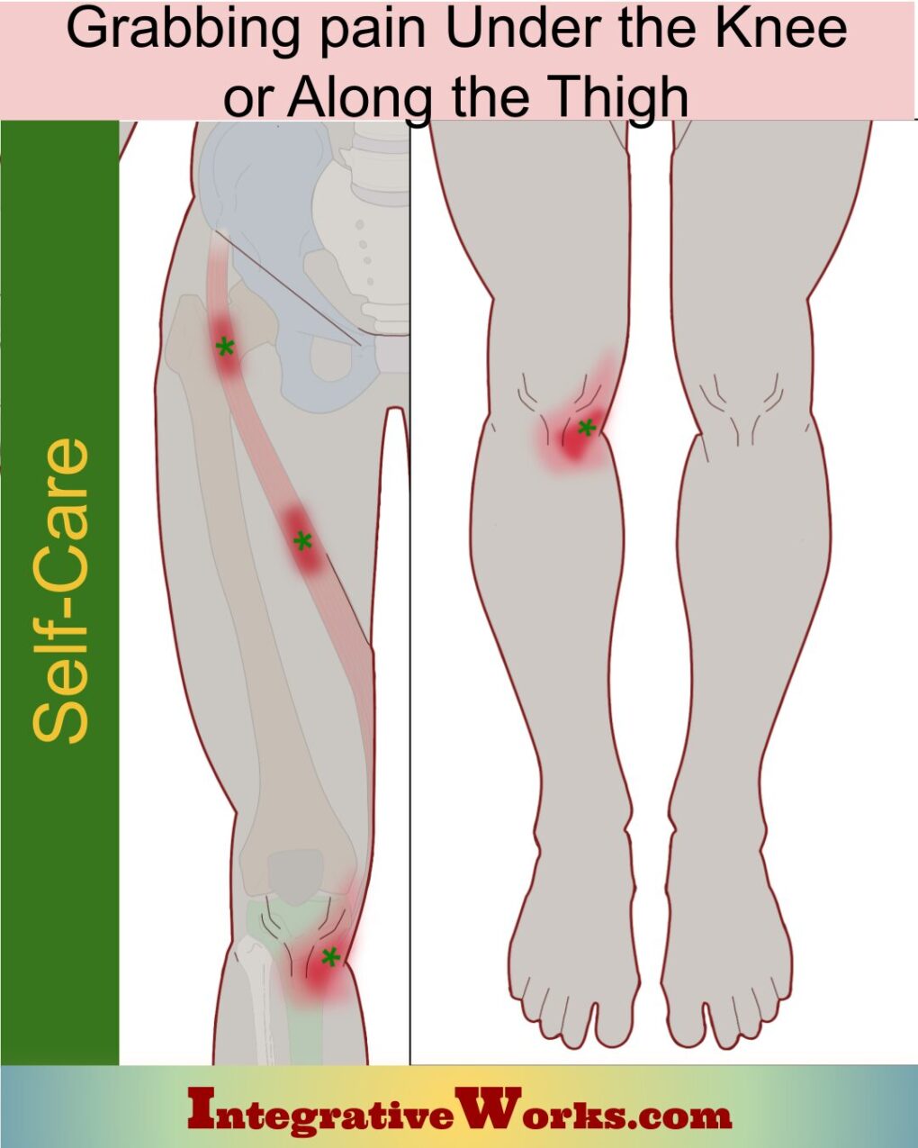
Sartorius, like latissimus dorsi, is a muscle that often hangs loosely and produces sudden pain when it becomes taut. Reflexively, one might avoid knee pain from this muscle by simply turning the foot out. Then, for example, when the foot turns inward to correct balance, the sartorius tightens and produces pain. In addition, several studies are related to injuries from sports that jerk the muscle creating injuries like avulsions.
Anomalies, Etc.
Many studies are focused on the tendon attachments at the pes anserine. Notably, there are some isolated instances of an accessory sartorius muscle.
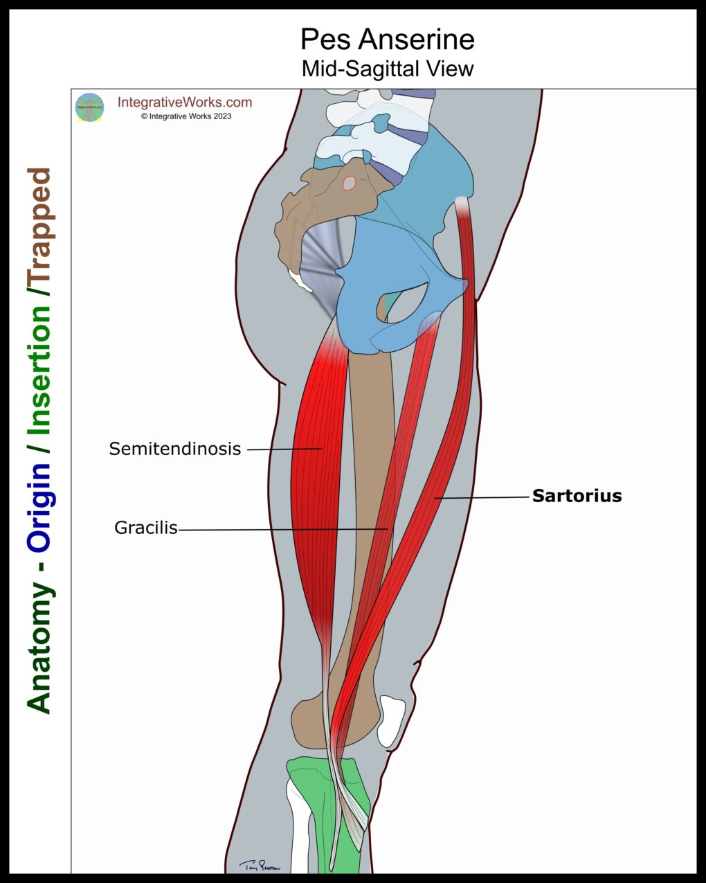
Pes Anserine
The pes anserine is the distal attachment of three iliotibial muscles on the medial tibia. They include the sartorius, gracilis and semitendinosus. Yet, this area tends to be complicated by a high degree of variability.
Typically, the sartorius attaches superiorly in a short wrapping that extends toward the tibial tuberosity. Inferiorly, the semitendinosus attaches and often extends for a short distance along the shaft of the medial tibia. Usually, gracilis attaches between those two tendons. But, again, these are generalities as the area is highly variable.
There are many studies of pes anserine. Naturally, most of them focus on the arrangement of semitendinosus, gracilis, and sartorius tendons. My illustration is based on a series of cadavers that were part of an extensive study on tendon arrangement but was modified as I explored other studies.
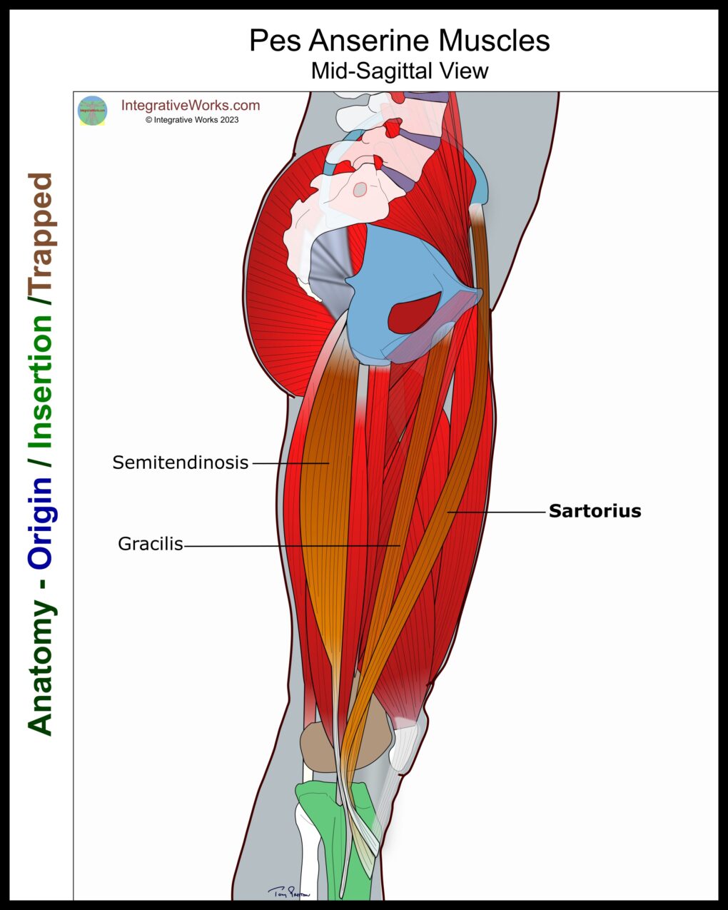
Variable Attachment
This study of Nigerian cadavers also found the area to be quite variable. Attachment tendons varied in number, length, and overlap. In general, the sartorius attached superiorly, and the semitendinosus attached most inferiorly. Notably, there are several helpful illustrations of variations with color coding.
Variable Bursa
Another study focused on the pes anserine bursa. Most of the time (64-67%), it was located between the tendons and the tibia. Additionally, about 20% of the time, it is located between the tendons and the medial collateral ligament. Finally, it developed among the tendons about 10% of the time. Conspicuously, there were no significant variations based on gender. However, the study has detailed charts based on gender, age, height, weight, and BMI.
Related Posts
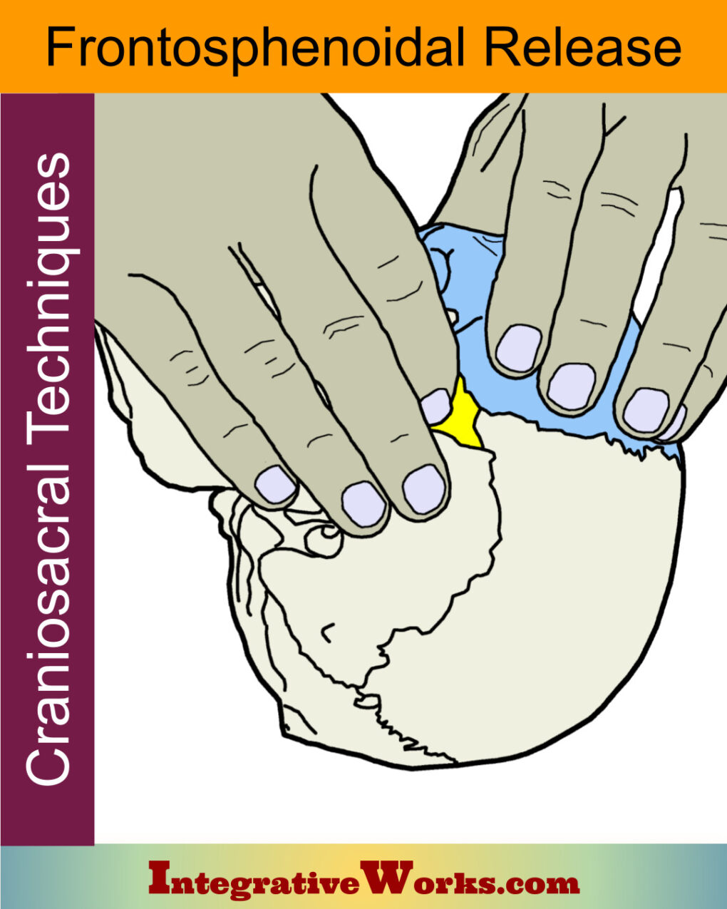
Frontosphenoidal Release – Cranisoacral
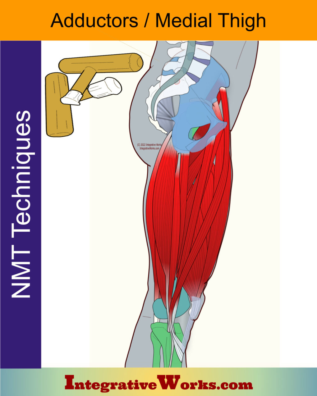
Neuromuscular Protocol – Adductors / Medial Thigh
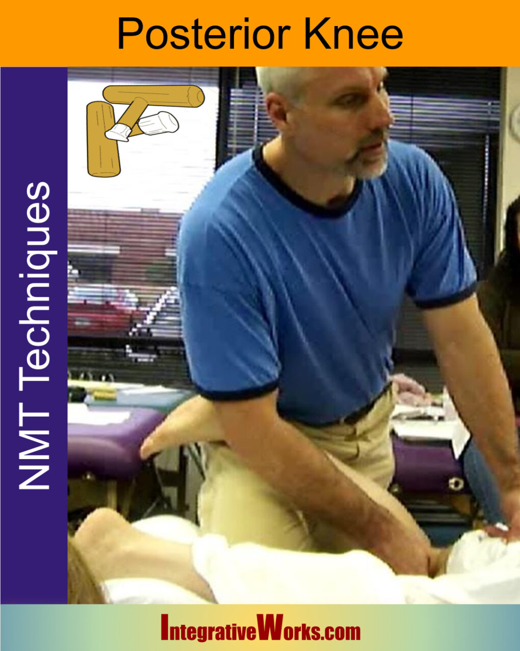
Neuromuscular Protocol – Posterior Knee
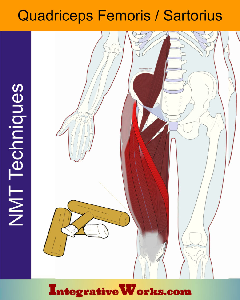
Neuromuscular Protocol – Quadriceps
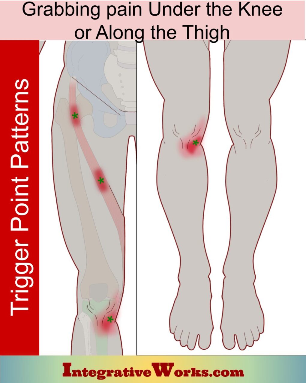
Pain Inside Knee. Sometimes Across Thigh
Sartorius – Massage Therapy Notes
Self Care – Kneeling Stretch for Hip Flexors
Self-Care – Pain Inside of Knee Sometime Across Thigh
Trigger Point Assessment Connects The Pain To The Problem
Support Integrative Works to
stay independent
and produce great content.
You can subscribe to our community on Patreon. You will get links to free content and access to exclusive content not seen on this site. In addition, we will be posting anatomy illustrations, treatment notes, and sections from our manuals not found on this site. Thank you so much for being so supportive.
Cranio Cradle Cup
This mug has classic, colorful illustrations of the craniosacral system and vault hold #3. It makes a great gift and conversation piece.
Tony Preston has a practice in Atlanta, Georgia, where he sees clients. He has written materials and instructed classes since the mid-90s. This includes anatomy, trigger points, cranial, and neuromuscular.
Question? Comment? Typo?
integrativeworks@gmail.com
Follow us on Instagram
*This site is undergoing significant changes. We are reformatting and expanding the posts to make them easier to read. The result will also be more accessible and include more patterns with better self-care. Meanwhile, there may be formatting, content presentation, and readability inconsistencies. Until we get older posts updated, please excuse our mess.

