Overview of Anatomy
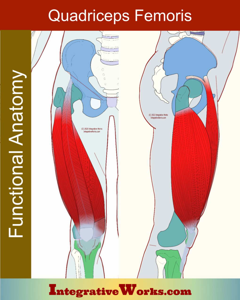
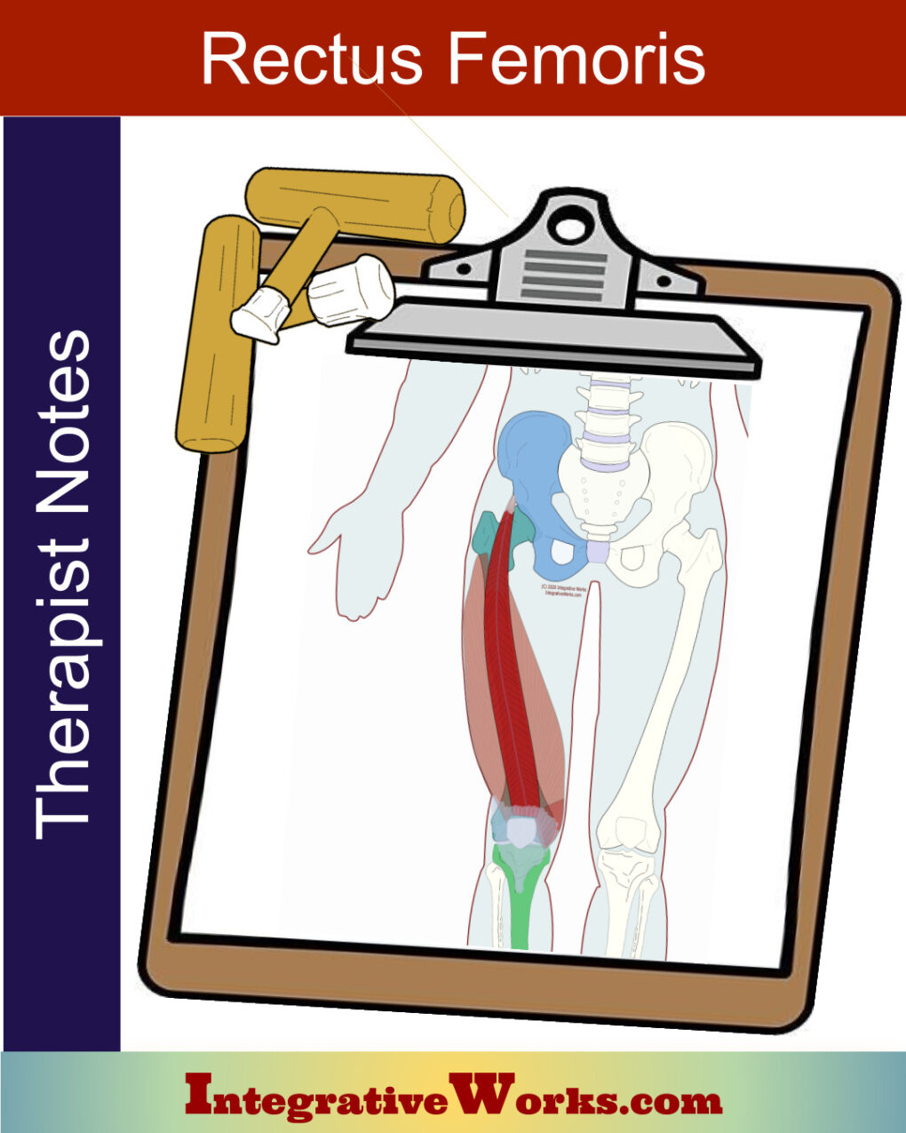
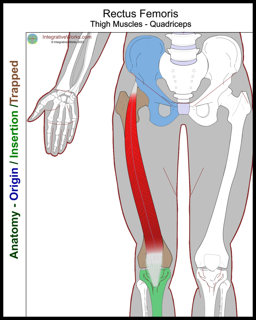
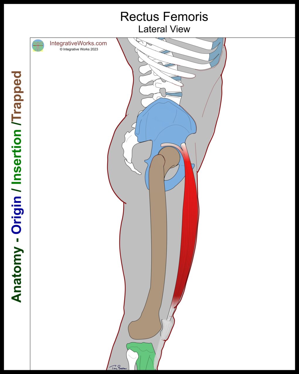
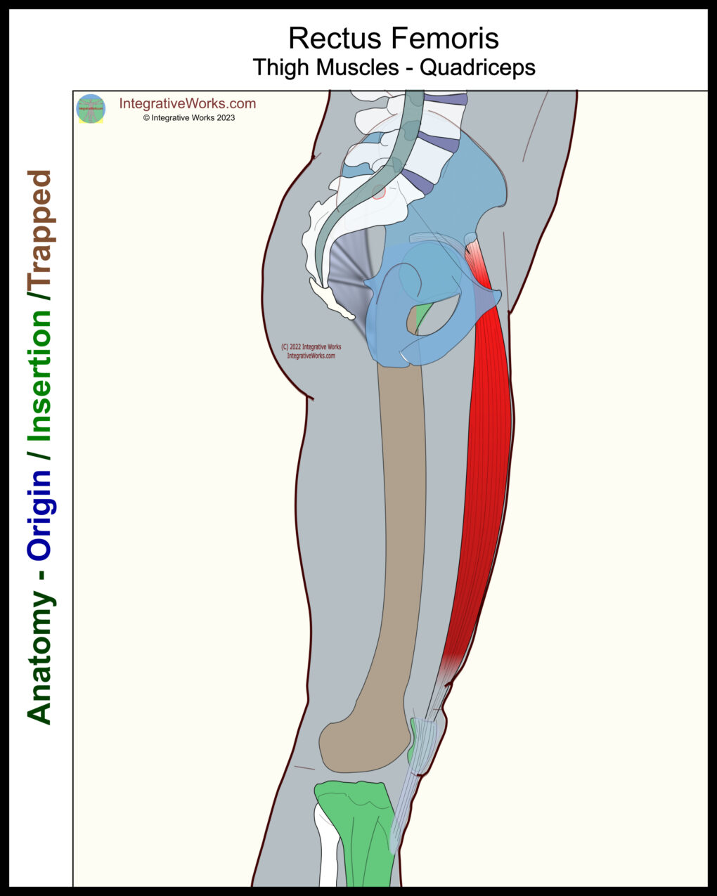
The rectus femoris is a superficial muscle on the anterior thigh. However, the functional anatomy of the rectus femoris can be more complex than expected. It is part of the quadriceps group. Notably, unlike the other quadriceps muscle, it attaches above the hip joint.
Origin
- Straight head – the anterior inferior iliac spine of the os coxae (ilium)
- Reflected head – supraacetabular groove
Insertion
- tibial tuberosity of the tibia via the patellar ligament
Function
- extension of the knee
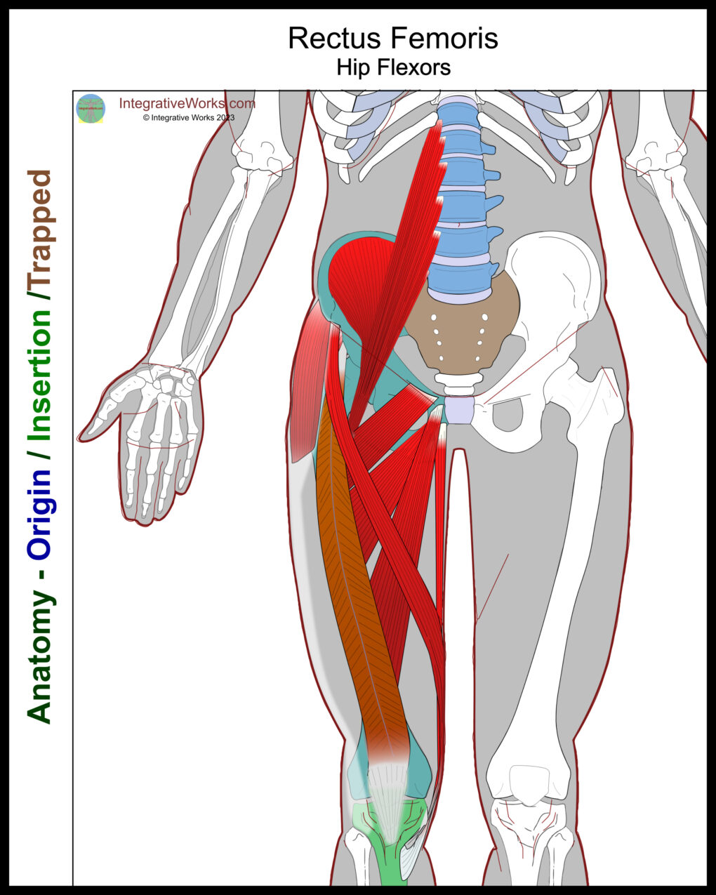
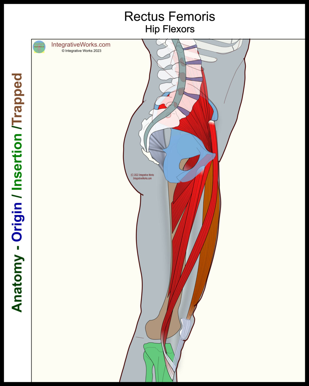
- flexion of the hip
Nerve
- the femoral nerve of the lumbar plexus (L2-L4)
Rectus Femoris Anatomy Evolution
The anatomy of the rectus femoris has evolved over the years. 30 years ago, it was seen as having a single head that may extend back toward the joint capsule. However, advancing technologies consistently defined more details of a second head and, occasionally, other variant heads. Many of those studies seem related to improving arthroscopy and surgical procedures. Also, later studies show more detail in separating the two main heads.
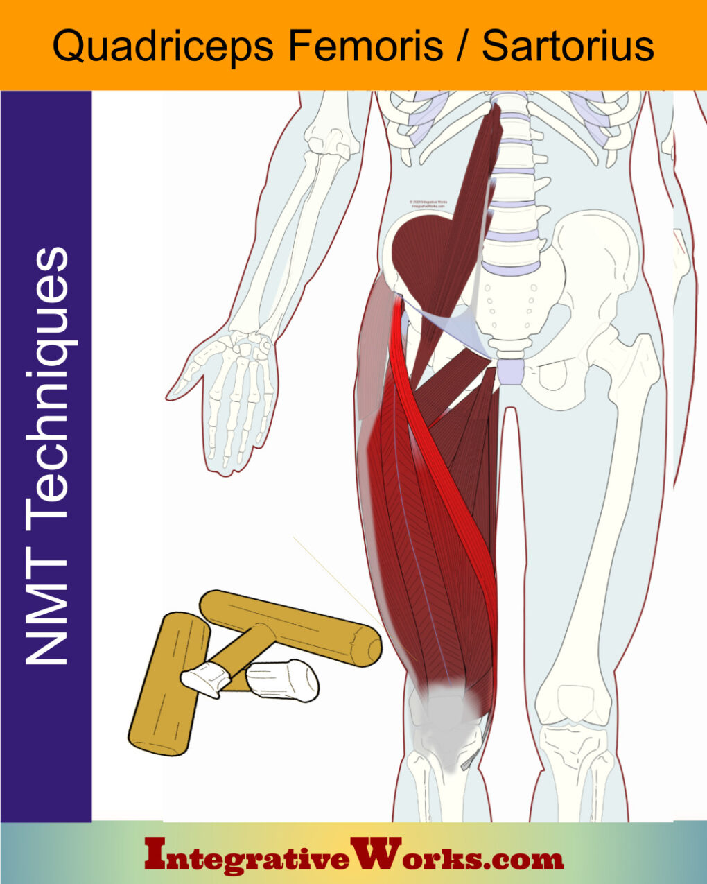
The superior portion lies deep to the tensor fascia lata and sartorius. Meanwhile, the main portion of the muscle lies just under the fascia lata, along the center of the anterior thigh.
Anatomy of the Main Heads
The first head attaches to the anterior inferior iliac spine. The tendon from this attachment extends into an aponeurosis along the proximal, anterior surface of the muscle.
The second reflected head has a short tendinous attachment that extends back to the upper rim of the acetabulum. Then, this tendon extends into the center of the muscle for about two-thirds of the muscle’s length.
Functional Considerations
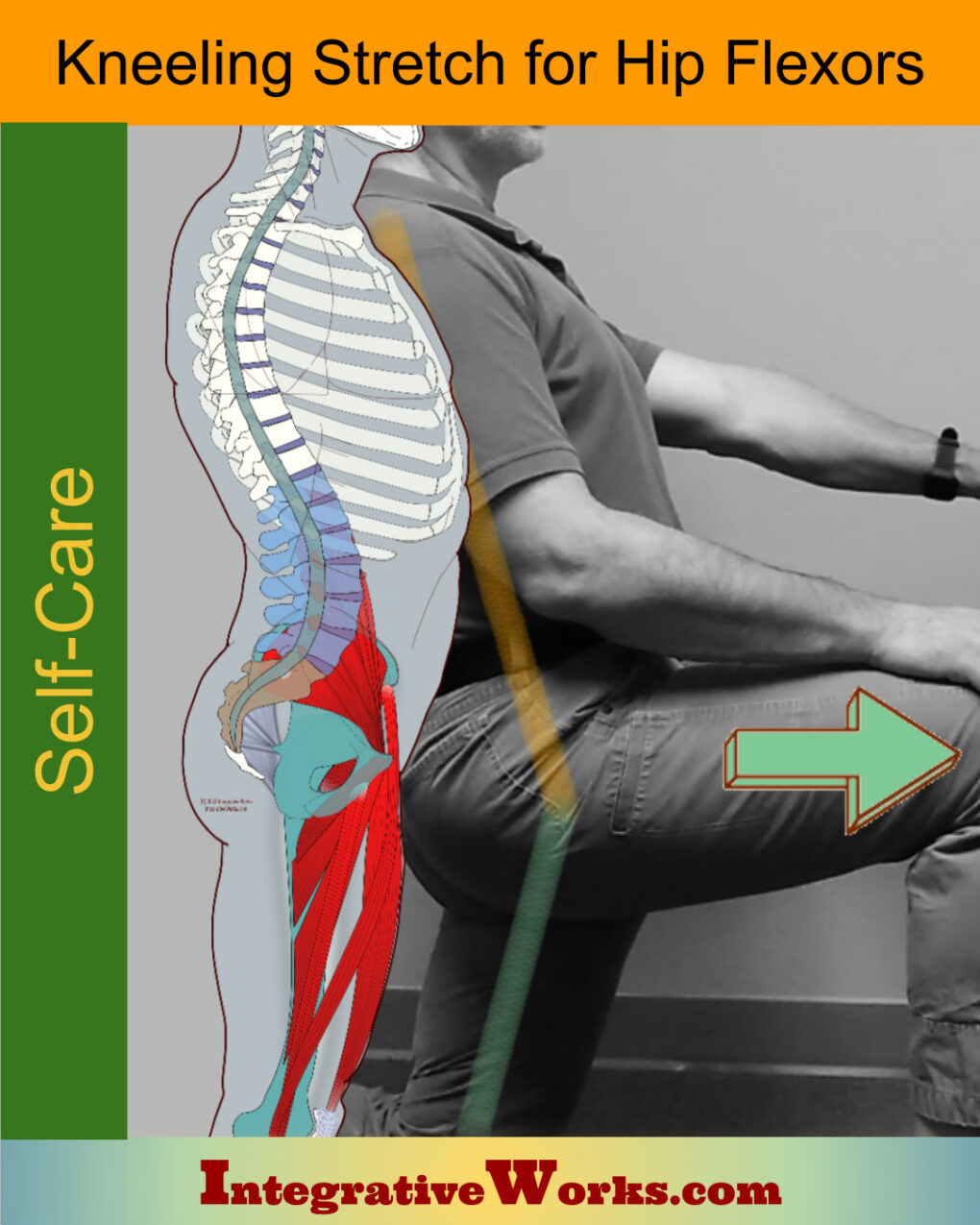
When shortened, the rectus femoris is effective as an antagonist of the hamstrings. This is a common factor in an anterior pelvic tilt.
The rectus femoris is a weak knee extender with the hip flexed. Conversely, it is an ineffective flexor of the hip with the knee extended.
Other studies
On the other hand, this study shows much stronger action potential in flexing the hip with the knee flexed. This is probably why the rectus femoris is known as the “soccer kick” muscle.
Another study shows the strongest potential at the beginning and end of knee and hip movement. Additionally, the same study shows that the rectus femoris potentials are greater in dorsal flexion of the foot and weaker in plantar flexion.
Anomalies, Etc.
Many studies refer to the anterior thigh muscles as having minimal variability. Additionally, most studies of variability cite one or two cases.
Studies Vary
However, studies like this one disagree. It reveals that a third head exists in 83% of cases. This muscle has inconsistencies, even within the same cadaver. It is a small head that attaches to the iliofemoral ligament and tendon of the gluteus minimus.
Another study suggests that variants include a slip of muscle to vastus lateralis. Additionally, it discusses variations in the attachment position on the anterior inferior iliac spine.
Some texts include the joint capsule of the hip as part of the origin. Also, some experts describe how the acetabular tendon attaches over the labrum, creating stress on the joint.
Related Posts
Support Integrative Works to
stay independent
and produce great content.
You can subscribe to our community on Patreon. You will get links to free content and access to exclusive content not seen on this site. In addition, we will be posting anatomy illustrations, treatment notes, and sections from our manuals not found on this site. Thank you so much for being so supportive.
Cranio Cradle Cup
This mug has classic, colorful illustrations of the craniosacral system and vault hold #3. It makes a great gift and conversation piece.
Tony Preston has a practice in Atlanta, Georgia, where he sees clients. He has written materials and instructed classes since the mid-90s. This includes anatomy, trigger points, cranial, and neuromuscular.
Question? Comment? Typo?
integrativeworks@gmail.com
Follow us on Instagram

*This site is undergoing significant changes. We are reformatting and expanding the posts to make them easier to read. The result will also be more accessible and include more patterns with better self-care. Meanwhile, there may be formatting, content presentation, and readability inconsistencies. Until we get older posts updated, please excuse our mess.

