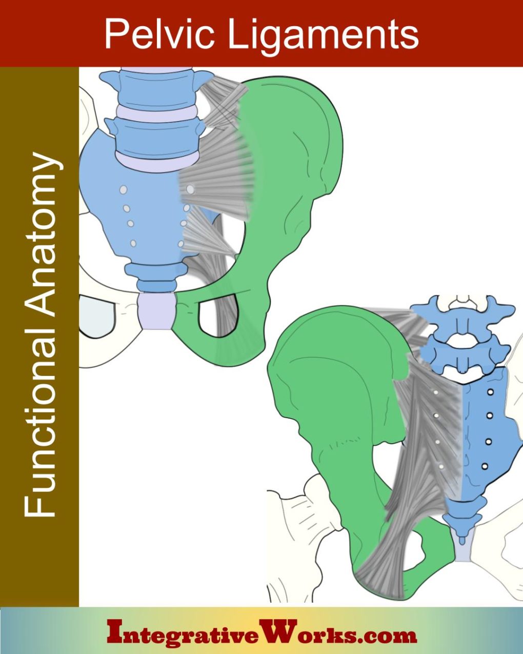
Here, I explore the anatomy of the pelvic ligaments, their structure, attachments, and how they mature through the decades of a person’s life.
Overview
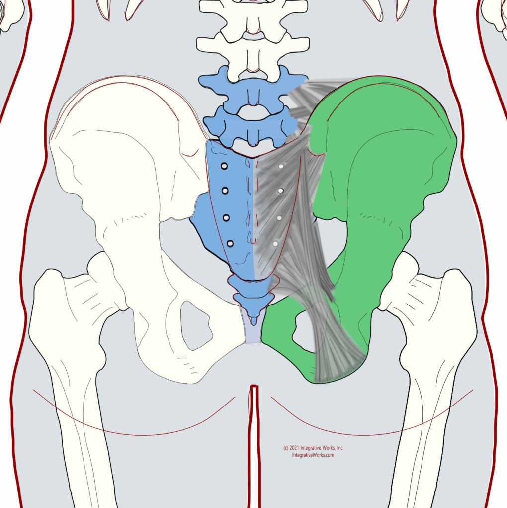
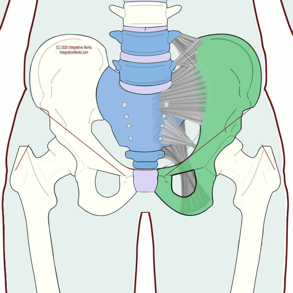
These pelvic ligaments bind the ox coxae of the lower extremity to the axial skeleton. They consist of:
- iliolumbar ligament
- sacroiliac ligament
- sacrotuberous ligament
- sacrospinous ligament
Progression of Fusion
A complex fascial structure wraps the major bony structures of the pelvis. Like those osseous structures, the ligaments also evolve as we age. As a result, we have more boney pieces held together by cartilaginous plates that fuse as decades pass in the first two or three decades. Eventually, in our fifth decade, pelvic joints, including the sacroiliac joint, often into one large pelvic structure. These ligaments also morph as the bones fuse.
It is larger in females than males.
In most illustrations, for clarity, they are shown as very separate structures. However, in reality, they appear more like thickened sections woven together into a larger whole.
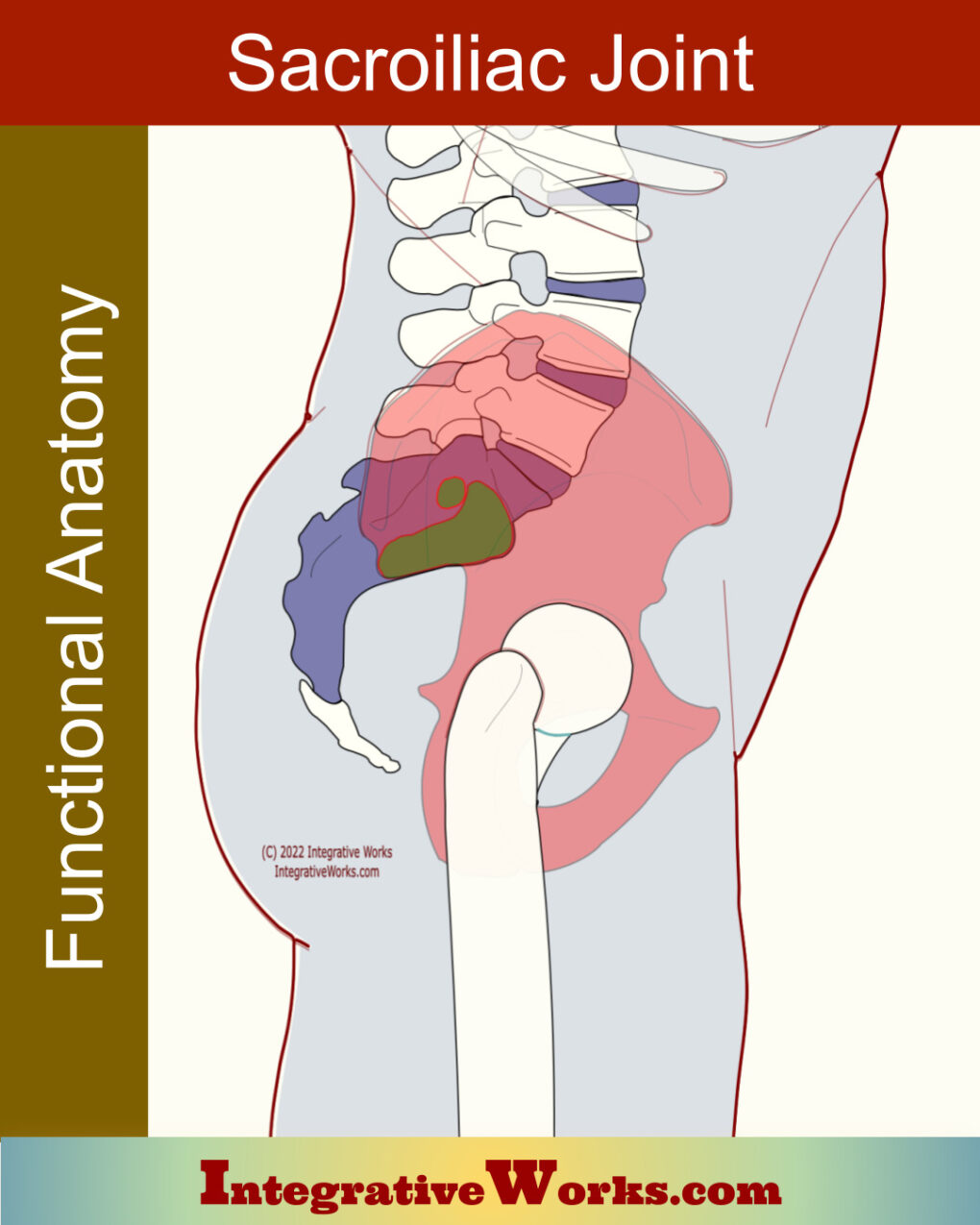
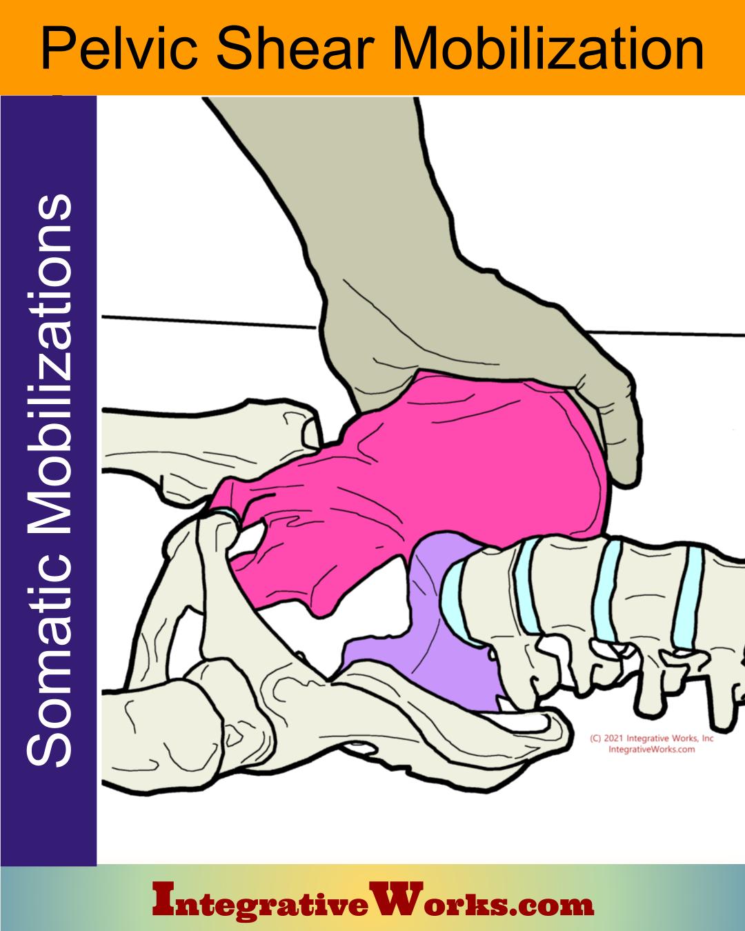
Sacroiliac Joint
Before looking at pelvic ligaments, review the complexities of the sacroiliac joint. It varies with gender, age, and race like several of the pelvic ligaments.
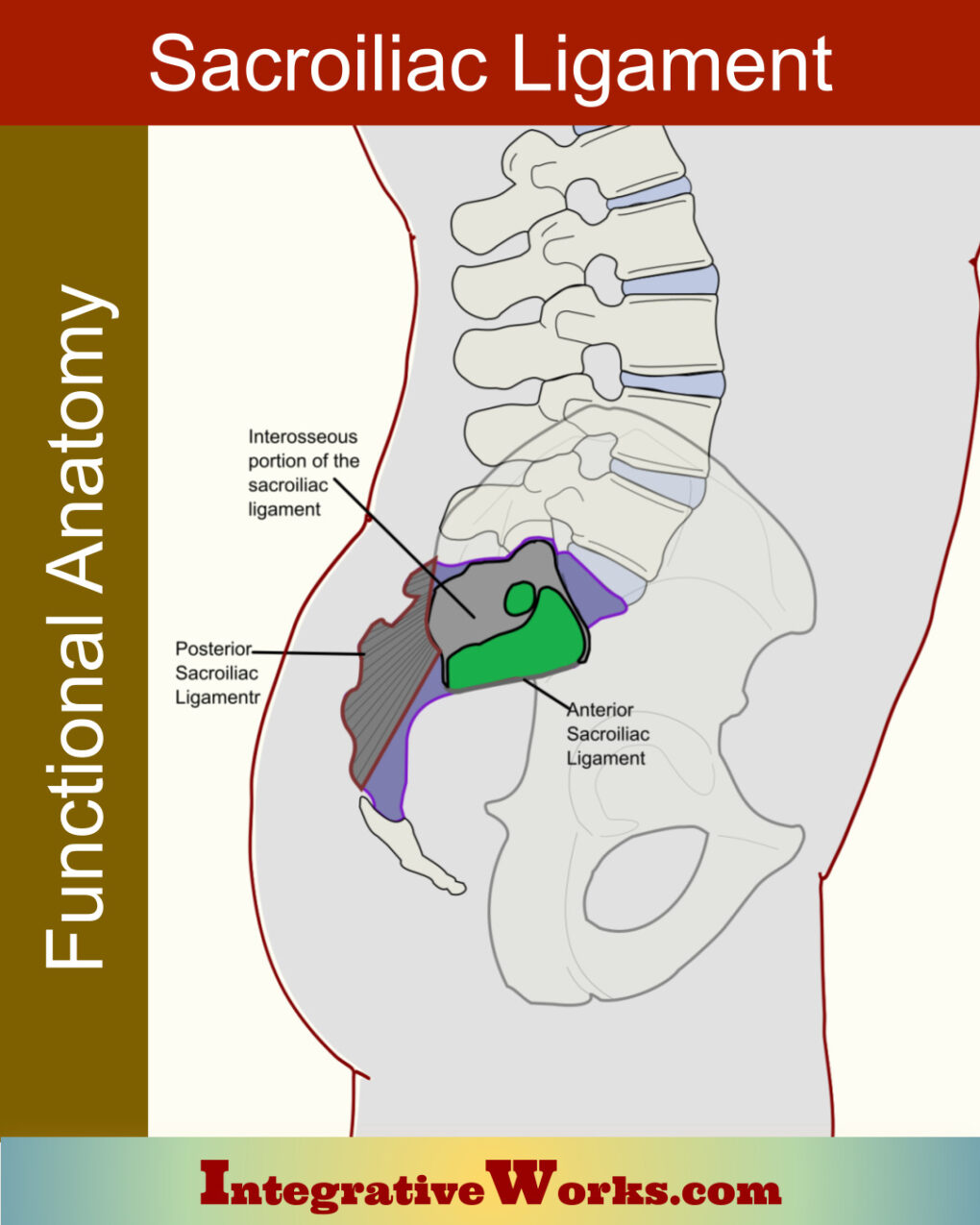
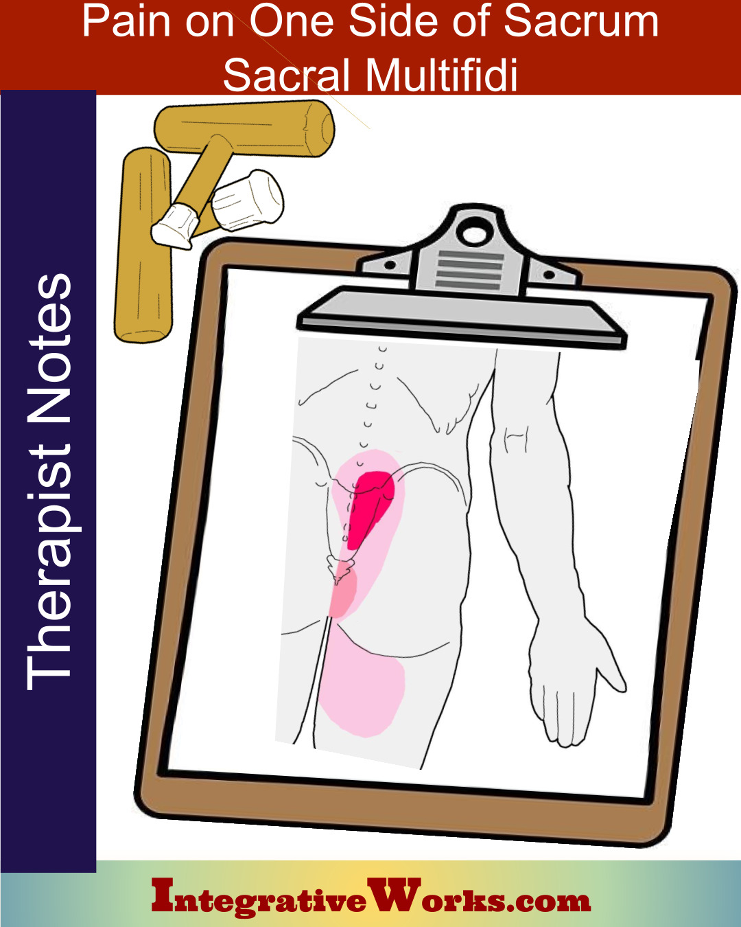
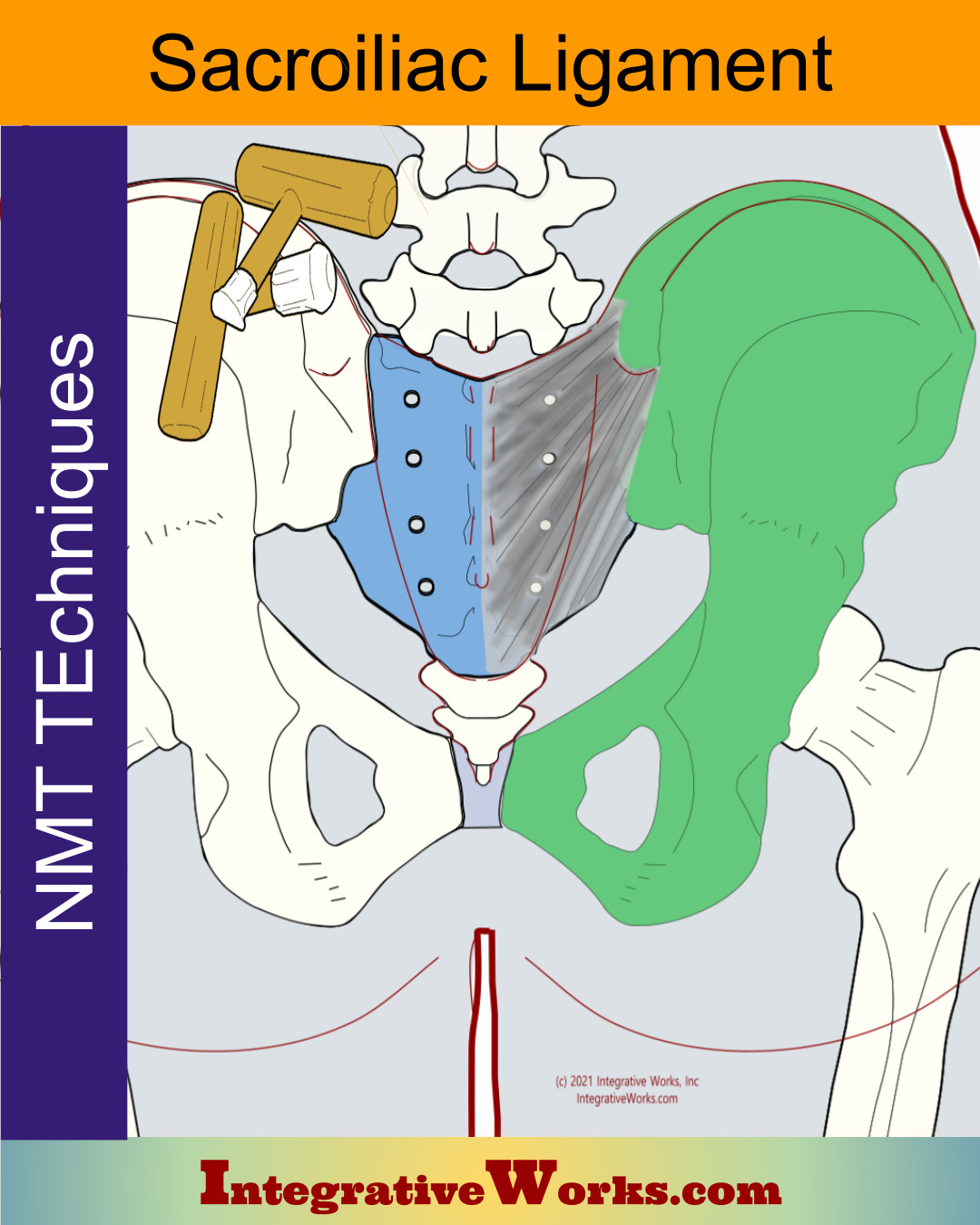
Sacroiliac Ligament
The most complex of the pelvic ligaments. The anterior, posterior, and interosseous sections all perform different functions. Also, each section has a notably different structure.
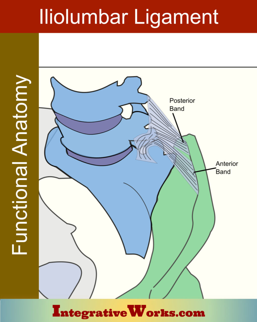
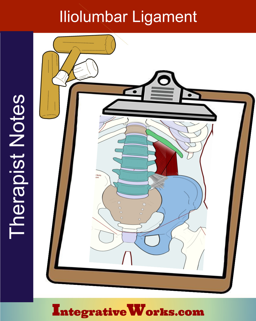
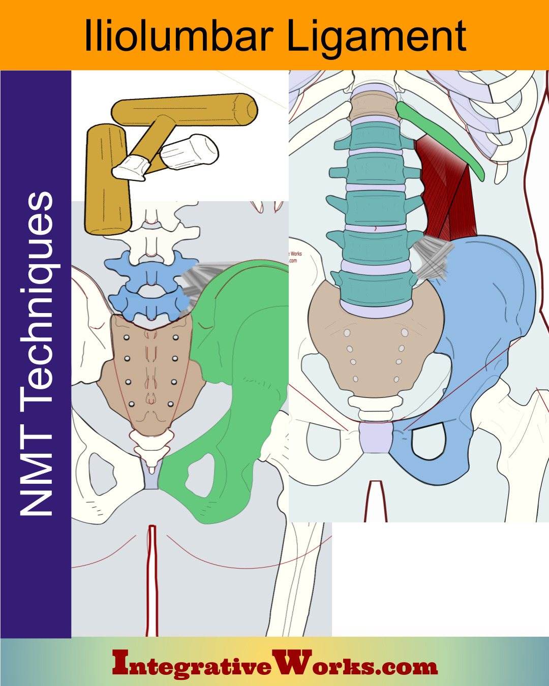
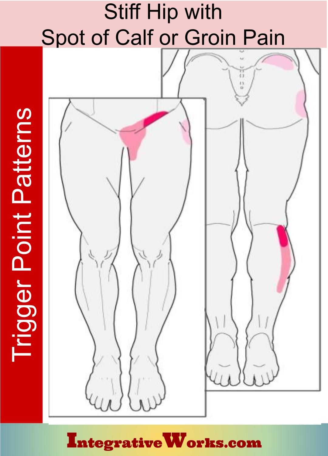
Iliolumbar Ligament
This ligament is unique to animals that support their weight on their caudal limbs. It varies by gender, race, and age. It is worth noting how those factors may impact your approach.
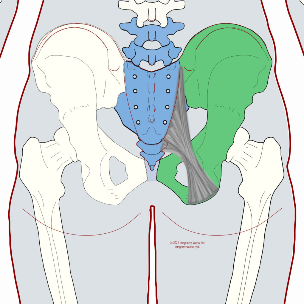
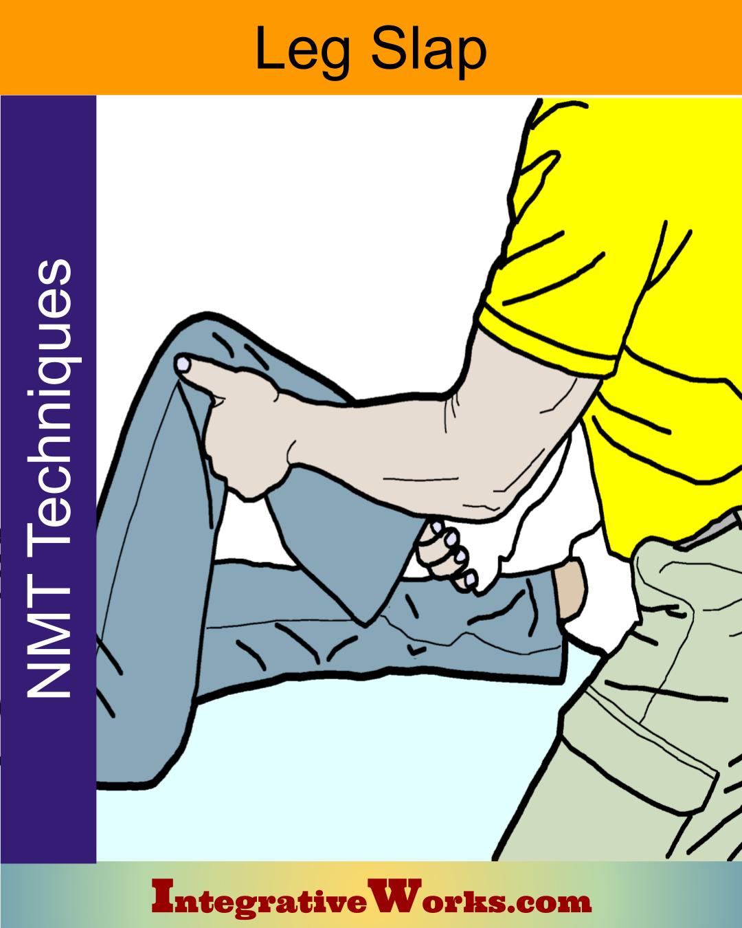
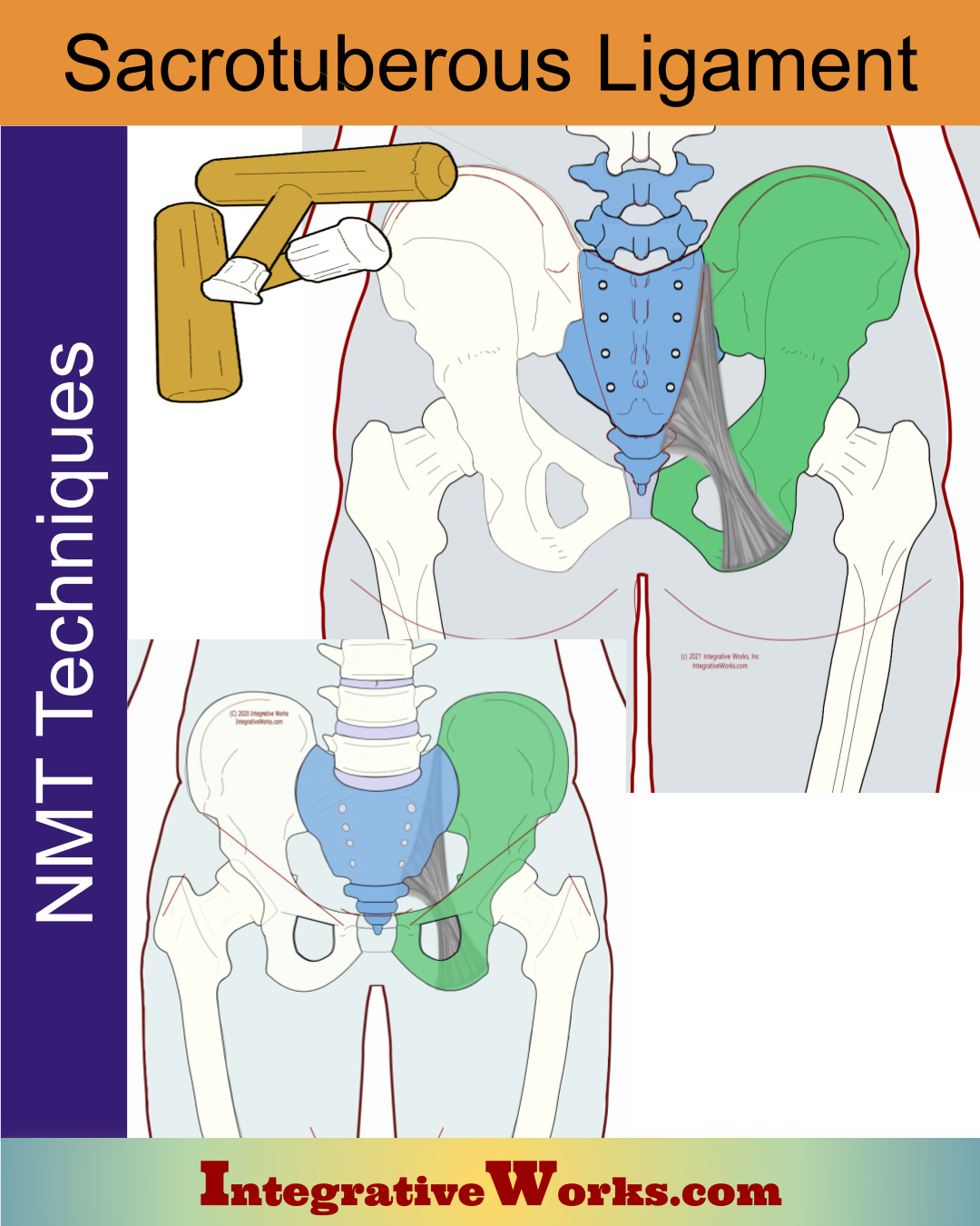
Sacrotuberous Ligament
The sacrotuberous ligament is attached broadly to the lateral aspect of the sacrum from the PSIS to the coccyx. The fibers converge and extend posteriorly to the ischial tuberosity. Fibers of this ligament pass over the ischial tuberosity to become the origin tendon of the biceps femoris.
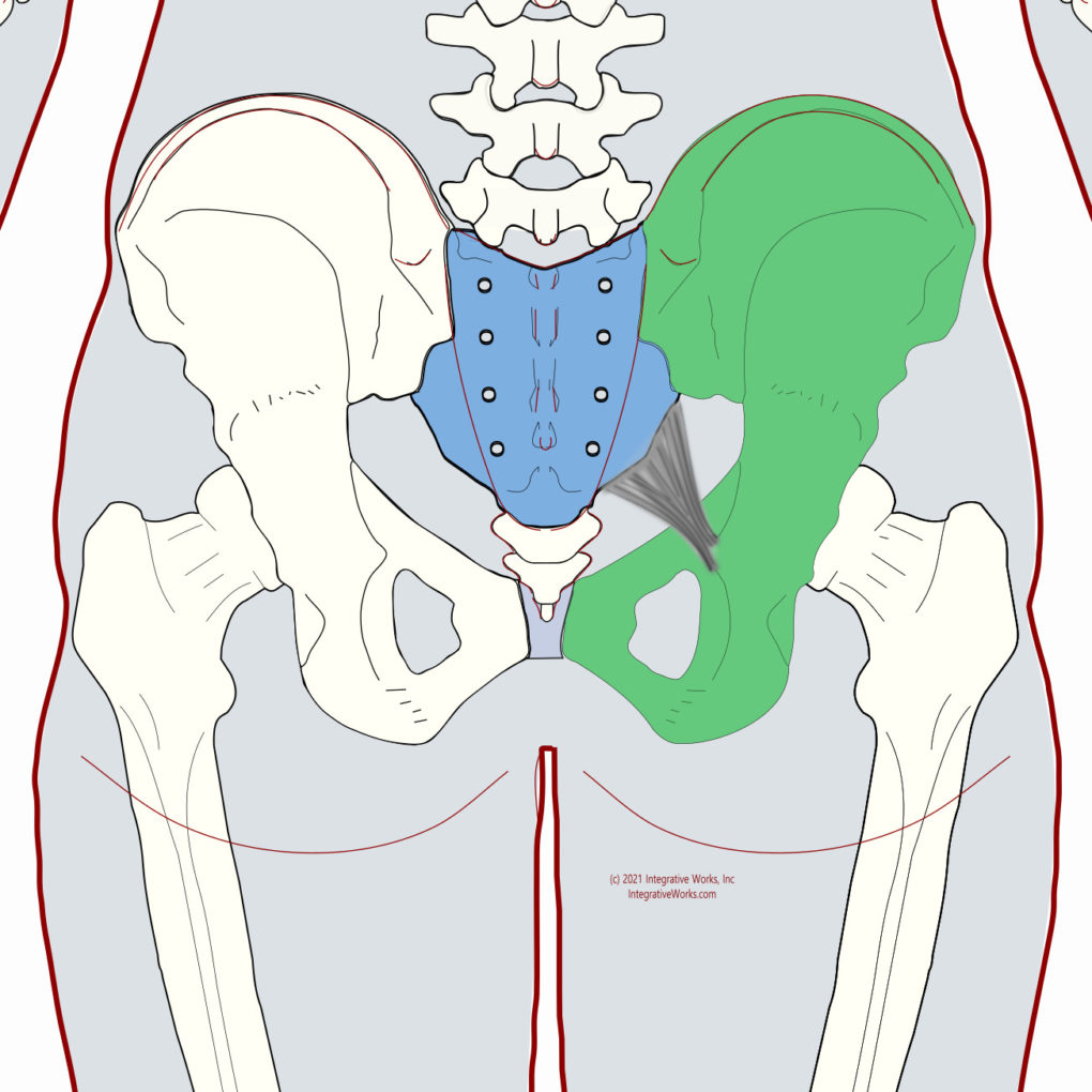
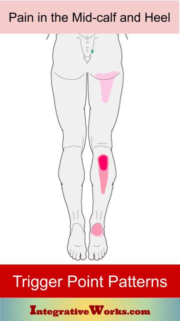
Sacrospinous Ligament
The sacrospinous ligament (small or anterior sacrosciatic ligament) is a thin, triangular ligament attached by its apex to the ischial spine and medially, by its broad base, to the lateral margins of the sacrum and coccyx, as it fuses with the sacrotuberous ligament. Its main function is to prevent posterior rotation of the ilium with relation to the sacrum. However, the laxity of this ligament, along with the sacrotuberous ligament, allows posterior rotation to occur.
Related Posts


Support Integrative Works to
stay independent
and produce great content.
You can subscribe to our community on Patreon. You will get links to free content and access to exclusive content not seen on this site. In addition, we will be posting anatomy illustrations, treatment notes, and sections from our manuals not found on this site. Thank you so much for being so supportive.
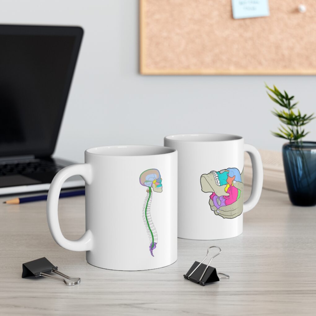
Cranio Cradle Cup
This mug has classic, colorful illustrations of the craniosacral system and vault hold #3. It makes a great gift and conversation piece.
Tony Preston has a practice in Atlanta, Georgia, where he sees clients. He has written materials and instructed classes since the mid-90s. This includes anatomy, trigger points, cranial, and neuromuscular.
Question? Comment? Typo?
integrativeworks@gmail.com
Follow us on Instagram

*This site is undergoing significant changes. We are reformatting and expanding the posts to make them easier to read. The result will also be more accessible and include more patterns with better self-care. Meanwhile, there may be formatting, content presentation, and readability inconsistencies. Until we get older posts updated, please excuse our mess.